Robert E. Marx - Oral Pathology in Clinical Dental Practice
Здесь есть возможность читать онлайн «Robert E. Marx - Oral Pathology in Clinical Dental Practice» — ознакомительный отрывок электронной книги совершенно бесплатно, а после прочтения отрывка купить полную версию. В некоторых случаях можно слушать аудио, скачать через торрент в формате fb2 и присутствует краткое содержание. Жанр: unrecognised, на английском языке. Описание произведения, (предисловие) а так же отзывы посетителей доступны на портале библиотеки ЛибКат.
- Название:Oral Pathology in Clinical Dental Practice
- Автор:
- Жанр:
- Год:неизвестен
- ISBN:нет данных
- Рейтинг книги:3 / 5. Голосов: 1
-
Избранное:Добавить в избранное
- Отзывы:
-
Ваша оценка:
Oral Pathology in Clinical Dental Practice: краткое содержание, описание и аннотация
Предлагаем к чтению аннотацию, описание, краткое содержание или предисловие (зависит от того, что написал сам автор книги «Oral Pathology in Clinical Dental Practice»). Если вы не нашли необходимую информацию о книге — напишите в комментариях, мы постараемся отыскать её.
Oral Pathology in Clinical Dental Practice — читать онлайн ознакомительный отрывок
Ниже представлен текст книги, разбитый по страницам. Система сохранения места последней прочитанной страницы, позволяет с удобством читать онлайн бесплатно книгу «Oral Pathology in Clinical Dental Practice», без необходимости каждый раз заново искать на чём Вы остановились. Поставьте закладку, и сможете в любой момент перейти на страницу, на которой закончили чтение.
Интервал:
Закладка:
2. African cutaneous Kaposi sarcoma: This type also occurs more commonly in men, specifically men 35 years or older, and is endemic in native black African men.
3. African lymphadenopathic Kaposi sarcoma: This type is endemic in black African children.
4. AIDS-related Kaposi sarcoma: This type is more common in adult men but also can occur in women and children.
Clinical features
The two most common types seen in the United States—classic Kaposi sarcoma and AIDS-related Kaposi sarcoma—appear as bluish-red submucosal or subdermal collections. Some will produce a soft tissue lobulated mass. The African cutaneous type is limited to the skin and is much more infiltrative, producing induration, and will spread rapidly in a proximal direction. The African lymphadenopathic type is a fulminant type involving lymph nodes, salivary glands, and internal organs, often leading to a rapid death.
Radiographic presentation
Usually none unless the mass invades bone; then it is usually only a superficial erosion.
Differential diagnosis
Many cases will mimic an area of ecchymosis from capillary fragility or platelet dysfunction. Those with a mass will suggest an angiosarcoma, rhabdomyosarcoma, hemangioma, lymphangioma, or a low-grade mucoepidermoid carcinoma.
Microscopic features
With some variability, there will be numerous blood-filled channels appearing like slits between spindle cells.
Suggested course of action
Refer to a cancer center or to a medical oncologist for workup.
Treatment
Isolated lesions are treated with intralesional injections of vinblastine 0.1 to 0.5 mg/mL or radiotherapy 1,800 to 2,400 cGy. Systemic treatment is via chemotherapy using vinblastine, etoposide, bleomycin, doxorubicin, and interferon alpha-2 as an adjunct.
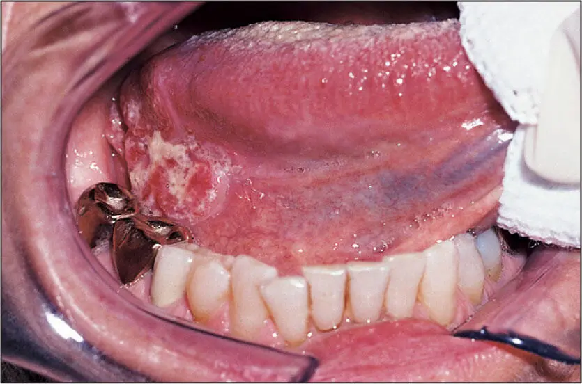
 Squamous cell carcinoma of the lateral border of the tongue.
Oral Squamous Cell Carcinoma
Nature of disease
An invasive epithelial malignancy arising from the basal cells of oral mucosa that may undergo a partial squamous differentiation. Many also develop the ability to metastasize to regional lymph nodes and a few others to distant organs such as the lungs via a vascular route.
Predilections
Adults, with a modest male predilection. More common on the lateral border of the tongue and floor of the mouth. No racial predilection is known. Since 2000, there is a trend toward younger adults (30 to 50 years) and an increased involvement of the tongue, as well as an increased incidence in patients who have never smoked.
Clinical features
May variably appear as a leukoplakia, erythroleukoplakia, erythroplakia, ulcer, verrucoid tissue mass, or indurated mass. Pain may be present but is not common. It may be associated with a palpable painless lymphadenopathy.
Radiographic presentation
If the squamous cell carcinoma invades into bone, it will be seen as an osteolytic area. Extensive invasion may cause a pathologic fracture.
Differential diagnosis
The entities that may be confused with oral squamous cell carcinoma include benign hyperkeratosis, epithelial dysplasias, carcinoma in situ, verrucous carcinoma, lichen planus, and candidiasis.
Microscopic features
Sheets and cords of dysplastic epithelial cells with pleomorphic nuclei, a high nuclear to cytoplasmic ratio, and some mitotic figures. These cells will be seen infiltrating the normal structures below the basement membrane. At times, keratin pearls will be seen. The infiltrations are frequently accompanied by inflammatory cells as well.
Suggested course of action
Note and document the size of the lesion in centimeters. Palpate the neck and document the findings (ie, number, approximate size, and anatomical location of palpable lymph nodes) and then biopsy. Or refer to an oral and maxillofacial surgeon for workup, biopsy, staging, and treatment.
Treatment
Most squamous cell carcinomas are treated with surgery to remove the primary tumor along with a neck dissection of one of many possible types. Lower-staged carcinomas may not require neck dissection. More advanced–staged carcinomas will be followed by radiation therapy or a chemoradiation therapy protocol.
Конец ознакомительного фрагмента.
Текст предоставлен ООО «ЛитРес».
Прочитайте эту книгу целиком, купив полную легальную версию на ЛитРес.
Безопасно оплатить книгу можно банковской картой Visa, MasterCard, Maestro, со счета мобильного телефона, с платежного терминала, в салоне МТС или Связной, через PayPal, WebMoney, Яндекс.Деньги, QIWI Кошелек, бонусными картами или другим удобным Вам способом.
Интервал:
Закладка:
Похожие книги на «Oral Pathology in Clinical Dental Practice»
Представляем Вашему вниманию похожие книги на «Oral Pathology in Clinical Dental Practice» списком для выбора. Мы отобрали схожую по названию и смыслу литературу в надежде предоставить читателям больше вариантов отыскать новые, интересные, ещё непрочитанные произведения.


Обсуждение, отзывы о книге «Oral Pathology in Clinical Dental Practice» и просто собственные мнения читателей. Оставьте ваши комментарии, напишите, что Вы думаете о произведении, его смысле или главных героях. Укажите что конкретно понравилось, а что нет, и почему Вы так считаете.