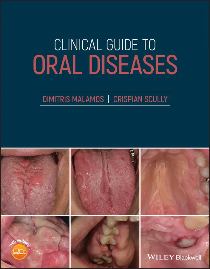
Crispian Scully - Clinical Guide to Oral Diseases
Здесь есть возможность читать онлайн «Crispian Scully - Clinical Guide to Oral Diseases» — ознакомительный отрывок электронной книги совершенно бесплатно, а после прочтения отрывка купить полную версию. В некоторых случаях можно слушать аудио, скачать через торрент в формате fb2 и присутствует краткое содержание. Жанр: unrecognised, на английском языке. Описание произведения, (предисловие) а так же отзывы посетителей доступны на портале библиотеки ЛибКат.
- Название:Clinical Guide to Oral Diseases
- Автор:
- Жанр:
- Год:неизвестен
- ISBN:нет данных
- Рейтинг книги:4 / 5. Голосов: 1
-
Избранное:Добавить в избранное
- Отзывы:
-
Ваша оценка:
Clinical Guide to Oral Diseases: краткое содержание, описание и аннотация
Предлагаем к чтению аннотацию, описание, краткое содержание или предисловие (зависит от того, что написал сам автор книги «Clinical Guide to Oral Diseases»). Если вы не нашли необходимую информацию о книге — напишите в комментариях, мы постараемся отыскать её.
Provides nearly 300 high-quality clinical photos and relevant questions to help lead readers to the proper diagnosis of common oral diseases Contains concise tables relevant to each chapter with a list of common oral lesions and conditions Offers MCQs of varying levels of difficulty to help readers test their knowledge in Oral Medicine Includes clinical flow charts according to the location and duration of oral lesions Incorporates the ICD-10 Codes of oral lesions and diseases
is a valuable reference for general dental and medical practitioners, undergraduate dental students, and postgraduate trainees in oral and maxillofacial surgery, oral medicine, oral pathology, periodontology as well as general pathology, dermatology or head and neck oncology.
Clinical Guide to Oral Diseases — читать онлайн ознакомительный отрывок
Ниже представлен текст книги, разбитый по страницам. Система сохранения места последней прочитанной страницы, позволяет с удобством читать онлайн бесплатно книгу «Clinical Guide to Oral Diseases», без необходимости каждый раз заново искать на чём Вы остановились. Поставьте закладку, и сможете в любой момент перейти на страницу, на которой закончили чтение.
Интервал:
Закладка:
Table of Contents
1 Cover
2 Title Page Clinical Guide to Oral Diseases Dimitris Malamos DDS, MSC, PhD, Dip. O.M Oral Medicine Specialist Diplomat of the European Association of Oral Medicine Head of the Oral Medicine Clinic at the National Organization for the Provision of Health Services 1st Health Region and at Iatrokosmos Athens, Greece Crispian Scully CBE, DSc, DChD, DMed (HC), Dhc (Multi), MD, PhD, PhD (HC), FMedSci, MDS, MRCS, BSc, FDSRCS, FDSRCPS, FFDRCSI, FDSRCSEd FRCPath, FHEA Emeritus Professor University College London, UK Content Editor Márcio Diniz Freitas Special Care Dentistry Unit University of Santiago de Compostela Spain
3 Copyright Page
4 Preface
5 Foreword
6 Acknowledgment
7 About the Companion Website
8 Section I 1 Bleeding 2 Blue and/or Black Lesions 3 Brown Lesions 4 Malodor 5 Muscle Deficits (Trismus/Paralysis) 6 Orofacial Pain 7 Red Lesions 8 Saliva Disturbances (Xerostomia/Sialorrhea) 9 Swellings (Diffuse/Lumps) 10 Taste Deficits 11 Ulcerations 12 Vesiculobullous Lesions 13 White Lesions 14 Yellow Lesions
9 Section II 15 Buccal Mucosa 16 Floor of the Mouth 17 Gingivae 18 Jaws 19 Lips 20 Neck 21 Palate 22 Salivary Glands (Minor/Major) 23 Teeth 23.1 Part A: Teeth Anomalies Related to their Number, Size, and Shape 23.2 Part B: Disorders of Teeth Structures 23.3 Part C: Diseases Affecting the Teeth's Components in Relation to the Adjacent Tissues 24 Tongue
10 Section III 25 Normal Variations 26 Oral Lesions According to Patient's Age 27 Clinical Tests, Signs and Phenomena
11 Abbreviations
12 Diagnostic Flow Charts According to the Location of Oral Lesions
13 Appendix: ICD‐10 Codes of Oral Diseases/Lesions
14 Index
15 End User License Agreement
List of Tables
1 Chapter 1 Table 1 Conditions related to oral bleeding.
2 Chapter 2 Table 2 The most common causes of blue and black lesions.
3 Chapter 3 Table 3 The most common oral brown lesions.
4 Chapter 4 Table 4 Common and important conditions associated with malodor
5 Chapter 5 Table 5 Common and important conditions related to muscle dysfunctions.
6 Chapter 6Table 6 Common and important conditions in orofacial pain.
7 Chapter 7Table 7 The common causes of oral red lesions.
8 Chapter 8Table 8.1 Drooling and sialorrhea: Common and important conditions.Table 8.2 Dryness: common and important conditions.
9 Chapter 9Table 9 The most important causes of swellings and lumps.
10 Chapter 10Table 10 The most important causes of taste changes.
11 Chapter 11Table 11 Ulcers: Common and important causes.
12 Chapter 12Table 12 Common and important vesiculobullous conditions.
13 Chapter 13Table 13 Common and important conditions of white lesions.
14 Chapter 14Table 14 Common and important conditions of yellow lesions.
15 Chapter 15Table 15 Buccal mucosa: Common and important lesions.
16 Chapter 16Table 16 Floor of the mouth: common and important lesions and conditions.
17 Chapter 17Table 17 Gingivae lesions: common and important conditions.
18 Chapter 18Table 18 Common and important conditions of occurring in the jaws.
19 Chapter 19Table 19 Common and important conditions of the lips.
20 Chapter 20Table 20 Neck: common and important lesions.
21 Chapter 21Table 21 Palate lesions.
22 Chapter 22Table 22 Common and important salivary gland lesions and diseases.
23 Chapter 23Table 23 Teeth: common and important changes.
24 Chapter 24Table 24 Common and important lesions of the tongue.
25 Chapter 25Table 25 Normal oral variations according to location.
26 Chapter 26Table 26 Common and important lesions according to patient's age.
27 Chapter 27Table 27 Clinical tests, signs, and phenomena commonly seen in oral medicine ...
List of Illustrations
1 Chapter 1 Figure 1.0 Tongue hematoma in a woman with seizures. Figure 1.1a Figure 1.1b Figure 1.2 Figure 1.3 Figure 1.4 Figure 1.5 Figure 1.6a Figure 1.6b Figure 1.7 Figure 1.8 Figure 1.9 Figure 1.10
2 Chapter 2 Figure 2.0a Blue lesion. Figure 2.0b Black lesion. Figure 2.1 Figure 2.2a Figure 2.2b Figure 2.3 Figure 2.4 Figure 2.5 Figure 2.6 Figure 2.7 Figure 2.8 Figure 2.9 Figure 2.10
3 Chapter 3 Figure 3.0a Brown discoloration of neck skin after radiation. Figure 3.0b Hydroxyurea‐induced oral brown pigmentation. Figure 3.1 Figure 3.2a Figure 3.2b Figure 3.3 Figure 3.4 Figure 3.5 Figure 3.6 Figure 3.7 Figure 3.8 Figure 3.9 Figure 3.10a Figure 3.10b
4 Chapter 4 Figure 4.0a Halitosis from a patient with advanced oro‐nasal carcinoma. Figure 4.0b Halitosis from a patient with a neglected mouth. Figure 4.1 Figure 4.2 Figure 4.3 Figure 4.4 Figure 4.5 Figure 4.6 Figure 4.7 Figure 4.8 Figure 4.9 Figure 4.10
5 Chapter 5 Figure 5.0a Trismus. Figure 5.0b Facial palsy. Figure 5.1 Figure 5.2 Figure 5.3 Figure 5.4 Figure 5.5 Figure 5.6Figure 5.7 Figure 5.8 Figure 5.9 Figure 5.10
6 Chapter 6Figure 6.0 Pain from the ulcerated oral mucosa of a patient with graft‐versu...Figure 6.1 Figure 6.2 Figure 6.3 Figure 6.4 Figure 6.5 Figure 6.6 Figure 6.7Figure 6.8 Figure 6.9 Figure 6.10
7 Chapter 7Figure 7.0 Erythematous candidiasis in the palate: induced by constant wear ...Figure 7.1 Figure 7.2 Figure 7.3 Figure 7.4 Figure 7.5 Figure 7.6 Figure 7.7 Figure 7.8 Figure 7.9 Figure 7.10
8 Chapter 8Figure 8.0a Increased saliva in a patient with graft versus host disease (Gv...Figure 8.0b Xerostomia.Figure 8.1 Figure 8.2 Figure 8.3 Figure 8.4 Figure 8.5 Figure 8.6 Figure 8.7 Figure 8.8 Figure 8.9 Figure 8.10
9 Chapter 9Figure 9.0 Traumatic fibroma.Figure 9.1 Figure 9.2a Figure 9.2b Figure 9.3 Figure 9.4 Figure 9.5 Figure 9.6 Figure 9.7 Figure 9.8 Figure 9.9 Figure 9.10
10 Chapter 10Figure 10.0 Testing sour taste with drops of lemon.Figure 10.1 Figure 10.2 Figure 10.3 Figure 10.4 Figure 10.5 Figure 10.6 Figure 10.7 Figure 10.8 Figure 10.9 Figure 10.10
11 Chapter 11Figure 11.0 Traumatic tongue ulcer in a healing process.Figure 11.1 Figure 11.2 Figure 11.3 Figure 11.4 Figure 11.5 Figure 11.6 Figure 11.7 Figure 11.8 Figure 11.9 Figure 11.10a Figure 11.10b
12 Chapter 12Figure 12.0 Intact bulla on the inner surface of the lower lip in a patient ...Figure 12.1 Figure 12.2a Figure 12.2b Figure 12.3 Figure 12.4 Figure 12.5 Figure 12.6a Figure 12.6b Figure 12.7a Figure 12.7b Figure 12.8a Figure 12.8b Figure 12.9a Figure 12.9b Figure 12.10
13 Chapter 13Figure 13.0 White plaque on the dorsum and lateral left margin of the tongue...Figure 13.1 Figure 13.2 Figure 13.3 Figure 13.4 Figure 13.5 Figure 13.6 Figure 13.7 Figure 13.8 Figure 13.9 Figure 13.10
14 Chapter 14Figure 14.0 Aphtha.Figure 14.1 Figure 14.2a Figure 14.2b Figure 14.3 Figure 14.4 Figure 14.5 Figure 14.6 Figure 14.7 Figure 14.8 Figure 14.9 Figure 14.10
15 Chapter 15Figure 15.0a Leukoedema.Figure 15.0b Oral carcinoma.Figure 15.1 Figure 15.2 Figure 15.3 Figure 15.4 Figure 15.5
16 Chapter 16Figure 16.0 Trauma of the duct of the right sublingual gland.Figure 16.1 Figure 16.2 Figure 16.3 Figure 16.4 Figure 16.5
17 Chapter 17Figure 17.0 Materia alba on the anterior lower gingivae of a patient receivi...Figure 17.1a Figure 17.1b Figure 17.2 Figure 17.3 Figure 17.4 Figure 17.5
18 Chapter 18Figure 18.0a Enlargement of the mandible from an odontogenic tumor (adenomat...Figure 18.0b Enlargement of the mandible from non‐odontogenic tumor (multipl...Figure 18.1 Figure 18.2 Figure 18.3 Figure 18.4a Figure 18.4b Figure 18.4c Figure 18.4d Figure 18.5a Figure 18.5b
19 Chapter 19Figure 19.0 Fordyce spots on the vermilion border of the upper lip.Figure 19.1a Figure 19.1b Figure 19.2a Figure 19.2bFigure 19.3 Figure 19.4a Figure 19.4bFigure 19.5
20 Chapter 20Figure 20.0 Lipoma of the neck.Figure 20.1 Figure 20.2 Figure 20.3 Figure 20.4 Figure 20.5
Читать дальшеИнтервал:
Закладка:
Похожие книги на «Clinical Guide to Oral Diseases»
Представляем Вашему вниманию похожие книги на «Clinical Guide to Oral Diseases» списком для выбора. Мы отобрали схожую по названию и смыслу литературу в надежде предоставить читателям больше вариантов отыскать новые, интересные, ещё непрочитанные произведения.


Обсуждение, отзывы о книге «Clinical Guide to Oral Diseases» и просто собственные мнения читателей. Оставьте ваши комментарии, напишите, что Вы думаете о произведении, его смысле или главных героях. Укажите что конкретно понравилось, а что нет, и почему Вы так считаете.