Crispian Scully - Clinical Guide to Oral Diseases
Здесь есть возможность читать онлайн «Crispian Scully - Clinical Guide to Oral Diseases» — ознакомительный отрывок электронной книги совершенно бесплатно, а после прочтения отрывка купить полную версию. В некоторых случаях можно слушать аудио, скачать через торрент в формате fb2 и присутствует краткое содержание. Жанр: unrecognised, на английском языке. Описание произведения, (предисловие) а так же отзывы посетителей доступны на портале библиотеки ЛибКат.
- Название:Clinical Guide to Oral Diseases
- Автор:
- Жанр:
- Год:неизвестен
- ISBN:нет данных
- Рейтинг книги:4 / 5. Голосов: 1
-
Избранное:Добавить в избранное
- Отзывы:
-
Ваша оценка:
Clinical Guide to Oral Diseases: краткое содержание, описание и аннотация
Предлагаем к чтению аннотацию, описание, краткое содержание или предисловие (зависит от того, что написал сам автор книги «Clinical Guide to Oral Diseases»). Если вы не нашли необходимую информацию о книге — напишите в комментариях, мы постараемся отыскать её.
Provides nearly 300 high-quality clinical photos and relevant questions to help lead readers to the proper diagnosis of common oral diseases Contains concise tables relevant to each chapter with a list of common oral lesions and conditions Offers MCQs of varying levels of difficulty to help readers test their knowledge in Oral Medicine Includes clinical flow charts according to the location and duration of oral lesions Incorporates the ICD-10 Codes of oral lesions and diseases
is a valuable reference for general dental and medical practitioners, undergraduate dental students, and postgraduate trainees in oral and maxillofacial surgery, oral medicine, oral pathology, periodontology as well as general pathology, dermatology or head and neck oncology.
Clinical Guide to Oral Diseases — читать онлайн ознакомительный отрывок
Ниже представлен текст книги, разбитый по страницам. Система сохранения места последней прочитанной страницы, позволяет с удобством читать онлайн бесплатно книгу «Clinical Guide to Oral Diseases», без необходимости каждый раз заново искать на чём Вы остановились. Поставьте закладку, и сможете в любой момент перейти на страницу, на которой закончили чтение.
Интервал:
Закладка:
3 Peripheral giant cell granuloma
4 Gingival hemangioma
5 Peripheral ossifying fibroma
Answers:
1 No
2 Pregnancy epulis is a localized hyperplastic hemorrhagic soft lesion on the upper and lower gingivae of pregnant women with decayed teeth and poor oral hygiene. The lesion grows slowly and reaches its largest size during the last trimester of pregnancy.
3 No
4 No
5 No
Comments: In contrary to pregnancy epulis the gingival hemangiomas are found earlier (at childhood); sarcoma Kaposi are usually associated with lymphadenopathy and have an aggressive course. The peripheral odontogenic fibroma has a firmer feel on palpation, while the peripheral giant cell epulis does not improve with the baby's birth and is associated with endocrinopathies.
Q2Which are the other oral conditions seen during pregnancy?
1 Melasma
2 Pregnancy gingivitis
3 Increased risk of caries
4 Erosions of teeth
5 Sialorrhea
Answers:
1 No
2 Pregnancy gingivitis is the commonest complication of pregnancy and can start even from the second month, reaching its peak on the eight month of pregnancy. This type of gingivitis is due rather to the action of increased female hormones on their gingival receptors rather than to microbial plaque.
3 Pregnant women tend to be at increased risk of caries as the number of cariogenic bacteria in the mouth, and the frequency of eating, especially sweet food as a means of coping with nausea, are increased.
4 Erosions on the palatal tooth surface and especially on the upper anterior teeth are common and are also attributed to the acidity of gastric juice that reaches the mouth during vomiting.
5 Sialorrhea is a common finding in pregnant women and caused by the increased nausea and vomiting recorded during their pregnancy.
Comments: Melasma or pregnancy mask as it is known, is characterized by a brown discoloration of the facial skin and lips, but is never seen within the mouth of pregnant women and those taking contraceptives or hormone replacement medications.
Q3Which conditions have been detected in babies, related to the periodontal status of their mothers?
1 Premature birth
2 Low weight
3 Vision or hearing deficits
4 Mental retardation
5 Dental anomalies
Answers:
1 Women with chronic inflammation of their gingivae seem to produce a number of inflammatory cytokines, some of which are responsible for the uterine muscle contractions which finally induce early labor.
2 Premature babies show incomplete growth and low weight.
3 Vision or hearing deficits are commonly seen in premature babies whose early birth may be associated with the periodontal problems of their mother.
4 No
5 No
Comments: The mental status of pregnant women may worsen their periodontal problems by increased secretion of cortisol and refusal of tooth brushing, while the periodontitis per se does not affect the mental status or dentition of their children.
Case 1.4
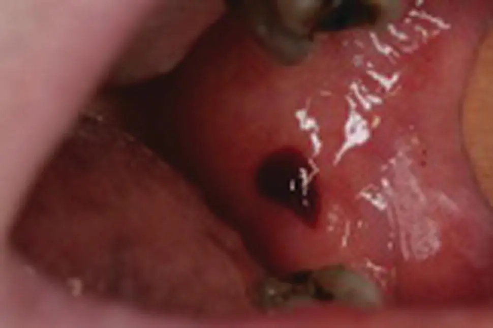
Figure 1.4
CO: A 42‐year‐old woman came in with a hemorrhagic bulla inside her cheek.
HPC: The bulla appeared three hours ago after eating a sandwich. No similar bullae were recorded in her or her close relatives in the past.
PMH: From her medical records a few episodes of allergic rhinitis controlled with antihistamine and steroids in crisis were recorded. No blood and other systemic diseases, other allergies or drug uptakes were recorded.
OE: Examination revealed a large bulla with hemorrhagic content on the left buccal mucosa at the occlusion level ( Figure 1.4). The bulla appeared during mastication and was easily broken during the examination's manipulations, leaving a painful superficial ulceration. No other bullae, ulcerations or petechiae and ecchymoses were seen on the oral and other mucosae or skin. Cervical lymphadenopathy was not seen.
Q1Which is the disease responsible for this bulla?
1 Thrombocytopenia
2 Burns
3 Mucous membrane pemphigoid
4 Angina bullosa hemorrhagica
5 Hemorrhagic mucocele
Answers:
1 No
2 No
3 No
4 Angina bullosa hemorrhagica (ABH) is an acute, benign condition characterized by the development of subepithelial bullae filled with blood that are not attributed to any systemic disorders. A chronic trauma, consumption of hot and spicy or abrasive foods, or difficulties in restorative or periodontal treatment are considered to be the commonest causes.
5 No
Comments: Hemorrhagic bullae are often seen in other conditions such as thrombocytopenia, mucous pemphigoids and burns, but the presence of specific elements indicated the diagnosis. The appearance of low platelets and ecchymoses, epistaxis, and gingival bleeding in thrombocytopenia, the presence of numerous bullae with clear fluid in mucous pemphigoid, and the lack of close contact with thermal, chemical, or electrical elements in burns help to exclude these conditions from the diagnosis. Mucoceles are thicker and more resistant, while the bullae in angina are thinner and more easily break.
Q2Which of the drugs below are usually related to the development of this condition?
1 NSAIDs
2 Antibiotics
3 Steroids
4 Anti‐diabetics
5 Bronchodilators
Answers:
1 No
2 No
3 Chronic use of steroids, especially inhalers, causes oral mucosa atrophy and decreases the submucosa's elastic fiber content, resulting in capillary breakdown locally, finally forming the characteristic hemorrhagic bullae.
4 No
5 No
Comments: Diabetes mellitus has also been associated with hemorrhagic bulla formation due to increased vascular fragility in these patients, and not to the drug for blood sugar reduction. Similar bullae could also be seen in patients who take antibiotics or bronchodilators and have various autoimmune diseases. As for NSAIDs, these drugs are responsible for peptic ulcers and bleeding from the intestine, but more rarely for skin bullous rash and fever.
Q3Which of the histopathological finding(s) is/or are characteristics of this condition?
1 Subepithelial bulla
2 Intra‐epithelial abscess of neutrophils
3 Mononuclear inflammatory infiltration of submucosa deep to the muscles
4 Direct immunofluorescence negative
5 Eosinophils accumulation in the corium
Answers:
1 The breakage of the epithelial–connective tissue junction due to topical agents leads to local capillary hemorrhage and subepithelial bulla formation.
2 No
3 No
4 Direct immune‐fluorescence is always negative. DI is always positivein pemphigus and other intra‐epithelial blistering diseases.
5 No
Comments: The inflammatory response in ABH is intense and located only at the superficial parts of submucosa, often containing neutrophils but not eosinophils and mast cells.
Case 1.5
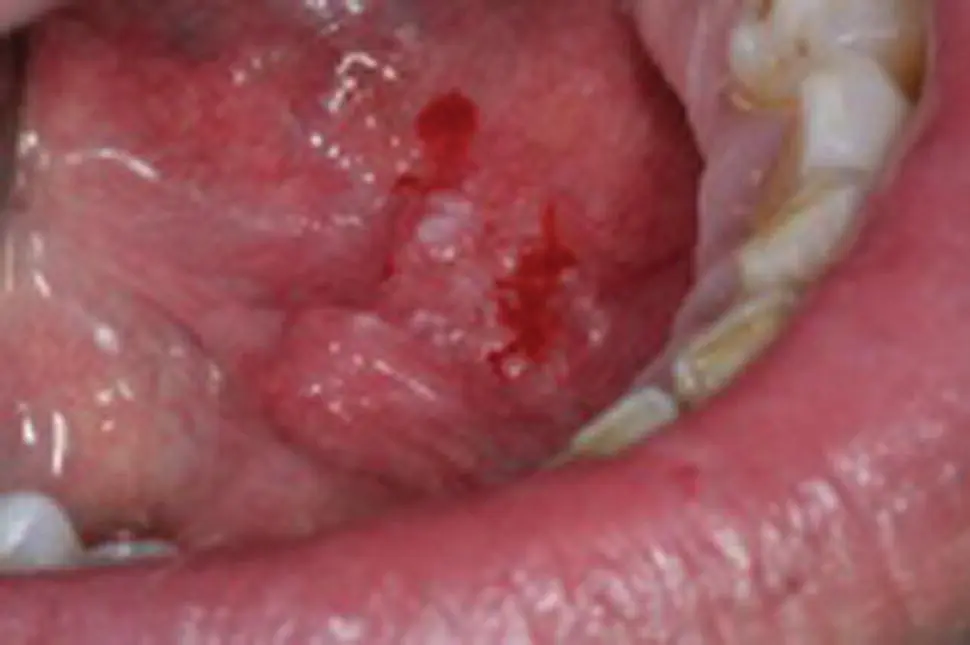
Figure 1.5
CO: A 48‐year old man was presented with a hemorrhagic lesion on the floor of his mouth.
HPC: The lesion appeared six months ago while the bleeding was noticed during eating three days ago.
Читать дальшеИнтервал:
Закладка:
Похожие книги на «Clinical Guide to Oral Diseases»
Представляем Вашему вниманию похожие книги на «Clinical Guide to Oral Diseases» списком для выбора. Мы отобрали схожую по названию и смыслу литературу в надежде предоставить читателям больше вариантов отыскать новые, интересные, ещё непрочитанные произведения.


Обсуждение, отзывы о книге «Clinical Guide to Oral Diseases» и просто собственные мнения читателей. Оставьте ваши комментарии, напишите, что Вы думаете о произведении, его смысле или главных героях. Укажите что конкретно понравилось, а что нет, и почему Вы так считаете.