Crispian Scully - Clinical Guide to Oral Diseases
Здесь есть возможность читать онлайн «Crispian Scully - Clinical Guide to Oral Diseases» — ознакомительный отрывок электронной книги совершенно бесплатно, а после прочтения отрывка купить полную версию. В некоторых случаях можно слушать аудио, скачать через торрент в формате fb2 и присутствует краткое содержание. Жанр: unrecognised, на английском языке. Описание произведения, (предисловие) а так же отзывы посетителей доступны на портале библиотеки ЛибКат.
- Название:Clinical Guide to Oral Diseases
- Автор:
- Жанр:
- Год:неизвестен
- ISBN:нет данных
- Рейтинг книги:4 / 5. Голосов: 1
-
Избранное:Добавить в избранное
- Отзывы:
-
Ваша оценка:
Clinical Guide to Oral Diseases: краткое содержание, описание и аннотация
Предлагаем к чтению аннотацию, описание, краткое содержание или предисловие (зависит от того, что написал сам автор книги «Clinical Guide to Oral Diseases»). Если вы не нашли необходимую информацию о книге — напишите в комментариях, мы постараемся отыскать её.
Provides nearly 300 high-quality clinical photos and relevant questions to help lead readers to the proper diagnosis of common oral diseases Contains concise tables relevant to each chapter with a list of common oral lesions and conditions Offers MCQs of varying levels of difficulty to help readers test their knowledge in Oral Medicine Includes clinical flow charts according to the location and duration of oral lesions Incorporates the ICD-10 Codes of oral lesions and diseases
is a valuable reference for general dental and medical practitioners, undergraduate dental students, and postgraduate trainees in oral and maxillofacial surgery, oral medicine, oral pathology, periodontology as well as general pathology, dermatology or head and neck oncology.
Clinical Guide to Oral Diseases — читать онлайн ознакомительный отрывок
Ниже представлен текст книги, разбитый по страницам. Система сохранения места последней прочитанной страницы, позволяет с удобством читать онлайн бесплатно книгу «Clinical Guide to Oral Diseases», без необходимости каждый раз заново искать на чём Вы остановились. Поставьте закладку, и сможете в любой момент перейти на страницу, на которой закончили чтение.
Интервал:
Закладка:
Dimitris Malamos
Athens, 2020
Foreword
It is an honor and an immense satisfaction for me to provide the prologue for this book for two main reasons: the experience of its authors and the originality of this proposed editorial. When reviewing this ‘’Clinical Guide to Oral Diseases”, one is surprised by the practical perspective with which its authors have imbued it, combining basic principles of problem‐based learning with excellent images and an accurate and essential critical overview for arguing the differential diagnosis of each injury. Throughout the 27 chapters, the authors conduct an exhaustive review of the most common oral injuries based on the discussion of case studies. This approach perfectly combines Dimitris Malamos’ clinical experience and Crispian Scully’s academic rigor.
All of these features make this book a cross‐sectional work, which can be useful both for undergraduate students, general dentists and specialists in the setting of medical‐surgical dentistry. In the end, the success of the clinical activity is not based on academic titles but rather on the knowledge and expertise of the observer. In some manner, Professor Scully had already promulgated this idea during his final years, because he tended to end his emails with a phrase attributed to Goethe, “One only sees what one looks for. One only looks for what one knows.”
Pedro Diz Dios
MD, DDS, PHD, EDOM, FRCSED ad hominem
Professor of Special Care Dentistry
Santiago de Compostela University, Spain
Acknowledgment
I would like to thank the individuals whose help made this Clinical Guide possible; particularly Mrs J. Saiprasad for being so helpful and co‐operative in determination to overcome delays in publication Mrs Carolyn Holleyman for checking the flowcharts and webcases, Mrs Susan Engelken for the cover of the book and especially Mr Vincent Rajan as Production Editor organizing and resolving problems before the publication and Mrs A. Argyropouloy for revising the text. I am grateful to my mentors Professor G. Laskaris (Greece) and Professor C. Scully (United Kingdom) as I truly appreciated their teaching and encouragement to me, to become seriously involved in Oral Medicine. Professor Scully's friendship, advice and expertise is still a part that I miss, and this book was written in his memory.
I wish also to express my sincere gratitude to Dr Pedro Diz Dios and to Dr Marcio Diniz Freitas for their help and advice throughout the preparation of this book. The participation of Dr Marcio Diniz Freitas as Content editor is precious.
Finally, I am especially indebted to my wife Vasiliki and my children Panagiotis and Katerina for their continuous love and support for all those years of my involvement in Oral Medicine, and during the preparation of this guide.
About the Companion Website
Don't forget to visit the companion website for this book:
www.wiley.com/go/malamos/clinical_guide 
There you will find valuable material designed to enhance your learning, including:
Clinical cases
Further reading
Scan this QR code to visit the companion website

1 Bleeding
Bleeding in the mouth may be a sign of various conditions related to the structure of blood vessels, the number or function of white blood cells and especially platelets, the deficiency or dysfunction of clotting factors or even interaction of various drugs. Some of these bleeding disorders appear at a very young age; some are also found among close relatives (inherited) while others are noticed later with a negative family history (acquired). The severity of the bleeding ranges from minor hemorrhages from gingivae and other parts of the oral mucosa with the formation of petechiae or ecchymosis ( Figure 1.0), to extensive bleeding in other parts of the body, causing severe blood loss, even jeopardizing the patient's life.
The more important causes of oral bleeding are seen in Table 1.
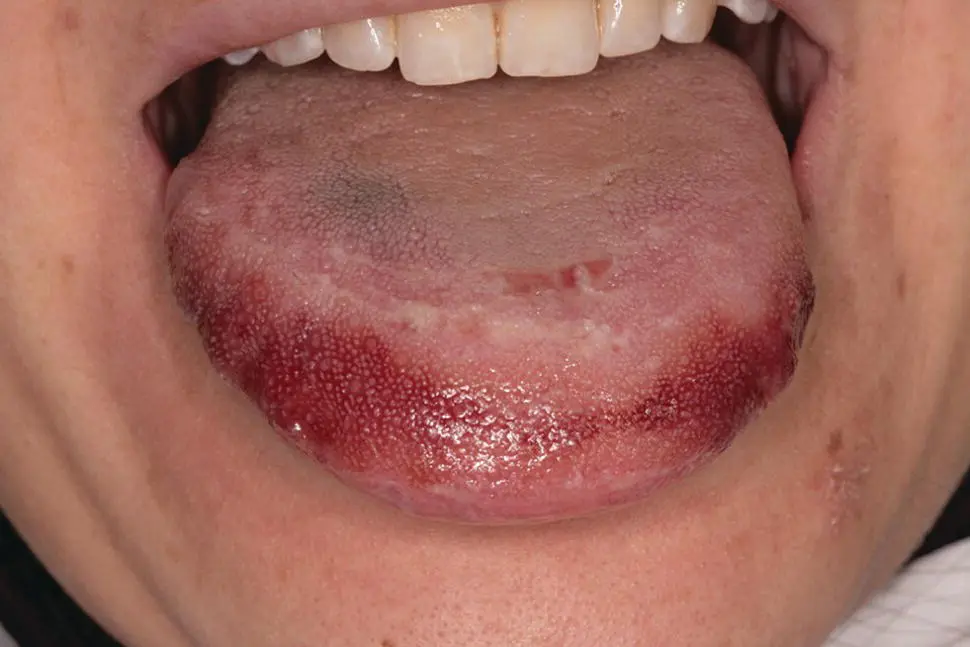
Figure 1.0 Tongue hematoma in a woman with seizures.
Table 1 Conditions related to oral bleeding.
| Common and important conditions |
| Local conditionsGingivitis/periodontitisGranuloma pyogenic/giant cellJaw fractureTraumaTumors invading blood vesselsSystemic conditionsCongenitalHemophilia A or BVon Willebrand's diseaseOther factor deficienciesGlanzman thrombastheniaAcquiredrelated to coagulationLiver diseaseVit. K deficiency, warfarin drug useDisseminated intravascular coagulationrelated to thrombocytopeniaIdiopathicDrug‐inducedCollagen vascular diseaseSarcoidosisHemolytic anemiaLeukemiaMyelomaWaldestmomrelated to platelet dysregulationAlcoholismChronic renal failureDrugsLiver diseaserelated to vascular disordersAngina bullosa hemorrhagicaAngiomasEhrler‐Danlos syndromeHereditary hemorrhagic telangiectasiaInfections from Ebola, HIV, HSV; EBV, RubellaMarfan syndromePurpuraScurvyrelated to fibrinolysisAmyloidosisStreptokinase treatment |
Case 1.1
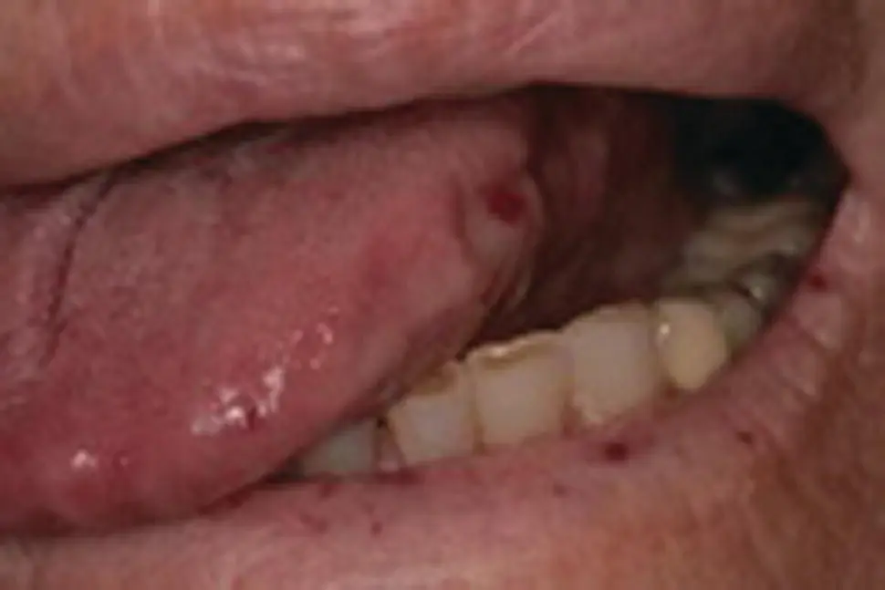
Figure 1.1a
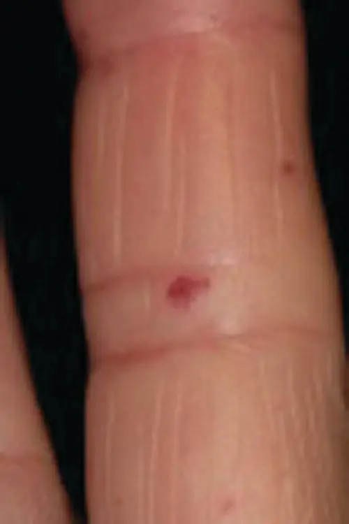
Figure 1.1b
CO: A 62‐year‐old woman was referred by her family doctor for evaluation of several red spots on her lips, mouth, and the skin of her fingers.
HPC: The red spots had been present since childhood, but had become greater on the surface of her face over the last five years causing cosmetic problems and patient’s concern.
PMH: Her medical history revealed a chronic iron deficiency anemia which still remained despite the fact that the patient was in the post‐menopause phase and had been treated occasionally with iron tablets. No other serious medical problems were recorded except for a few episodes of nose and gut bleeding which had caused her to ask for medical advice. She was a non‐smoker and non‐drinker.
OE: The examination revealed numerous red vascular papules, variable in size, ranging from pin head‐like lesions to small red plaques at the vermilion border of her lips, and on the tongue and buccal mucosae ( Figure 1.1a). A few asteroid‐like red lesions, were also seen on the skin of her fingers ( Figure 1.1b) and inside her nose which were responsible for her episodes of epistaxis.
Q1Which is the possible cause of her red spots?
1 Crest syndrome
2 Sjogren syndrome
3 Rendu‐Osler‐Weber syndrome
4 Rosacea
Ataxia ‐ telangiectasia
Answers:
1 No
2 No
3 Rendu‐Osler‐Weber syndrome or hereditary hemorrhagic telangiectasia (HHT) is a rare autosomal dominant condition that affects blood vessels throughout the body (telangiectasia; arteriovenous malformations) with a tendency for bleeding. This vascular dysplasia is commonly seen in oral, nasopharynx, lung, liver, spleen, gastrointestinal and urinary tracts, conjunctiva and the skin of arms and fingers.
Читать дальшеИнтервал:
Закладка:
Похожие книги на «Clinical Guide to Oral Diseases»
Представляем Вашему вниманию похожие книги на «Clinical Guide to Oral Diseases» списком для выбора. Мы отобрали схожую по названию и смыслу литературу в надежде предоставить читателям больше вариантов отыскать новые, интересные, ещё непрочитанные произведения.


Обсуждение, отзывы о книге «Clinical Guide to Oral Diseases» и просто собственные мнения читателей. Оставьте ваши комментарии, напишите, что Вы думаете о произведении, его смысле или главных героях. Укажите что конкретно понравилось, а что нет, и почему Вы так считаете.