Robert E. Marx - Oral Pathology in Clinical Dental Practice
Здесь есть возможность читать онлайн «Robert E. Marx - Oral Pathology in Clinical Dental Practice» — ознакомительный отрывок электронной книги совершенно бесплатно, а после прочтения отрывка купить полную версию. В некоторых случаях можно слушать аудио, скачать через торрент в формате fb2 и присутствует краткое содержание. Жанр: unrecognised, на английском языке. Описание произведения, (предисловие) а так же отзывы посетителей доступны на портале библиотеки ЛибКат.
- Название:Oral Pathology in Clinical Dental Practice
- Автор:
- Жанр:
- Год:неизвестен
- ISBN:нет данных
- Рейтинг книги:3 / 5. Голосов: 1
-
Избранное:Добавить в избранное
- Отзывы:
-
Ваша оценка:
Oral Pathology in Clinical Dental Practice: краткое содержание, описание и аннотация
Предлагаем к чтению аннотацию, описание, краткое содержание или предисловие (зависит от того, что написал сам автор книги «Oral Pathology in Clinical Dental Practice»). Если вы не нашли необходимую информацию о книге — напишите в комментариях, мы постараемся отыскать её.
Oral Pathology in Clinical Dental Practice — читать онлайн ознакомительный отрывок
Ниже представлен текст книги, разбитый по страницам. Система сохранения места последней прочитанной страницы, позволяет с удобством читать онлайн бесплатно книгу «Oral Pathology in Clinical Dental Practice», без необходимости каждый раз заново искать на чём Вы остановились. Поставьте закладку, и сможете в любой момент перейти на страницу, на которой закончили чтение.
Интервал:
Закладка:
Treatment
It is a relief to patients to hear that their benign hyperkeratosis lesion is not considered to be a cancer. However, incisional and even excisional biopsy cannot predict either regrowth or a sampling error that may result in a squamous cell carcinoma developing later. Therefore, a 6-month recall schedule is recommended even though no specific treatment is required.
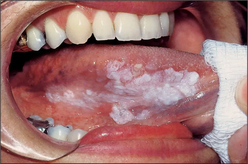
 Leukoplakia.
Leukoplakia
Nature of disease
A white patch on the oral mucosa. Three conditions can present as a clinically apparent white patch: acanthosis/hyperkeratosis, Candida , or fibrin.
Predilections
Can occur at any age but is more often seen in adults. There is no sex or racial predilection.
Clinical features
A white patch of oral mucosa most commonly noted on the buccal mucosa, lateral border of the tongue, or floor of the mouth.
Radiographic presentation
None.
Differential diagnosis
Clinical leukoplakia may only represent an ulcer with a fibrin base or Candida and sometimes lichen planus by virtue of its acanthosis and hyperkeratosis. However, after benign hyperkeratosis, the most serious considerations are premalignant dyplasias, carcinoma in situ, verrucous carcinoma, and invasive squamous cell carcinoma.
Microscopic features
The three entities that cause a clinical leukoplakia will appear different:
1. Fibrin: Thin strands of light eosinophilic staining over a wound base with inflammation.
2. Candida : Vertically positioned hyphae with prominent periodic acid-Schiff (PAS) or silver staining on an epithelial surface.
3. Acanthosis/hyperkeratosis:
a. Acanthosis/benign hyperkeratosis: A thickened keratinocyte layer without cellular atypia but with surface keratin.
b. Premalignant dysplasia: Various degrees of epithelial atypia above an intact basement membrane.
c. Carcinoma in situ: Severe epithelial dysplasia with nuclear pleomorphism and abnormal mitotic figures from the basal cell layer to the surface.
d. Verrucous carcinoma: A significant exophytic proliferation as well as a blunted endophytic proliferation of epithelial cells but with an intact basement membrane beneath which most often resides a dense inflammatory response.
e. Invasive carcinoma: Atypical epithelial cells forming bundles and cords through the basement membrane into the underlying tissues.
Suggested course of action
Biopsy and/or refer to an oral and maxillofacial surgeon.
Treatment
Treatment will vary according to the biopsy result. For:
♦ Fibrin: Wound care and treatment of the underlying etiology, as well as observation and surveillance for acanthosis/hyperkeratosis.
♦ Premalignant dysplasia: Excision with frozen section control.
♦ Carcinoma in situ: Excision with frozen section control.
♦ Verrucous carcinoma: Excision with frozen section control.
♦ Invasive carcinoma: Excision with frozen section control and evaluation for radiotherapy and/or chemotherapy.
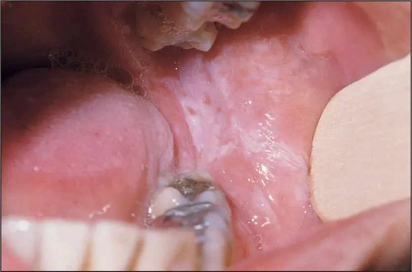
Smokeless tobacco keratosis from chewing tobacco.
Non–Betel Nut/Slaked Lime Smokeless Tobacco Keratosis
Nature of disease
A reactive nondysplastic hyperkeratosis of the mucosa due to inflammation and stimulation from various smokeless tobacco products.
Predilections
May occur in anyone who uses such products but has a higher incidence in the western United States and the Scandinavian countries due to cultural traditions. More common in men also because of cultural acceptance. No racial predilection is known.
Clinical features
The area corresponding to the placement of the tobacco product (usually the mandibular attached gingiva and mucosa of the vestibule) will appear as an irregular and often leathery white patch referred to as a “snuffle dippers patch.” It is usually nonpainful unless the area is inflamed.
Radiographic presentation
None.
Differential diagnosis
The clinician is advised to ask the patient about use of betel nut/slaked lime use versus true smokeless tobacco products. Betel nut/slaked lime keratosis is premalignant, whereas other true tobacco-containing, topical smokeless tobacco products are not. Additionally, clinicians should also consider epithelial dysplasia, carcinoma in situ, invasive squamous cell carcinoma, and even lichen planus and hypertrophic candidiasis.
Microscopic features
Nondysplastic epithelium will be present with a thick layer of pale-staining cells occupying the superficial half of the keratinocyte layer along with significant hyperkeratosis.
Suggested course of action
Confirm that the patch is not the result of betel nut/slaked lime use. Incisional biopsy is indicated to document the absence of dysplasia. Counsel the patient about tobacco cessation, particularly as it relates to erosion of the mucosa and gingiva as well as staining of the teeth.
Treatment
No specific treatment other than cessation of smokeless tobacco use.
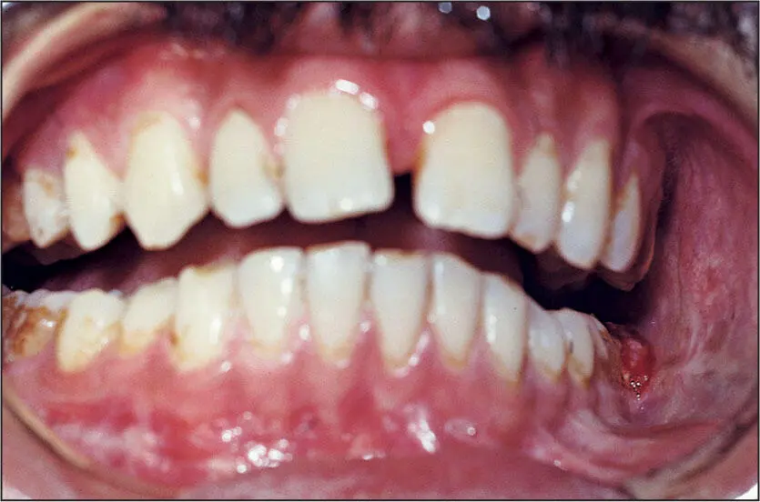
Trismus, buccal mucosa hyperkeratosis, and fibrosis from betel nut/slaked lime use.
Betel Nut/Slaked Lime Keratosis
Nature of disease
A dysplastic premalignant transformation of mucosal epithelial cells due to the carcinogenic effects of slaked lime. Additionally, the Areca catechu chemical in the betel nut often causes a concomitant submucosal fibrosis.
Predilections
Mostly seen in individuals from India, Sri Lanka, and East Asian countries, where the produce is called quid or pan and used for oral gratification much like chewing gum in the United States.
Clinical features
A white patch is usually seen on either buccal mucosa. There is often a limited jaw opening due to the submucosal fibrosis. Induration around the white patch is common.
Radiographic presentation
None.
Differential diagnosis
The clinician is advised to ask the patient specifically about betel nut/slaked lime use versus use of smokeless tobacco products, as the latter are not carcinogenic to a significant degree. Other considerations include squamous cell carcinoma, actinomycosis, lichen planus, and hypertrophic candidiasis.
Microscopic features
Various degrees of epithelial dysplasia up to invasive carcinoma may be seen together with hyperkeratosis. Dense, poorly cellular collagen is usually seen in the submucosa. Inflammatory cells are variably present.
Читать дальшеИнтервал:
Закладка:
Похожие книги на «Oral Pathology in Clinical Dental Practice»
Представляем Вашему вниманию похожие книги на «Oral Pathology in Clinical Dental Practice» списком для выбора. Мы отобрали схожую по названию и смыслу литературу в надежде предоставить читателям больше вариантов отыскать новые, интересные, ещё непрочитанные произведения.


Обсуждение, отзывы о книге «Oral Pathology in Clinical Dental Practice» и просто собственные мнения читателей. Оставьте ваши комментарии, напишите, что Вы думаете о произведении, его смысле или главных героях. Укажите что конкретно понравилось, а что нет, и почему Вы так считаете.