Complications in Equine Surgery
Здесь есть возможность читать онлайн «Complications in Equine Surgery» — ознакомительный отрывок электронной книги совершенно бесплатно, а после прочтения отрывка купить полную версию. В некоторых случаях можно слушать аудио, скачать через торрент в формате fb2 и присутствует краткое содержание. Жанр: unrecognised, на английском языке. Описание произведения, (предисловие) а так же отзывы посетителей доступны на портале библиотеки ЛибКат.
- Название:Complications in Equine Surgery
- Автор:
- Жанр:
- Год:неизвестен
- ISBN:нет данных
- Рейтинг книги:3 / 5. Голосов: 1
-
Избранное:Добавить в избранное
- Отзывы:
-
Ваша оценка:
Complications in Equine Surgery: краткое содержание, описание и аннотация
Предлагаем к чтению аннотацию, описание, краткое содержание или предисловие (зависит от того, что написал сам автор книги «Complications in Equine Surgery»). Если вы не нашли необходимую информацию о книге — напишите в комментариях, мы постараемся отыскать её.
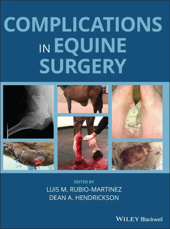
Offers the first resource specifically focused on complications encountered in equine surgery Takes a helpful format organized by body system Provides consistently formatted chapters for ease of use Covers clinically relevant information for dealing with technical and post-operative complications Presents more than 350 color images to illustrate the concepts described Written for general practitioners and specialists,
is an essential resource to decreasing morbidity and mortality and increasing surgical success in horses.
Complications in Equine Surgery — читать онлайн ознакомительный отрывок
Ниже представлен текст книги, разбитый по страницам. Система сохранения места последней прочитанной страницы, позволяет с удобством читать онлайн бесплатно книгу «Complications in Equine Surgery», без необходимости каждый раз заново искать на чём Вы остановились. Поставьте закладку, и сможете в любой момент перейти на страницу, на которой закончили чтение.
Интервал:
Закладка:
35 Chapter 47Figure 47.1 CT image of septic osteitis of the distal phalanx demonstrating ...Figure 47.2 Defect in the solar surface of the hoof must be large enough to ...Figure 47.3 Type III fracture of the distal phalanx, 8 months post repair vi...Figure 47.4 Use of medical maggots to assist in the debridement of necrotic ...Figure 47.5 Partial hoof wall resection for the treatment of a keratoma in t...
36 Chapter 48Figure 48.1 Dorsopalmar radiographic images of both carpi of a foal after bi...Figure 48.2 Examples of complications from single screw transphyseal bridgin...Figure 48.3 Dorsopalmar radiographic image of the right metacarpophalangeal ...Figure 48.4 Dorsopalmar radiographic image of a metacarpophalangeal joint of...Figure 48.5 Cosmetic complications associated with the use of single transph...Figure 48.6 Dorsopalmar radiographic image of the right metacarpophalangeal ...Figure 48.7 The arrow indicates an example of a seroma over the medial physi...
37 Chapter 49Figure 49.1 A superficial bandage sore over the dorsolateral aspect of the l...Figure 49.2 A sloughed hoof as a result of an over tight bandage.Figure 49.3 An active tension‐extension splint applied to a foal's front lim...Figure 49.4 Swan‐necked shoe.Figure 49.5 Lateromedial radiographic image showing radiolucency at the dors...Figure 49.6 Devitalized tissue and necrotic bone are being removed at the do...Figure 49.7 A fragment of the third phalanx that was removed surgically.
38 Chapter 50Figure 50.1 (a) Dorsal 45‐degree lateral‐plantaromedial oblique radiographic...Figure 50.2 Dorsal 60‐degree lateral‐plantaromedial oblique radiographic pro...Figure 50.3 (a) Dorsal 45‐degree lateral‐palmaromedial oblique radiographic ...
39 Chapter 51Figure 51.1 Latero‐lateral radiograph of the rostral aspect of the head of a...Figure 51.2 Laterolateral radiograph of the rostral aspect of the head of th...Figure 51.3 Right‐dorsal‐left‐ventral‐oblique radiographic view of the skull...Figure 51.4 (a) Photograph of the ventral aspect of the head of a horse with...
40 Chapter 52Figure 52.1 Ultrasound guided injection of superficial digital flexor tendon...Figure 52.2 Non‐weight‐bearing transverse image showing a defect created thr...Figure 52.3 Iatrogenically created damage to the surface of the deep digital...Figure 52.4 (a) Damage visible postoperatively in the suspensory ligament af...Figure 52.5 Superficial digital flexor tendon lesion (a) arrowed, treated wi...Figure 52.6 Arthroscopic portal complications. (a) Appearance of the proxima...Figure 52.7 Iatrogenically‐induced tendon mineralization after intra‐lesiona...Figure 52.8 Neuroma formation post tenoscopy. This longitudinal ultrasound i...Figure 52.9 Adhesion formation after palmar annular ligament desmotomy. (a) ...Figure 52.10 Sudden exacerbation (arrows) of proximal suspensory desmitis po...Figure 52.11 Distal interphalangeal joint subluxation (solid arrow) as a rar...Figure 52.12 Tendon rupture after protected loading. (a) shows an acquired f...Figure 52.13 Fragmentation of the apex of the patella (arrow) – a common com...
41 Chapter 53Figure 53.1 Placement of closed suction drains in a horse following the modi...Figure 53.2 Dehiscence of a pectoral wound closure caused by excessive tensi...
42 Chapter 56Figure 56.1 (a) Migration of orbital prosthetic. (b) Rejection of orbital pr...Figure 56.2 (a) Image of an orbital implant. (b) Orbital implant infection....Figure 56.3 Dorsal orbital rim fracture.Figure 56.4 Foal lower lid laxity and entropion.Figure 56.5 Superior lid suture malposition causing corneal ulcer.Figure 56.6 (a) Extensive SCC of the eyelids and periocular region OS. (b) S...Figure 56.7 (a) Cisplatin beads prior to injection. (b) Immediate postoperat...Figure 56.8 (a) Preoperative periocular sarcoid. (b) Week’s postoperative ci...Figure 56.9 (a) Lower lid SCC pre‐excision. (b) Debulking of lower lid SCC. ...Figure 56.10 Inferomedial SPL placement, corneal fibrosis post‐SCC keratecto...Figure 56.11 Dorsal corneal ulcer from superior lid SPL endplate slippage....Figure 56.12 (a) Seidel positive fungal ulcer needing conjunctival pedicle g...Figure 56.13 (a) Retraction and thickening of graft due to aqueous humor mic...Figure 56.14 Corneal SCC pretreatment.Figure 56.15 (a) Granulation tissue post‐keratectomy + beta. (b) Fibrosis an...Figure 56.16 Corneal edema 3 days post‐DLEK.Figure 56.17 (a) Corneal fibrosis and vascularization some weeks post‐DLEK. ...Figure 56.18 (a) SCCED/indolent ulcer pretreatment. (b) Thermal cautery unit...Figure 56.19 Melting corneal ulcer post‐thermal cautery procedure.Figure 56.20 (a) Corneal fibrosis and edema immediately postoperatively. (b)...Figure 56.21 Fibrin and cataract in chronic ERU eye.Figure 56.22 Conjunctival and episcleral hemorrhage post‐cyclosporine implan...Figure 56.23 Migration/wandering implant visible in anterior chamber.Figure 56.24 (a) Uveal melanoma near corpora nigra. (b) Introcular hemorrhag...Figure 56.25 (a) Post‐phaco lens fragment in anterior chamber. (b) Post‐phac...Figure 56.26 (a) Incisional fibrosis, PCO, and melanin on anterior lens caps...Figure 56.27 Retinal detachment post‐phacoemulsification.
43 Chapter 58Figure 58.1 Radiograph showing the central Kerf Cut Cylinder backing out of ...Figure 58.2 Radiograph showing the caudal Kerf Cut Cylinder backing out of t...Figure 58.3 Radiograph showing the treaded profile of the “Seattle Slew” imp...Figure 58.4 Necropsy image of vertebra showing Kerf Cut Cylinder with a cros...Figure 58.5 Radiograph showing the straight profile of the “Bagby Basket” im...
44 Chapter 59Figure 59.1 Dystrophic mineralization on a horse 24 months after surgery wit...Figure 59.2 Cranial rotation of the DSP 12 in a horse after wedge ostectomy ...Figure 59.3 Postoperative radiograph of a horse undergoing wedge resection o...Figure 59.4 Focal depressions at the surgical sites after wedge resection os...Figure 59.5 Presence of white hairs at the surgical sites after desmotomy of...
45 Chapter 60Figure 60.1 A non‐painful neuroma found during a second neurectomy in the mi...Figure 60.2 Two segments of nerve that have been removed during a second neu...Figure 60.3 Lateromedial radiographic images of the front digits of a horse ...Figure 60.4 A facial laceration that has severed the facial nerve resulting ...
Guide
1 Cover
2 Table of Contents
3 Begin Reading
Pages
1 iii
2 iv
3 v
4 xi
5 xii
6 xiii
7 xix
8 xv
9 xvi
10 xvii
11 xviii
12 xix
13 1
14 2
15 3
16 4
17 5
18 6
19 7
20 8
21 9
22 10
23 11
24 12
25 13
26 14
27 15
28 16
29 17
30 18
31 19
32 20
33 21
34 22
35 23
36 24
37 25
38 26
39 27
40 28
41 29
42 30
43 31
44 32
45 33
46 34
47 35
48 36
49 37
50 38
51 39
52 40
53 41
54 42
55 43
56 44
57 45
58 46
59 47
60 48
61 49
62 50
63 51
64 52
65 53
66 54
67 55
68 56
69 57
70 58
71 59
72 60
73 61
74 62
75 63
76 64
77 65
78 66
79 67
80 68
81 69
82 70
83 71
84 72
85 73
86 74
87 75
88 76
89 77
90 78
91 79
92 80
93 81
94 82
95 83
96 84
97 85
98 86
99 87
100 88
101 89
102 90
103 91
104 92
105 93
106 94
107 95
108 96
109 97
110 98
111 99
112 100
113 101
114 102
115 103
116 104
Читать дальшеИнтервал:
Закладка:
Похожие книги на «Complications in Equine Surgery»
Представляем Вашему вниманию похожие книги на «Complications in Equine Surgery» списком для выбора. Мы отобрали схожую по названию и смыслу литературу в надежде предоставить читателям больше вариантов отыскать новые, интересные, ещё непрочитанные произведения.


Обсуждение, отзывы о книге «Complications in Equine Surgery» и просто собственные мнения читателей. Оставьте ваши комментарии, напишите, что Вы думаете о произведении, его смысле или главных героях. Укажите что конкретно понравилось, а что нет, и почему Вы так считаете.