Complications in Equine Surgery
Здесь есть возможность читать онлайн «Complications in Equine Surgery» — ознакомительный отрывок электронной книги совершенно бесплатно, а после прочтения отрывка купить полную версию. В некоторых случаях можно слушать аудио, скачать через торрент в формате fb2 и присутствует краткое содержание. Жанр: unrecognised, на английском языке. Описание произведения, (предисловие) а так же отзывы посетителей доступны на портале библиотеки ЛибКат.
- Название:Complications in Equine Surgery
- Автор:
- Жанр:
- Год:неизвестен
- ISBN:нет данных
- Рейтинг книги:3 / 5. Голосов: 1
-
Избранное:Добавить в избранное
- Отзывы:
-
Ваша оценка:
Complications in Equine Surgery: краткое содержание, описание и аннотация
Предлагаем к чтению аннотацию, описание, краткое содержание или предисловие (зависит от того, что написал сам автор книги «Complications in Equine Surgery»). Если вы не нашли необходимую информацию о книге — напишите в комментариях, мы постараемся отыскать её.
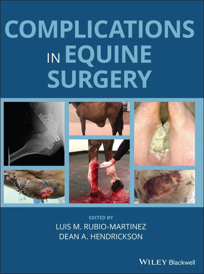
Offers the first resource specifically focused on complications encountered in equine surgery Takes a helpful format organized by body system Provides consistently formatted chapters for ease of use Covers clinically relevant information for dealing with technical and post-operative complications Presents more than 350 color images to illustrate the concepts described Written for general practitioners and specialists,
is an essential resource to decreasing morbidity and mortality and increasing surgical success in horses.
Complications in Equine Surgery — читать онлайн ознакомительный отрывок
Ниже представлен текст книги, разбитый по страницам. Система сохранения места последней прочитанной страницы, позволяет с удобством читать онлайн бесплатно книгу «Complications in Equine Surgery», без необходимости каждый раз заново искать на чём Вы остановились. Поставьте закладку, и сможете в любой момент перейти на страницу, на которой закончили чтение.
Интервал:
Закладка:
26 Chapter 36Figure 36.1 Transverse section of the interior of the left guttural pouch to...Figure 36.2 Parotid salivary duct (a) is part of duct exposed on lateral asp...Figure 36.3 (a) Dorsal view of the normal anatomy of the terminal portions o...Figure 36.4 Laterolateral radiograph taken the day after insertion of a nond...Figure 36.5 Landmarks used to locate the internal carotid artery in the gutt...Figure 36.6 Accidental penetration of the defect in the internal carotid art...Figure 36.7 Role of the “steal phenomenon” to cause blindness after some, bu...Figure 36.8 Anatomy of the left lateral aspect of the stylohyoid apparatus, ...
27 Chapter 38Figure 38.1 Left hemithorax thoracic tube placed within the 13th intercostal...
28 Chapter 39Figure 39.1 This horse developed a hematoma in the sutured scrotum after cas...Figure 39.2 Intraoperative image of a horse anesthetized and in dorsal recum...Figure 39.3 Sands emasculator. This emasculator is similar to a Reimer emasc...Figure 39.4 Close‐up view of the Henderson Equine Castrating Instrument (Sto...Figure 39.5 The Henderson Equine Castrating Instrument (Stone Manufacturing ...Figure 39.6 A Stille kidney clamp has been attached to the end of the severe...Figure 39.7 This horse suffered eventration 4 hours after being castrated wh...Figure 39.8 Prolapse of the greater omentum from the abdomen and inguinal ca...Figure 39.9 Excised infected spermatic cords of a horse that developed bilat...Figure 39.10 Intraoperative image of a pony anesthetized and in dorsal recum...Figure 39.11 An accumulation of fluid within the scrotum caused by formation...Figure 39.12 Intraoperative image of a horse anesthetized and in dorsal recu...Figure 39.13 Intraoperative image of a horse anesthetized and in dorsal recu...Figure 39.14 Intraoperative image of a horse anesthetized and in dorsal recu...Figure 39.15 Intraoperative image of a horse anesthetized and in dorsal recu...Figure 39.16 The ligament of the tail of the epididymis (LTE) connects the v...Figure 39.17 The proper ligament of the testis of an abdominal testis is som...Figure 38.18 (a) An inguinal approach has been made to the inguinal canal on...
29 Chapter 40Figure 40.1 Perineal urethrotomy or a spongiotomy can be performed to resolv...Figure 40.2 A complication of partial phallectomy is hemorrhage from a corpo...Figure 40.3 Stenosis of the urethral stoma after partial phallectomy perform...Figure 40.4 Reappearance of a carcinoma after partial phallectomy performed ...Figure 40.5 Dehiscence of the sutured internal lamina of the prepuce of a do...Figure 40.6 Intraoperative image of a horse anesthetized and in dorsal recum...Figure 40.7 This diagram shows the transverse section of the penile shaft. A...
30 Chapter 41Figure 41.1 An écraseur is used to cut and crush the ovarian pedicle when pe...Figure 41.2 Intraoperative image of a mare undergoing ovariectomy while anes...Figure 41.3 Intraoperative image of a mare undergoing hysterectomy using a c...Figure 41.4 Intraoperative image of a mare undergoing Cesarean section. The ...
31 Chapter 42Figure 42.1 Vaginoscopic examination of a standing mare showing urine in the...Figure 42.2 The Pouret procedure to resolve vesicovaginal reflux. Separating...Figure 42.3 (a–e) The Brown procedure to resolve vesicovaginal reflux. The u...Figures 42.4 The Shires and Kaneps procedure to resolve vesicovaginal reflux...Figure 42.5 The McKinnon and Beldon procedure to resolve vesicovaginal reflu...Figure 42.6 Urethral extension with a fistula. The blue‐stained balloon of a...Figure 42.7 Vaginoscopic examination of a standing mare showing a cervical l...Figure 42.8 This mare developed pyometra when luminal adhesions developed in...Figure 42.9 Endoscopic picture of a healed cervix after a wedge resection to...Figure 42.10 Repair of an acute rectovestibular laceration is seldom success...Figure 42.11 To reconstruct the rectovestibular tissue, submucosa between th...Figure 42.12 Rectovestibular fistulae. A fistula of this size is best conver...Figure 42.13 Prolapse of the bladder of a mare caused by cystitis resulting ...
32 Chapter 44Figure 44.1 (a) Lateromedial radiographic view of the left front foot of a h...Figure 44.2 Dorsolateroplantaromedial oblique radiographic view of the right...
33 Chapter 45Figure 45.1 Damage to the tip of a 30‐degree rigid endoscope. Note multiple ...Figure 45.2 Arthroscopic images of a dull Ferris–Smith rongeur (a) and curet...Figure 45.3 Arthroscopic image of a 2 × 10 mm Ferris–Smith rongeur. Note tha...Figure 45.4 Arthroscopic image of the cranial medial femorotibial joint demo...Figure 45.5 Arthroscopic image of a large osteochondral fragment in the dors...Figure 45.6 (a) Photograph of an assembled 30‐degree rigid endoscope that is...Figure 45.7 Lateral intraoperative radiograph of a right stifle. The arthros...Figure 45.8 Arthroscopic image demonstrating an obstructed view of a Ferris–...Figure 45.9 Arthroscopic image in a dorsal pouch of a metacarpophalangeal jo...Figure 45.10 Arthroscopic image of fluid being pumped into the extrasynovial...Figure 45.11 Arthroscopic image in a tarsocrural joint after laceration of t...Figure 45.12 Arthroscopic image from the same joint shown in Figure 45.10 af...Figure 45.13 (a) Arthroscopic image of hypertrophic and hyperplastic synovia...Figure 45.14 Arthroscopic image of the plantar pouch of a fetlock. The image...Figure 45.15 Arthroscopic image demonstrating small metallic debris (arrow) ...Figure 45.16 Arthroscopic image of a broken #11 blade in the palmar aspect o...Figure 45.17 Arthroscopic image of a synovial resector within the synovial c...Figure 45.18 Arthroscopic image of a large free‐floating fragment in a femor...Figure 45.19 Arthroscopic image of a new portal created in the metacarpophal...Figure 45.20 Intraoperative radiograph of a right stifle after removal of OC...Figure 45.21 Arthroscopic image of a fragment that is too large for the port...Figure 45.22 Intraoperative radiograph of a tarsus with the arthroscope plac...Figure 45.23 Arthroscopic image of a large loose fragment that was identifie...Figure 45.24 Arthroscopic image of a tarsocrural joint after debridement of ...Figure 45.25 Arthroscopic image of needle insertion into a septic joint. Not...Figure 45.26 Arthroscopic image of hair debris (arrows) being introduced int...
34 Chapter 46Figure 46.1 (a) Ceretom (Samsung Neurologica, Danvers, MA) portable 8‐slice ...Figure 46.2 (a) Diagram of a horse demonstrating important anatomic/biomecha...Figure 46.3 (a) Arthroscopic reduction of a displaced lateral condylar fract...Figure 46.4 (a) Lateral‐medial radiograph of the third metacarpus with a bro...Figure 46.5 (a) Synthes screw removal set containing instruments required fo...Figure 46.6 Depiction of counter‐sinking a screw placed in the proximal phal...Figure 46.7 Two broken screws (arrow and circle) in an imperfectly stable fe...Figure 46.8 (a) A long screw was placed during repair of a spiraling medial ...Figure 46.9 Thermal injury to bone is evidenced by osteolysis around the scr...Figure 46.10 Inadequate implants usually result in failure. (a) Any fracture...Figure 46.11 (a) Radiographs demonstrating different configurations and comp...Figure 46.12 (a) Inaccurate positioning of lag screws and inadequate compres...Figure 46.13 (a) Dorsoplantar radiograph, (b) frontal plane CT image, and (c...Figure 46.14 Wedge‐shaped fragments at the proximal margin of third carpal s...Figure 46.15 Small (3.5‐mm) cortical screws placed too distally through the ...Figure 46.16 (a) Arthroscopic image showing the tip edge of a #10 scalpel bl...Figure 46.17 Some common errors in P1 screw placement. (a) This proximal scr...Figure 46.18 (a) An ulnar fracture repaired with single 4.5‐mm screw placed ...Figure 46.19 Radiographs of several different pastern arthrodeses. (a) Radio...Figure 46.20 Placing lag screws through the metacarpus into the proximal ses...Figure 46.21 Some errors with fetlock arthrodesis. (a) An LCP placed in the ...Figure 46.22 Fluoroscopic images used to avoid drilling through the cable. (...Figure 46.23 (a) Cast sores on the mid‐dorsal metacarpus and the palmar fetl...Figure 46.24 (a) Radiograph of comminuted proximal phalangeal fracture repai...Figure 46.25 (a) An inverted “T” incision 2 weeks following a pastern arthro...Figure 46.26 (a) Antibiotic‐impregnated polymethylmethacrylate (PMMA) being ...
Читать дальшеИнтервал:
Закладка:
Похожие книги на «Complications in Equine Surgery»
Представляем Вашему вниманию похожие книги на «Complications in Equine Surgery» списком для выбора. Мы отобрали схожую по названию и смыслу литературу в надежде предоставить читателям больше вариантов отыскать новые, интересные, ещё непрочитанные произведения.


Обсуждение, отзывы о книге «Complications in Equine Surgery» и просто собственные мнения читателей. Оставьте ваши комментарии, напишите, что Вы думаете о произведении, его смысле или главных героях. Укажите что конкретно понравилось, а что нет, и почему Вы так считаете.