Complications in Equine Surgery
Здесь есть возможность читать онлайн «Complications in Equine Surgery» — ознакомительный отрывок электронной книги совершенно бесплатно, а после прочтения отрывка купить полную версию. В некоторых случаях можно слушать аудио, скачать через торрент в формате fb2 и присутствует краткое содержание. Жанр: unrecognised, на английском языке. Описание произведения, (предисловие) а так же отзывы посетителей доступны на портале библиотеки ЛибКат.
- Название:Complications in Equine Surgery
- Автор:
- Жанр:
- Год:неизвестен
- ISBN:нет данных
- Рейтинг книги:3 / 5. Голосов: 1
-
Избранное:Добавить в избранное
- Отзывы:
-
Ваша оценка:
Complications in Equine Surgery: краткое содержание, описание и аннотация
Предлагаем к чтению аннотацию, описание, краткое содержание или предисловие (зависит от того, что написал сам автор книги «Complications in Equine Surgery»). Если вы не нашли необходимую информацию о книге — напишите в комментариях, мы постараемся отыскать её.
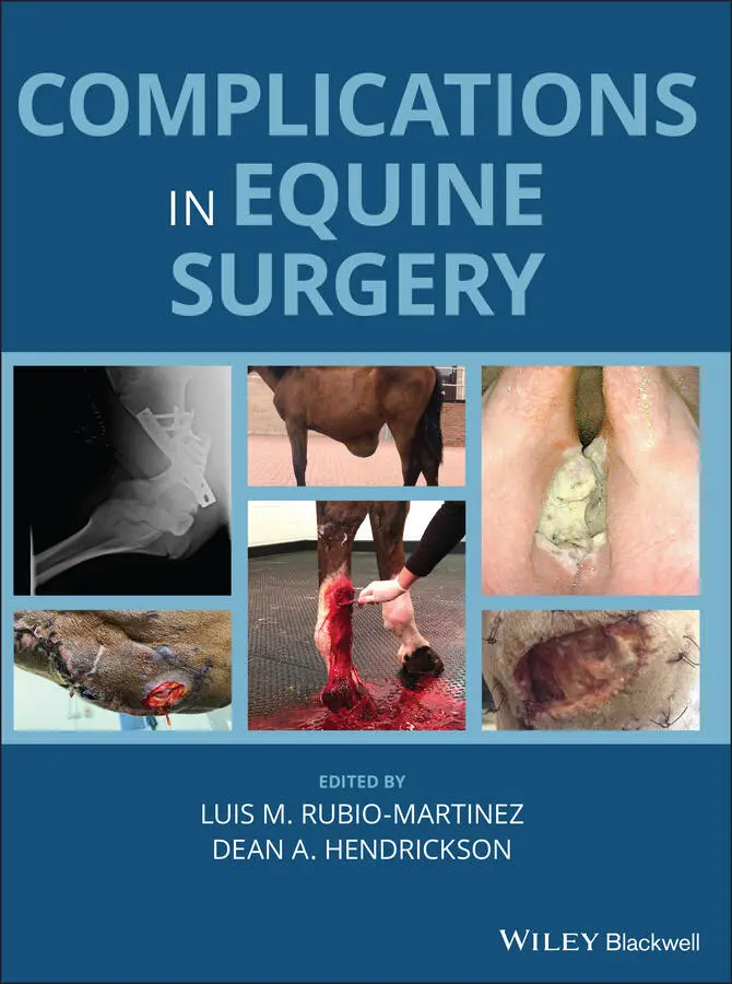
Offers the first resource specifically focused on complications encountered in equine surgery Takes a helpful format organized by body system Provides consistently formatted chapters for ease of use Covers clinically relevant information for dealing with technical and post-operative complications Presents more than 350 color images to illustrate the concepts described Written for general practitioners and specialists,
is an essential resource to decreasing morbidity and mortality and increasing surgical success in horses.
Complications in Equine Surgery — читать онлайн ознакомительный отрывок
Ниже представлен текст книги, разбитый по страницам. Система сохранения места последней прочитанной страницы, позволяет с удобством читать онлайн бесплатно книгу «Complications in Equine Surgery», без необходимости каждый раз заново искать на чём Вы остановились. Поставьте закладку, и сможете в любой момент перейти на страницу, на которой закончили чтение.
Интервал:
Закладка:
64 56 Complications of Equine Ophthalmic Surgery Overview List of Complications Associated with Equine Ophthalmic Surgery Globe and Orbit Adnexal Surgery Ocular Surgery Intraocular Surgery References
65 57 Complications of Diagnostic Procedures of the Nervous System Overview List of Complications Associated with Diagnostic Procedures of the Nervous System Patient or Personnel Injury Increased Intracranial Pressure Complications Associated with Cerebrospinal Fluid Centesis Complications Associated with Cervical Myelography Complications Associated with Myeloscopy and Epiduroscopy Complications Associated with Cervical Articular Process Joint Injection Complications Associated with Nerve and Muscle Biopsy Complications Associated with Electrodiagnostics References
66 58 Complications of Anterior Cervical Fusion Overview List of Complications Associated with Anterior Cervical Fusion Intraoperative and Preoperative Planning: Fusing the Incorrect Site Intraoperative: Insecure Implant Neuropathy Hematoma/Seroma Infection Traumatic Recovery/Fracture Fractures in the Rehabilitation Stage
67 59 Complications of Surgery for Impingement of Dorsal Spinous Processes Overview List of Complications Associated with Surgery for Impingement of Dorsal Spinous Processes Ostectomy of the DSP Desmotomy of the Interspinous Ligament References
68 60 Complications of Peripheral Nerve Surgery Overview List of Complications Associated with Peripheral Nerve Surgery Anatomy and Pathophysiology Neurectomy Procedures Palmar or Plantar Digital Neurectomy References
69 Index
70 End User License Agreement
List of Tables
1 Chapter 10 Table 10.1 Summary of bone graft donor sites
2 Chapter 11 Table 11.1 Complications related to cryosurgery
3 Chapter 12 Table 12.1 Common laser techniques and considerations [9].
4 Chapter 16 Table 16.1 Recovery methods reported by 34 equine practices from universities...
5 Chapter 17Table 17.1 Antisepsis versus asepsisTable 17.2 Wound class definitionsTable 17.3 Wound contamination definitionsTable 17.4 Wound closure definitionsTable 17.5 Wound infection definitionsTable 17.6 Specific organ infection criteria. Source: Based and adapted from ...Table 17.7 Basic SSI risk index calculation factors [5].Table 17.8 Surveillance systemsTable 17.9 Reported SSI rates in Equine Surgery [17].Table 17.10 Intraoperative measures for SSI prevention [129].
6 Chapter 28Table 28.1 Numeric rating scale that can be used to evaluate the postoperativ...Table 28.2 Composite pain score that can be used to evaluate postoperative co...Table 28.3 Phases of intestinal motility in the small intestine [157].Table 28.4 Prokinetic drugs used in horses with postoperative ileus.Table 28.5 Nomenclature for systemic conditions affecting postoperative colic...Table 28.6 Obel grade for laminitis [317].
7 Chapter 44Table 44.1 Complications of perineural anesthetic techniques. The complicatio...Table 44.2 Complications of intra‐synovial/thecal anesthetic techniques. The ...Table 44.3 Complications of diagnostic imaging techniques.
8 Chapter 60Table 60.1 List of complications related to a neurectomy.Table 60.2 List of complications related to restoration of nerves.
List of Illustrations
1 Chapter 3 Figure 3.1 Photograph of a left jugular catheter insertion site associated w... Figure 3.2 Transverse ultrasound image of the jugular vein shown in Figure 3... Figure 3.3 Local abscessation of a jugular thrombophlebitis with complete th... Figure 3.4 Polyurethane catheter removed from a jugular vein 48 hours after ... Figure 3.5 Lateral radiograph of the cranial cervical region (cranial to the...
2 Chapter 4 Figure 4.1 Photograph showing large segment (spanning the 160 cm to 205 cm g...
3 Chapter 5 Figure 5.1 Lateral radiograph of the pharyngeal region of a miniature horse ... Figure 5.2 Lateral radiograph of the thorax of a neonatal foal to document t... Figure 5.3 Intraoperative photograph showing the removal of a large nasogast...
4 Chapter 6 Figure 6.1 Frothy nasal discharge due to pulmonary edema from fluid overload... Figure 6.2 Ventral edema as a consequence of fluid overload in a horse. Figure 6.3 Chemosis as a consequence of fluid overload in a horse. Figure 6.4 Urticaria due to an immunological reaction after plasma transfusi... Figure 6.5 Blood from a patient with severe hyperlipemia as a consequence of...
5 Chapter 7 Figure 7.1 A horse is positioned in reverse Trendelenburg in preparation for... Figure 7.2 A tourniquet is applied over the metatarsophalangeal joint to lim... Figure 7.3 Transabdominal ultrasound image showing cellular echogenic free f...
6 Chapter 10 Figure 10.1 Loosely arranged cancellous bone graft in blood‐soaked sponge fo...
7 Chapter 11 Figure 11.1 A self‐made PVC cup is used to confine the sprayed liquid nitrog... Figure 11.2 Equine sarcoid on the medial aspect of the right elbow of a hors... Figure 11.3 Sloughing of the cryonecrotic eschar 3 weeks after cryosurgery o... Figure 11.4 (a) Excessive tissue necrosis occurring at the dorsal aspect of ... Figure 11.5 Very extensive slough of skin after cryosurgery of an equine sar...
8 Chapter 12 Figure 12.1 Wavelengths of surgical lasers. Wavelengths in common veterinary... Figure 12.2 Tissue absorption common surgical laser wavelengths. The visible... Figure 12.3 Power density profoundly affects rate of tissue effect and colla... Figure 12.4 Power density decreases with the square of the increase in spot ... Figure 12.5 CO 2laser handpiece with focusing lens. The stylus indicates the... Figure 12.6 Absorption length of various wavelengths of surgical lasers in u... Figure 12.7 Range of tissue changes from laser beam. With sufficient power d... Figure 12.8 Pulsed laser energy compared to continuous laser energy. Pulsing... Figure 12.9 (a) Typical higher‐powered CO 2laser delivered through an articu... Figure 12.10 (a) Preoperative image of large mixed sarcoid covering the scap... Figure 12.11 (a) Bare quartz fibers (1,000‐μ) for use with Nd:YAG or diode l... Figure 12.12 Operating facility for standing endoscopic surgery. The video e... Figure 12.13 Incomplete ventricular mucosal ablation left buried viable mucu... Figure 12.14 (a) Preoperative laser fiber inspection in a darkened room with...
9 Chapter 16 Figure 16.1 Horses exhibiting signs of weakness that may lead to ataxia and ... Figure 16.2 Use of a sling to support a horse to stand up and/or to remain s... Figure 16.3 Modalities of free recovery with the horse directly on the floor... Figure 16.4 Use of a hydropool recovery system. Figure 16.5 Use of a pool‐raft recovery system. Figure 16.6 Use of tilt‐table recovery system.
10 Chapter 17Figure 17.1 State‐of‐the‐art presurgical hand asepsis preparation method as ...Figure 17.2 Perioperative antimicrobial prophylaxis based on use of crystall...Figure 17.3 Prevention of healthcare associated infections and surgical site...
11 Chapter 18Figure 18.1 Surgical reconstruction of a chronic non‐healing wound on the la...Figure 18.2 a and b: A non‐healing wound on the dorsal aspect of the fetlock...Figure 18.3 a and b: The flap of the failed reconstruction in Figure 18.2 co...
12 Chapter 19Figure 19.1 (a) A wound on the dorsal aspect of the tarsus showing the typic...
13 Chapter 20Figure 10.1 Mast cell tumor in the metacarpal region of a horse (a). MRI and...Figure 20.2 Areas of irregular tissue at the site of previous laser removal ...Figure 20.3 Horse that developed severe hemorrhage following standing surgic...Figure 20.4 Horse presented for ear reconstruction following laser removal o...Figure 20.5 (a–c) Removal of a melanoma on the lower eyelid. Use of a slidin...
14 Chapter 21Figure 21.1 Initially successful Meek grafting but subsequent graft failure ...Figure 21.2 (a) Meek graft recipient site 9 days after grafting. Note the di...Figure 21.3 (a) Appearance of a wound dorsal to the fetlock that had been gr...Figure 21.4 (a) An example of a poor cosmetic outcome in a grafted wound ove...Figure 21.5 The selection of the donor site is important to provide a good c...
Читать дальшеИнтервал:
Закладка:
Похожие книги на «Complications in Equine Surgery»
Представляем Вашему вниманию похожие книги на «Complications in Equine Surgery» списком для выбора. Мы отобрали схожую по названию и смыслу литературу в надежде предоставить читателям больше вариантов отыскать новые, интересные, ещё непрочитанные произведения.


Обсуждение, отзывы о книге «Complications in Equine Surgery» и просто собственные мнения читателей. Оставьте ваши комментарии, напишите, что Вы думаете о произведении, его смысле или главных героях. Укажите что конкретно понравилось, а что нет, и почему Вы так считаете.