Complications in Equine Surgery
Здесь есть возможность читать онлайн «Complications in Equine Surgery» — ознакомительный отрывок электронной книги совершенно бесплатно, а после прочтения отрывка купить полную версию. В некоторых случаях можно слушать аудио, скачать через торрент в формате fb2 и присутствует краткое содержание. Жанр: unrecognised, на английском языке. Описание произведения, (предисловие) а так же отзывы посетителей доступны на портале библиотеки ЛибКат.
- Название:Complications in Equine Surgery
- Автор:
- Жанр:
- Год:неизвестен
- ISBN:нет данных
- Рейтинг книги:3 / 5. Голосов: 1
-
Избранное:Добавить в избранное
- Отзывы:
-
Ваша оценка:
Complications in Equine Surgery: краткое содержание, описание и аннотация
Предлагаем к чтению аннотацию, описание, краткое содержание или предисловие (зависит от того, что написал сам автор книги «Complications in Equine Surgery»). Если вы не нашли необходимую информацию о книге — напишите в комментариях, мы постараемся отыскать её.
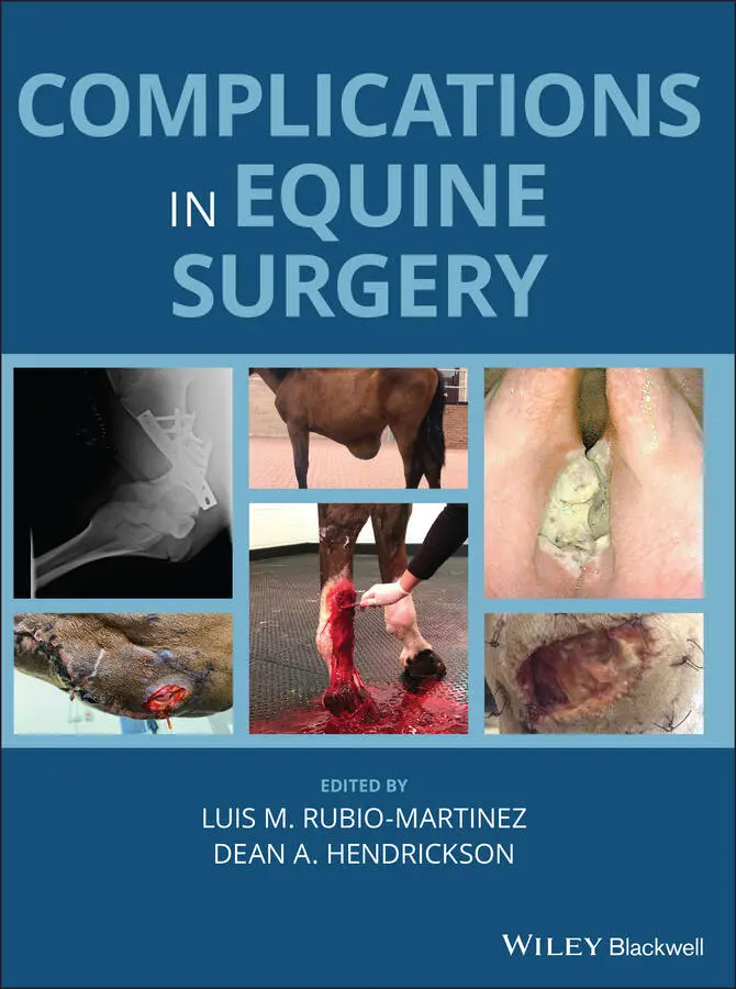
Offers the first resource specifically focused on complications encountered in equine surgery Takes a helpful format organized by body system Provides consistently formatted chapters for ease of use Covers clinically relevant information for dealing with technical and post-operative complications Presents more than 350 color images to illustrate the concepts described Written for general practitioners and specialists,
is an essential resource to decreasing morbidity and mortality and increasing surgical success in horses.
Complications in Equine Surgery — читать онлайн ознакомительный отрывок
Ниже представлен текст книги, разбитый по страницам. Система сохранения места последней прочитанной страницы, позволяет с удобством читать онлайн бесплатно книгу «Complications in Equine Surgery», без необходимости каждый раз заново искать на чём Вы остановились. Поставьте закладку, и сможете в любой момент перейти на страницу, на которой закончили чтение.
Интервал:
Закладка:
15 Chapter 22Figure 22.1 Laceration on the left side of the tongue following an inferior ...Figure 22.2 Transverse CT image at level of caudal aspect of mandibular Tria...Figure 22.3 Gingival ulceration following removal of the first prosthesis fo...Figure 22.4 Gingival hyperplasia and wound dehiscence with food pocketing 10...Figure 22.5 Surgical extraction of a developmentally displaced 201.Figure 22.6 Inadvertent exposure of the 5th pulp horn (dotted ellipse) of to...Figure 22.7 Open 6th pulp horn in tooth 106, 3 years following “bit seating....Figure 22.8 Small apical (caudal root) fragment (arrow) radiographically evi...Figure 22.9 Thick alveolar sequestrae removed 4 weeks following oral extract...Figure 22.10 (a) Oral aspect of an oro‐maxillary fistula following repulsion...Figure 22.11 Sinoscopic removal of feed material from the rostral maxillary ...Figure 22.12 Endoscopy of the right nasal cavity of a horse with an oro‐nasa...Figure 22.13 Compression of tooth 206 into its alveolus by a fulcrum during ...Figure 22.14 Chronic salivary leakage (solid arrow and rostral to it) follow...
16 Chapter 23Figure 23.1 Endoscopic appearance of esophageal stricture. The discolored ar...Figure 23.2 Esophageal stricture (arrows) diagnosed with contrast radiograph...Figure 23.3 Transcutaneous ultrasonographic image through the 9 thleft inter...Figure 23.4 Transnasal endoscopic appearance of the larynx of a horse with l...
17 Chapter 25Figure 25.1 Laparoscopic image with an arrow indicating location of splenic ...Figure 25.2 Laparoscopic image from a horse undergoing standing exploratory ...Figure 25.3 Ultrasonographic image of the left dorsolateral abdomen of a hor...Figure 25.4 Laparoscopic image from a horse undergoing standing exploratory ...Figure 25.5 Laparoscopic image from a horse undergoing standing nephrospleni...
18 Chapter 26Figure 26.1 Marked incisional edema surrounding an equine ventral midline ce...Figure 26.2 A hernia belt has been applied to a horse healing from incisiona...Figure 26.3 Appropriate closure of the linea alba following ventral midline ...Figure 26.4 An incise drape has been applied to cover an equine ventral midl...Figure 26.5 Ventral midline celiotomy incisional dehiscence treated with ful...Figure 26.6 Ventral midline celiotomy incisional dehiscence treated with ful...Figure 26.7 A Friesen horse that has developed a large hernia associated wit...Figure 26.8 A horse that has developed multiple small hernias associated wit...Figure 26.9 Mesh hernioplasty of a single large hernia that developed follow...
19 Chapter 27Figure 27.1 Portion of stomach wall sutured to a plastic drape to prevent ab...Figure 27.2 Mesenteric hematoma associated with a strangulating lipoma. A si...Figure 27.3 Method of occluding colonic mesenteric vessels with the TA‐90 TM...Figure 27.4 After large colon resection, as the anastomosis is allowed to re...Figure 27.5 Method of leaving an edge of mesentery (white arrow) that is suf...Figure 27.6 Selection of a site for mesenteric transection for an anastomosi...Figure 27.7 Diaphragmatic hernia in a horse with much of the large colon in ...Figure 27.8 (a) Rent in the duodenojejunal mesentery that strangulated small...Figure 27.9 Avulsion of the mesocolon from the most distal part of the small...Figure 27.10 Method of using the thumb forceps as a backstop and to elevate ...Figure 27.11 Method of using the back of the hand to retain any loops of int...
20 Chapter 28Figure 28.1 Monitoring (a) and treatment (b) used in the management of posto...Figure 28.2 Postoperative transabdominal ultrasonographic image of an 18‐yea...Figure 28.3 Postoperative transabdominal ultrasonographic image of a 30‐year...Figure 28.4 (a) Postoperative transabdominal ultrasonographic image from the...Figure 28.5 Postoperative transabdominal ultrasonographic evaluation of a 6‐...Figure 28.6 Transabdominal ultrasonographic image (obtained at the right ing...Figure 28.7 Cytology of peritoneal fluid from a horse with septic peritoniti...Figure 28.8 Schematic outlining the pathophysiology associated with pyrexia....Figure 28.9 Excessive per‐incisional edema can be an indication of a surgica...Figure 28.10 Ultrasonographic image of a ventral midline surgical site 10 da...Figure 28.11 Trans‐thoracic ultrasonographic appearance of a horse with pneu...Figure 28.12 Transtracheal wash kits are available and human central venous ...Figure 28.13 A 10‐year‐old Thoroughbred broodmare had a Cesarean section for...Figure 28.14 Photograph shows a horse that developed what appeared to be an ...Figure 28.15 Drainage of purulent material from the incision.Figure 28.16 Ultrasonographic image of a body wall defect in a 30‐year‐old p...Figure 28.17 Ultrasonographic image of the abdominal ventral midline of a 13...Figure 28.18 Diagnosis of a body wall hernia is typically made by the owner ...Figure 28.19 Abdominal hernia belt (CM Equine Products). Note that the herni...Figure 28.20 Pressure necrosis of the withers associated with an improperly ...Figure 28.21 Hernia formation should not affect athletic activity (horse fro...Figure 28.22 Horse with bilateral septic jugular vein thrombophlebitis; (a) ...Figure 28.23 Ultrasonographic appearance of the horse from Figure 28.23. Not...Figure 28.24 Mesenteric hemorrhage identified at repeat celiotomy in a 19‐ye...Figure 28.25 Ultrasonographic appearance of a hemoperitoneum. Note the large...Figure 28.26 Horse that had undergone a previous jejunoileostomy that develo...Figure 28.27 Ultrasonographic appearance of the intramural cecal abscess in ...Figure 28.28 (a) Bowel‐to‐bowel fibrinous adhesions: with fibrinous adhesion...Figure 28.29 Severe laminitis with rotation of the distal phalanx through th...
21 Chapter 30Figure 30.1 Laparoscopic incisional mesh hernioplasty: (a) Preoperative appe...Figure 30.2 The appearance of a seroma occupying the original hernia sac 24 ...Figure 30.3 Clinical appearance of chronic mesh infection. Three of several ...Figure 30.4 Laparoscopic image of a composite mesh prosthesis in position, i...Figure 30.5 Open mesh incisional hernioplasty. Preoperative (a) and 90 days ...Figure 30.6 Laparoscopic appearance of failed open mesh hernioplasty conduct...
22 Chapter 32Figure 32.1 Intraoperative videoendoscopic image of the ventral aspect of th...Figure 32.2 Postoperative videoendoscopic image of the ventral aspect of the...Figure 32.3 An epiglottic hook is being used elevate the aryepiglottic fold ...Figure 32.4 Postoperative videoendoscopic image of the ventral aspect of the...Figure 32.5 Granulation tissue mass associated with aryepiglottic fold follo...Figure 32.6 Postoperative videoendoscopic image of the epiglottis showing su...Figure 32.7 Videoendoscopic image of the corniculate processes of the laryng...
23 Chapter 33Figure 33.1 Anatomy of the sinuses. (a) Close‐up image of the sinus compartm...Figure 33.2 Photograph of the dorsal aspect of the head of a horse after fro...Figure 33.3 (a) Photograph of the left side of the head of a horse at approx...
24 Chapter 34Figure 34.1 Intraoperative view on a horse under general anesthesia and dors...Figure 34.2 Intraoperative view during an LTF procedure. Rostral is to the l...Figure 34.3 Radiographic assessment of the stability of the sutures after a ...Figure 34.4 (a) Endoscopic view of the pharynx of a horse that underwent a s...Figure 34.5 Endoscopic view of the nasopharynx of a horse 1 month after lase...
25 Chapter 35Figure 35.1 Latex arterial injection anatomical dissection showing the left ...Figure 35.2 (a) Tie back being performed on a horse under standing sedation....Figure 35.3 Left side (a) rostral to the left of the image and dorso‐rostral...Figure 35.4 (a) Tie back being performed on a horse under standing sedation....Figure 35.5 Tie back being performed on a horse under standing sedation. Ros...Figure 35.6 Endoscopic view of the lumen of the larynx and trachea showing p...Figure 35.7 Dorsal (a and b) and lateral (c) views of an anatomic specimen w...Figure 35.8 Resting endoscopic view of the larynx of a horse 48 hours after ...Figure 35.9 Dynamic endoscopic view of the larynx of a horse with dynamic up...Figure 35.10 Retrograde swallowing test. (a) The endoscope is introduced thr...Figure 35.11 Endoscopic view of the larynx of a horse that was coughing and ...Figure 35.12 Laser ventriculo‐cordectomy. (a) Fiber is placed inside the lef...Figure 35.13 Partial arytenoidectomy (PA). (a) Dysphagia after excessive tis...Figure 35.14 Laser transection of epiglottic entrapment. A low power (6 to 8...Figure 35.15 Permanent displacement of the soft palate associated with infla...
Читать дальшеИнтервал:
Закладка:
Похожие книги на «Complications in Equine Surgery»
Представляем Вашему вниманию похожие книги на «Complications in Equine Surgery» списком для выбора. Мы отобрали схожую по названию и смыслу литературу в надежде предоставить читателям больше вариантов отыскать новые, интересные, ещё непрочитанные произведения.


Обсуждение, отзывы о книге «Complications in Equine Surgery» и просто собственные мнения читателей. Оставьте ваши комментарии, напишите, что Вы думаете о произведении, его смысле или главных героях. Укажите что конкретно понравилось, а что нет, и почему Вы так считаете.