Johan P. Reyneke - Essentials of Orthognathic Surgery
Здесь есть возможность читать онлайн «Johan P. Reyneke - Essentials of Orthognathic Surgery» — ознакомительный отрывок электронной книги совершенно бесплатно, а после прочтения отрывка купить полную версию. В некоторых случаях можно слушать аудио, скачать через торрент в формате fb2 и присутствует краткое содержание. Жанр: unrecognised, на английском языке. Описание произведения, (предисловие) а так же отзывы посетителей доступны на портале библиотеки ЛибКат.
- Название:Essentials of Orthognathic Surgery
- Автор:
- Жанр:
- Год:неизвестен
- ISBN:нет данных
- Рейтинг книги:4 / 5. Голосов: 1
-
Избранное:Добавить в избранное
- Отзывы:
-
Ваша оценка:
Essentials of Orthognathic Surgery: краткое содержание, описание и аннотация
Предлагаем к чтению аннотацию, описание, краткое содержание или предисловие (зависит от того, что написал сам автор книги «Essentials of Orthognathic Surgery»). Если вы не нашли необходимую информацию о книге — напишите в комментариях, мы постараемся отыскать её.
Essentials of Orthognathic Surgery — читать онлайн ознакомительный отрывок
Ниже представлен текст книги, разбитый по страницам. Система сохранения места последней прочитанной страницы, позволяет с удобством читать онлайн бесплатно книгу «Essentials of Orthognathic Surgery», без необходимости каждый раз заново искать на чём Вы остановились. Поставьте закладку, и сможете в любой момент перейти на страницу, на которой закончили чтение.
Интервал:
Закладка:
The clinician should be aware that in McNamara analysis, the effective lengths of the midface and the mandibular length are related and are expressed as small , medium , and large . The maxillomandibular differential is determined by subtracting the midfacial length from the mandibular length. In small individuals (mixed dentition stage), the difference should be 20 to 23 mm. In medium-sized individuals, there should be a difference of 27 to 30 mm, and in large individuals, the difference should be 30 to 33 mm (Fig 2-58).
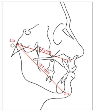
Fig 2-58Maxillomandibular anteroposterior relationship. The maxillomandibular differential of 24 mm shown here would be ideal for a small to medium individual. The maxillomandibular ratio of 1:1.24, however, indicates a discrepancy in the relationship between the jaws. According to McNamara’s normative standards (see Table 2-11), a midfacial length of 97 mm would be better related to a mandibular length of 126 to 129 mm. This may indicate a slight anteroposterior deficiency of the mandible.
The graph in Fig 2-59 illustrates the relationship between midfacial length, mandibular length, and lower anterior facial height. A discrepancy greater or smaller than the normative values would indicate a disharmonious relationship between the maxilla and mandible. Alternative analysis should be used to identify which jaw is at fault. The midfacial length is measured from Co to A-point, whereas the mandibular length is measured from Co to the anatomical Gn. The linear relationship between the midfacial length and the length of the mandible is called the ratio of effective maxillary to mandibular length . Any specific maxillary (midfacial) length will correspond to a specific mandibular length within a given range (see Table 2-11). The normal adult ratio should be maxilla (Co-A) to mandible (Co-Gn) = 1:1.3.
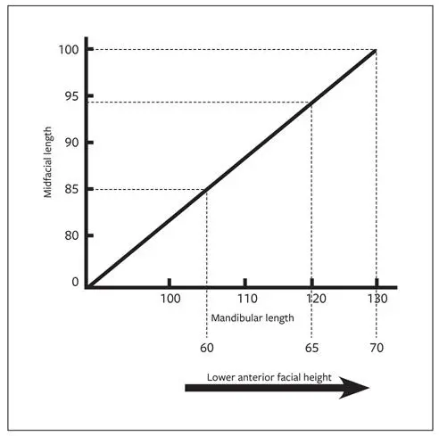
Fig 2-59The relationship between midfacial (maxillary) length and mandibular length is generally linear and dependent on size rather than on age or sex. An individual with a maxillary length of 100 mm should have an effective mandibular length of 130 mm. The difference between the maxillary and mandibular lengths in this instance would be 30 mm. (Adapted from McNamara and Brudon, 1993, with permission).
During normal growth, the ratio decreases from 1:1.25 for an 8-year-old child to the adult value. The effective mandibular length increases faster than the length of the maxilla, at the rate of 0.005 mm per year (ratios of 1:1.26 at 10 years of age, 1:1.27 at 12 years, 1:1.28 at 14 years, and 1:1.29 at 16 years). The clinician should be aware that a change in this value may indicate disproportionate growth, but it does not indicate which jaw is at fault.
The esthetic effect of the effective maxillary and mandibular lengths is closely related to the anterior facial height and, therefore, the angle formed between A-point, Co, and Gn. This angle normally decreases as an individual ages (Fig 2-60).
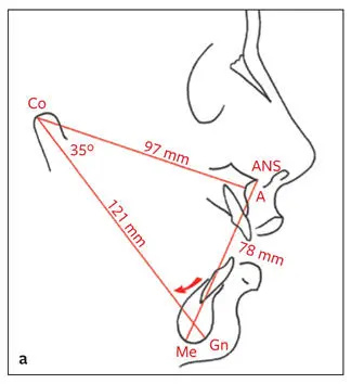
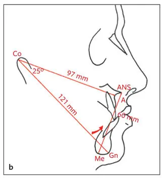
Fig 2-60The esthetic effect of the change in lower anterior facial height and backward (a) and forward (b) rotation of the chin. In both a and b , the midfacial lengths (97 mm) and mandibular lengths (121 mm) are the same. In a , the lower anterior facial height (ANS-Me) is 78 mm, whereas in b , it is 60 mm. The angle between Co-A and Co-Gn is 35 degrees in a but 25 degrees in b .
Skeletal anteroposterior relationships are summarized in Table 2-12.
Table 2-12| Summary of skeletal anteroposterior relationships
| Anteroposterior relationship | Normal value | |
| Maxilla | To anterior cranial base (SNA) To mandible (ANB) To mandible (Wits appraisal) Maxillary depth: A-point to N perpendicular to FH To mandibular length: Co-A:Co-Gn | 82 degrees 2 degrees AH 1 mm behind BO (males) AO and BO coincide (females) 0 mm 1:1.3 |
| Mandible | Mandibular plane angle S-N to Go-Gn To anterior cranial base (SNB) To maxilla (ANB) To maxilla (Wits appraisal) To maxillary length: Co-Gn:Co-A | 32 degrees 80 degrees 2 degrees BO 1 mm ahead of AO (males) BO and AO coincide (females) 1.3:1 |
Skeletal vertical relationships
Midface to lower face skeletal height
Skeletal vertical relationships are measured from N to ANS and from ANS to Me. A vertical line is drawn perpendicular to the FH anterior to the face. In turn, perpendicular lines are drawn to the vertical line from N, ANS, and Me, and the distance is measured from N to ANS and from ANS to Me (Fig 2-61). The normal values are 53 mm from N to ANS and 65 mm from ANS to Me. However, the relationship between the vertical heights is more important than the measurement. A ratio of 5:6 is normal. In most individuals with vertical dentofacial deformities, the lower measurement (ANS-Me) will be affected, which in turn will affect the relationship with the upper measurement (N-ANS). The ANS-Me distance will be increased in individuals with vertical maxillary excess, vertical mandibular excess, and open bites. The lower measurement (ANS-Me) will be decreased in individuals with vertical maxillary deficiency, closed bites, deep bites, and vertical mandibular deficiency.
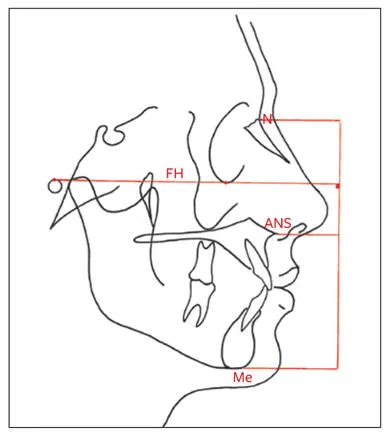
Fig 2-61Midface and lower face skeletal heights. N-ANS:ANS-Me = 5:6.
Analysis of dental relationships
Maxillary incisor evaluation
Maxillary incisor position
Steiner analysis. According to the Steiner analysis, the relative location of the maxillary incisor is determined by relating the angulation of the incisor to the N-A line. The most anterior point of the maxillary incisors should be 4 mm ahead of N-A, with an axial inclination of 22 degrees to this line (Fig 2-62a).
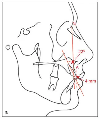
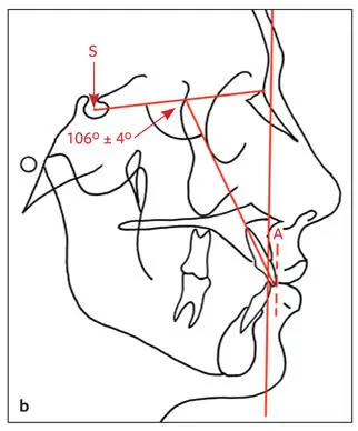
Fig 2-62 (a) Maxillary incisor position (Steiner): 22 degrees to N-A, 4 mm ahead of N-A. (b) An alternative measurement of maxillary incisor angulation. The angle between the cranial base (S-N) and maxillary incisor (apex to incisor tip) should be 106 ± 4 degrees.
The linear relationship of the incisor tip to N-A provides information on the anteroposterior position of the incisor to the maxilla, but not to the entire facial complex. In orthognathic cases, this measurement can be helpful in determining the preoperative positioning of the tooth if the relation of the maxilla to the cranial base is also considered. In individuals with dentofacial deformities, the N-A line is often not a reliable basis to assess the incisor position because of the skeletal disharmony between cranial base and maxilla. In these cases, a vertical line through A-point should be substituted for N-A (Fig 2-62b). A helpful additional measurement is the angulation between a line through the maxillary incisor (apex to incisor tip) and a line through the anterior cranial base (S-N), which should be 106 ± 4 degrees (see Fig 2-62b).
Читать дальшеИнтервал:
Закладка:
Похожие книги на «Essentials of Orthognathic Surgery»
Представляем Вашему вниманию похожие книги на «Essentials of Orthognathic Surgery» списком для выбора. Мы отобрали схожую по названию и смыслу литературу в надежде предоставить читателям больше вариантов отыскать новые, интересные, ещё непрочитанные произведения.


Обсуждение, отзывы о книге «Essentials of Orthognathic Surgery» и просто собственные мнения читателей. Оставьте ваши комментарии, напишите, что Вы думаете о произведении, его смысле или главных героях. Укажите что конкретно понравилось, а что нет, и почему Вы так считаете.