Point-of-Care Ultrasound Techniques for the Small Animal Practitioner
Здесь есть возможность читать онлайн «Point-of-Care Ultrasound Techniques for the Small Animal Practitioner» — ознакомительный отрывок электронной книги совершенно бесплатно, а после прочтения отрывка купить полную версию. В некоторых случаях можно слушать аудио, скачать через торрент в формате fb2 и присутствует краткое содержание. Жанр: unrecognised, на английском языке. Описание произведения, (предисловие) а так же отзывы посетителей доступны на портале библиотеки ЛибКат.

- Название:Point-of-Care Ultrasound Techniques for the Small Animal Practitioner
- Автор:
- Жанр:
- Год:неизвестен
- ISBN:нет данных
- Рейтинг книги:5 / 5. Голосов: 1
-
Избранное:Добавить в избранное
- Отзывы:
-
Ваша оценка:
Point-of-Care Ultrasound Techniques for the Small Animal Practitioner: краткое содержание, описание и аннотация
Предлагаем к чтению аннотацию, описание, краткое содержание или предисловие (зависит от того, что написал сам автор книги «Point-of-Care Ultrasound Techniques for the Small Animal Practitioner»). Если вы не нашли необходимую информацию о книге — напишите в комментариях, мы постараемся отыскать её.
New chapters in
cover ultrasound-guided nerve blocks, musculoskeletal, brain imaging, and applications of focused ultrasound techniques in cats, exotics and marine mammals—making it an essential purchase for veterinarians wanting to incorporate point-of-care ultrasound techniques into their veterinary practices.
Presents a standardized approach to point-of-care ultrasound as an extension of the physical exam, including trauma, non-trauma, and monitoring applications Includes coverage of new techniques for focused ultrasound exams, including lung, anesthesia and ultrasound guided nerve blocks, transcranial brain imaging, musculoskeletal, volume status evaluation, and rapid assessment for treatable forms of shock Adds cats, exotic and wildlife mammals, and marine mammals to the existing canine coverage Emphasizes the integration of point-of-care ultrasound techniques for optimizing patient care and accurate patient assessment Offers access to a companion website with supplementary video clips showing many clinically relevant didactic examples The second edition of
is an excellent resource for veterinary practitioners, ranging from the general practitioner to nearly all clinical specialists, including internal medicine, oncology, cardiology, emergency and critical care, anesthesiology, ophthalmology, exotics, and zoo medicine specialists, and veterinary students.
Point-of-Care Ultrasound Techniques for the Small Animal Practitioner — читать онлайн ознакомительный отрывок
Ниже представлен текст книги, разбитый по страницам. Система сохранения места последней прочитанной страницы, позволяет с удобством читать онлайн бесплатно книгу «Point-of-Care Ultrasound Techniques for the Small Animal Practitioner», без необходимости каждый раз заново искать на чём Вы остановились. Поставьте закладку, и сможете в любой момент перейти на страницу, на которой закончили чтение.
Интервал:
Закладка:
Air‐filled Colon
Air causing interference is usually not problematic since dogs and cats in right lateral recumbency often have their colon (by gravity) fall adequately away from the SR view. However, it is not uncommon for the air‐filled colon to dirty shadow through the far‐field and is typically present caudally or to the screen's right. Study the presence of the colon and its dirty shadowing in Figures 6.17– 6.19.
False Mirror Image in Cats
Commonly in cats and less so in small dogs, both kidneys are imaged in the same field of view at the SR view. This is unlikely to be a mirror image artifact, which requires a strong air–soft tissue interface ( Figure 6.22) (see Chapter 39).
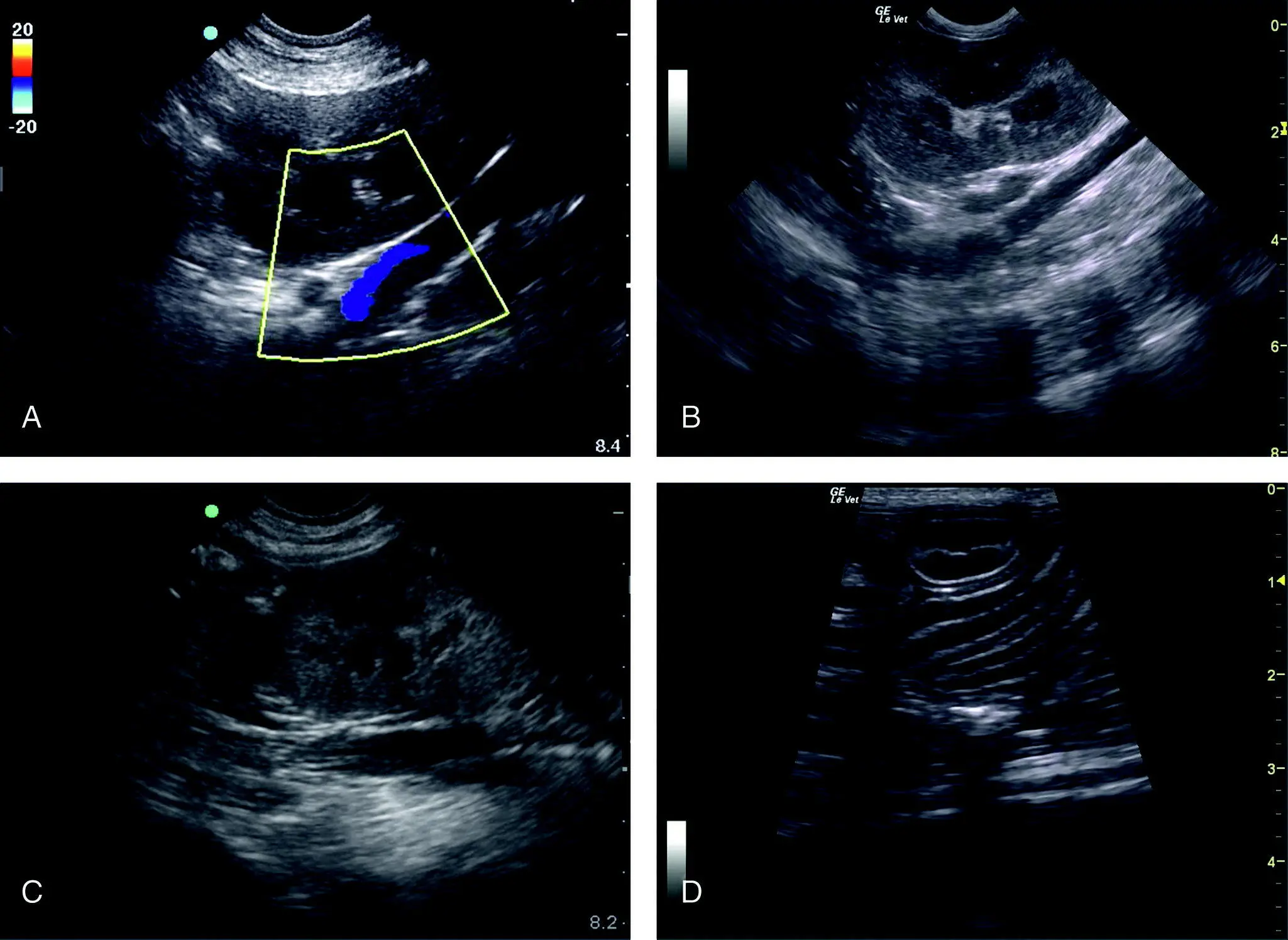
Figure 6.22. Pitfalls at the SR view and linear stripes are not free fluid. In (A) and (B) are similar images with color flow Doppler showing the great vessels (CVC and abdominal aorta) that are clearly linear and anechoic in B‐mode in (B). In (C) it is difficult to tell if the anechoic (hypoechoic) linear stripe is great vessels or small intestine; however, when fanning as a general rule, a great vessel will be an anechoic tube with pulsation and the small intestine will have sonographically layered linear stripes as in (D). The author refers to small intestinal wall layering in transverse orientation as appearing like “hamburgers” (oval with a line through its center) and in longitudinal (sagittal) orientation as “highways” (median stripe and two shoulders of the road). Note both planes of orientation are in the image.
Source: Reproduced with permission of Dr Gregory Lisciandro, Hill Country Veterinary Specialists and FASTVet.com, Spicewood, TX.
Edge Shadowing
Any reflection of the beam (echoes) from a curved soft tissue surface will often extend a hypoechoic to anechoic line off the margin of the curved surface through the far‐field. In haste, the artifact can mimic free fluid (see Figure 6.20and Figure 3.2).
False Positives
Linear Anechoic Stripes
These rarely represent free fluid and are more likely small intestine (intraabdominal) or the great vessels (retroperitoneal). Color flow Doppler is rarely needed to differentiate between these anatomical structures and free fluid, but may be used if in B‐mode the determination cannot be made using combinations of longitudinal and transverse orientations and observing for vessel pulsation (see Figure 6.22).
Pearl:Remember classic positives at the SR view are anechoic (black) triangles, not linear stripes.
Retroperitoneal Fluid versus Peritoneal Fluid
The problem posed in people differs from our veterinary patients which are less obese and have a suspended (versus attached) colon. In lateral recumbency, retroperitoneal fluid will remain static and not fall away from the field of view (see Figure 6.19). Increasing your depth will also help interrogate the two spaces, as well as considering the findings at the other AFAST views. The SR view is rarely the only positive view in intraabdominal effusions because it is the least gravity dependent. See examples in Chapter 10.
False Negatives
Serial AFAST Examinations Increase Sensitivity
Don’t sweat questionable small pockets of free fluid; stabilize the patient and do a serial (repeat) AFAST and assign an abdominal fluid score. Minimally, a second AFAST four hours later should be performed, and sooner if the patient is questionable or unstable (acep.org; Lisciandro et al. 2009; Blackbourne et al. 2004; Boysen and Lisciandro 2013).
AFAST Cysto‐Colic View
| Questions Asked at the CC View | |
| Is there any free fluid in the abdominal (peritoneal) cavity? | Yes or no |
| How much free fluid is at the CC view using the AFAST AFS system? | 0, 1/2, 1 |
| What does the urinary bladder look like? a | Unremarkable or abnormal |
| Is the patient intact reproductively? a | Yes or no |
| Could I be misinterpreting an artifact or pitfall as pathology? | Know pitfalls and artifacts |
aIt is important to know that the AFAST target organ approach for parenchymal abnormalities is binary as “unremarkable” or “abnormal” to capture the case for additional imaging and confirmatory testing. More interpretative skills may be gained through experience, and additional ultrasound study and training.
Source: Reproduced with permission of Dr Gregory Lisciandro, Hill Country Veterinary Specialists and FASTVet.com, Spicewood, TX.
The classic CC view includes imaging the urinary bladder (when present) against the abdominal wall in the far‐field by directing the probe externally placed dorsolateral from the midline and the scanning plane directed toward the tabletop into the most gravity‐dependent pouch formed between the bladder and the gravity‐dependent body wall ( Figures 6.23and 6.24). The most gravity‐dependent region at the CC view is referred to as the “CC pouch” (Lisciandro 2011, 2014a; Boysen and Lisciandro 2013). The CC view is slightly a misnomer because of its target organs; only the urinary bladder is specifically imaged whereas the colon is not (Lisciandro 2014a). However, an air‐filled colon will do the following.
Obscure imaging of the “CC pouch” by dirty shadowing.
May get between the urinary bladder and body wall, mimicking urine sediment and calculi by its dirty shadowing (see Figure 6.27and Chapter 11).
A stool‐filled colon may mimic pathology and thus a digital rectal examination and/or caudal abdominal palpation or both as part of a good physical exam can prove helpful along with additional imaging.
Place the probe dorsolateral to midline and direct toward the tabletop into the most gravity‐dependent region of the CC view called the “CC pouch” where the apical urinary bladder and the abdominal wall in the far‐field meet. If the urinary bladder is not in view, slide the probe toward the pubis with some fanning as needed. The process should be a slide and fan, followed by a slide and fan toward the pubis looking for the urinary bladder.
If the colon is creating dirty shadowing, then sweep the probe closer to midline while directing the ultrasound beam into the “CC pouch.”
When the urinary bladder is located, then fan through it in both directions in longitudinal (sagittal) orientation with the same methodology used for the gallbladder and left kidney previously at the DH and SR views, respectively. It is very important to apply minimal probe pressure over the urinary bladder because it is easily deformed by excessive compression of the probe pushing into the patient's body wall. The sonographer can easily appreciate this “artifact” by seeing the urinary bladder appear as a “dumbbell” or its wall flattened out in the far‐field against the body wall in the “CC pouch” (see Figure 6.26).
After interrogating the urinary bladder, return to your starting point by rocking back to the “CC pouch” for one final look in this most gravity‐dependent region for free intraabdominal fluid.
Serial AFAST provides the opportunity to image and interrogate the urinary bladder, which is expected to fill with urine during fluid resuscitation, and if the urinary bladder is not seen then interventional and therapeutic decisions are made depending on the patient.
Urine production may be estimated using our AFAST CC formula of length (cm) × width (cm) × height (cm) × 0.625 = estimation of urinary bladder volume in milliliters (Lisciandro and Fosgate 2017). With serial measurements over time, urine output may be estimated. Excessive probe pressure will distort the urinary bladder, leading to erroneous measurements.
Читать дальшеИнтервал:
Закладка:
Похожие книги на «Point-of-Care Ultrasound Techniques for the Small Animal Practitioner»
Представляем Вашему вниманию похожие книги на «Point-of-Care Ultrasound Techniques for the Small Animal Practitioner» списком для выбора. Мы отобрали схожую по названию и смыслу литературу в надежде предоставить читателям больше вариантов отыскать новые, интересные, ещё непрочитанные произведения.


Обсуждение, отзывы о книге «Point-of-Care Ultrasound Techniques for the Small Animal Practitioner» и просто собственные мнения читателей. Оставьте ваши комментарии, напишите, что Вы думаете о произведении, его смысле или главных героях. Укажите что конкретно понравилось, а что нет, и почему Вы так считаете.