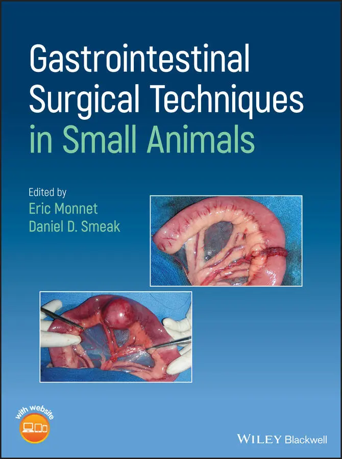
Gastrointestinal Surgical Techniques in Small Animals
Здесь есть возможность читать онлайн «Gastrointestinal Surgical Techniques in Small Animals» — ознакомительный отрывок электронной книги совершенно бесплатно, а после прочтения отрывка купить полную версию. В некоторых случаях можно слушать аудио, скачать через торрент в формате fb2 и присутствует краткое содержание. Жанр: unrecognised, на английском языке. Описание произведения, (предисловие) а так же отзывы посетителей доступны на портале библиотеки ЛибКат.
- Название:Gastrointestinal Surgical Techniques in Small Animals
- Автор:
- Жанр:
- Год:неизвестен
- ISBN:нет данных
- Рейтинг книги:5 / 5. Голосов: 1
-
Избранное:Добавить в избранное
- Отзывы:
-
Ваша оценка:
Gastrointestinal Surgical Techniques in Small Animals: краткое содержание, описание и аннотация
Предлагаем к чтению аннотацию, описание, краткое содержание или предисловие (зависит от того, что написал сам автор книги «Gastrointestinal Surgical Techniques in Small Animals»). Если вы не нашли необходимую информацию о книге — напишите в комментариях, мы постараемся отыскать её.
Describes techniques for canine and feline gastrointestinal surgery in detail Presents the state of the art for GI surgery in dogs and cats Includes access to a companion website with video clips demonstrating techniques
is an essential resource for small animal surgeons and veterinary residents.
Gastrointestinal Surgical Techniques in Small Animals — читать онлайн ознакомительный отрывок
Ниже представлен текст книги, разбитый по страницам. Система сохранения места последней прочитанной страницы, позволяет с удобством читать онлайн бесплатно книгу «Gastrointestinal Surgical Techniques in Small Animals», без необходимости каждый раз заново искать на чём Вы остановились. Поставьте закладку, и сможете в любой момент перейти на страницу, на которой закончили чтение.
Интервал:
Закладка:
Table of Contents
1 Cover
2 List of Contributors List of Contributors Chad Lothamer, DVM, DAVDC Dentistry and Oral Surgery College of Veterinary Medicine University of Tennessee Knoxville, TN, USA Eric Monnet, DVM, PhD, DACVS, DECVS Department of Clinical Sciences College of Veterinary Medicine and Biomedical Sciences Colorado State University Fort Collins, CO, USA Jennifer Rawlinson, DVM, DAVDC Department of Clinical Sciences College of Veterinary Medicine and Biomedical Sciences Colorado State University Fort Collins, CO, USA Bernard Séguin, DVM, MS, DACVS Department of Clinical Sciences College of Veterinary Medicine and Biomedical Sciences Colorado State University Fort Collins, CO, USA Daniel D. Smeak, DVM, DACVS Department of Clinical Sciences College of Veterinary Medicine and Biomedical Sciences Colorado State University Fort Collins, CO, USA
3 Preface Preface Small animal general surgery has evolved rapidly in the past decade. Procedures have become increasingly complex, including the expansion of minimally invasive surgery options. Gastrointestinal conditions requiring surgical therapy are very common in small animal practice. Therefore, we have created this gastrointestinal surgery textbook to serve as a current and comprehensive reference tool for general practitioners, surgery residents in training, and surgeons. In this textbook, current techniques for gastrointestinal surgery are thoroughly described with detailed illustrations embedded in the text. With this in mind we chose to work closely with Molly Pullen ( www.mborman.com ) for her expert pen‐and‐ink illustrations. In addition, when necessary, surgical techniques are supplemented with intraoperative images. For each technique described, tips are included by the authors to explain the surgeon's preferences among all the techniques depicted, and to give details to help the reader perform the procedures effectively. Our intent for adding tips was to allow chapter authors the ability to express their preferences in technique, many of which are not evidence‐based. In this textbook we placed a strong emphasis on description of surgical techniques. We chose not to delve heavily in the pathophysiology or the diagnostic workup for the different surgical conditions mentioned because those topics are well described in other textbooks. We would like to thank all the authors who assisted us during the arduous process of developing this new textbook. In the future, this textbook will be updated as needed to follow the progress and advances in veterinary gastrointestinal surgery.
4 About the Companion Website About the Companion Website Don’t forget to visit the companion website for this book: www.wiley.com/go/monnet/gastrointestinal There you will find valuable material designed to enhance your learning, including: Illustrations Video clips Scan this QR code to visit the companion website
5 Section I: General Concepts Section I General Concepts
1 Gastrointestinal Healing 1 Gastrointestinal Healing Eric Monnet and Daniel D. Smeak Department of Clinical Sciences, College of Veterinary Medicine and Biomedical Sciences, Colorado State University, Fort Collins, CO, USA The gastrointestinal tract heals as any other tissue in an orderly manner, with an inflammatory phase, a debridement phase, a granulation phase, and a maturation phase. However, there are some characteristics very specific to the gastrointestinal tract that sets it apart from other healing tissues. The healing process of the gastrointestinal tract should not only re‐establish the anatomical integrity of the tract but also its function. Healing should happen with minimal scaring and stricture formation that could impede the motility of the gastrointestinal tract. Additionally, the formation of adhesions, even though rare in small animal surgery, could deteriorate gastrointestinal motility and should be minimized.
1.1 Anatomy 1.1 Anatomy The wall of the gastrointestinal tract has a mucosa, a submucosa, a muscularis, and a serosa, except the esophagus and the distal rectum. The mucosa has three distinct layers: the epithelium, the lamina propria and the muscularis mucosa. The lamina propria layer is made of vessels, lymphatics, and mesenchymal cells whereas the muscularis mucosa is a thin muscle layer. After completing the anastomosis, the mucosa heals very fast by epithelial cells migration over the defect providing a rapid barrier from the intestinal content. For intestinal healing to occur, a good surgical apposition of layers is required; everting or inverting patterns interfere with mucosal healing. The submucosa layer, incorporating the bulk of the collagen, is the holding layer in intestinal surgery. Type I collagen (68%) predominates in the submucosa, followed by type III (20%) and finally collagen type V (12%) (Thornton and Barbul 1997). It is a loose connective tissue with lymphatic, nerve fibers, ganglia, and blood vessels that should be preserved during surgery. The muscularis layer consists of an inner circular muscle layer, a longitudinal outer muscle layer and collagen fibers. The serosal surface is made of connective tissue with mesothelial cells, lymphatics, and blood vessels. The serosa is important in the healing process because it helps prevent leakage of the gastrointestinal content in the immediate post‐operative period.
1.2 Phases of Wound Healing 1.2 Phases of Wound Healing 1.2.1 Partial Thickness Injury A partial thickness injury affecting only the mucosa or the serosa heals with epithelial cells and mesothelial cells proliferation without scaring. A full‐thickness trauma of the gastrointestinal tract results in an inflammatory reaction and a non‐epithelial cell proliferation that can result in scaring secondary to fibroblast activity (Thornton and Barbul 1997; Thompson et al. 2006).
1.3 Factors Affecting Gastrointestinal Tract Healing 1.3 Factors Affecting Gastrointestinal Tract Healing 1.3.1 Ischemia and Tissue Perfusion Ischemia interferes with healing of any tissue and especially the gastrointestinal tract. The oxygen delivery to the peripheral tissue depends on the anatomy of the capillaries, vasomotor control, and oxygen saturation. The tissue perfusion depends on the amount of soft tissue trauma and especially trauma to the blood supply of the loop of intestine. The placement of tight sutures interferes with tissue perfusion and may increase the risk of dehiscence, especially in the colon and esophagus (Shikata et al. 1982; Chung 1987; Jonsson and Hogstrom 1992; Thornton and Barbul 1997). Hypovolemia and hypotension are critical factors that divert blood flow to essential organs, and oxygen delivery is also very important for collagen synthesis. A partial pressure of oxygen of at least 40 mmHg is required for collagen synthesis. During a hypovolemic event, the gastrointestinal tract downregulates its own blood flow. Anemia does not interfere with healing as long as the patient has a good cardiac output to compensate (Thompson et al. 2006).
References 2 Suture Materials, Staplers, and Tissue Apposition Devices 2.1 Suture Materials 2.2 Staplers, Linear, Circular, Skin Staples References 3 Suture Patterns for Gastrointestinal Surgery 3.1 One‐ or Two‐Layer Closure 3.2 Tissue Inversion, Eversion, or Apposition 3.3 Stapled or Hand‐Sutured Anastomosis 3.4 Appositional Suture Patterns 3.5 Inverting Suture Patterns 3.6 Special Supplementary Patterns: Purse‐String Bibliography 4 Feeding Tubes 4.1 Nasoesophageal and Nasogastric Tubes 4.2 Esophagostomy Tube 4.3 Gastrostomy Tube 4.4 Jejunostomy Tube 4.5 Gastrojejunostomy Tube References 5 Drainage Techniques for the Peritoneal Space 5.1 Indications 5.2 Techniques 5.3 Tips 5.4 Complications and Aftercare References
6 Section II: Oral Cavity 6 Maxillectomy and Mandibulectomy 6.1 Indications 6.2 Surgical Techniques 6.3 Tips 6.4 Complications and Post‐Operative Care References 7 Glossectomy 7.1 Indications 7.2 Technique 7.3 Tips 7.4 Complications References 8 Tonsillectomy 8.1 Indications 8.2 Technique 8.3 Tips 8.4 Complications References 9 Palatal and Oronasal Defects 9.1 Indications 9.2 Surgical Techniques 9.3 Tips 9.4 Complications References 10 Salivary Gland Surgery 10.1 Indications 10.2 Techniques 10.3 Tips 10.4 Complications and Outcome Bibliography
Читать дальшеИнтервал:
Закладка:
Похожие книги на «Gastrointestinal Surgical Techniques in Small Animals»
Представляем Вашему вниманию похожие книги на «Gastrointestinal Surgical Techniques in Small Animals» списком для выбора. Мы отобрали схожую по названию и смыслу литературу в надежде предоставить читателям больше вариантов отыскать новые, интересные, ещё непрочитанные произведения.


Обсуждение, отзывы о книге «Gastrointestinal Surgical Techniques in Small Animals» и просто собственные мнения читателей. Оставьте ваши комментарии, напишите, что Вы думаете о произведении, его смысле или главных героях. Укажите что конкретно понравилось, а что нет, и почему Вы так считаете.