Point-of-Care Ultrasound Techniques for the Small Animal Practitioner
Здесь есть возможность читать онлайн «Point-of-Care Ultrasound Techniques for the Small Animal Practitioner» — ознакомительный отрывок электронной книги совершенно бесплатно, а после прочтения отрывка купить полную версию. В некоторых случаях можно слушать аудио, скачать через торрент в формате fb2 и присутствует краткое содержание. Жанр: unrecognised, на английском языке. Описание произведения, (предисловие) а так же отзывы посетителей доступны на портале библиотеки ЛибКат.

- Название:Point-of-Care Ultrasound Techniques for the Small Animal Practitioner
- Автор:
- Жанр:
- Год:неизвестен
- ISBN:нет данных
- Рейтинг книги:5 / 5. Голосов: 1
-
Избранное:Добавить в избранное
- Отзывы:
-
Ваша оценка:
Point-of-Care Ultrasound Techniques for the Small Animal Practitioner: краткое содержание, описание и аннотация
Предлагаем к чтению аннотацию, описание, краткое содержание или предисловие (зависит от того, что написал сам автор книги «Point-of-Care Ultrasound Techniques for the Small Animal Practitioner»). Если вы не нашли необходимую информацию о книге — напишите в комментариях, мы постараемся отыскать её.
New chapters in
cover ultrasound-guided nerve blocks, musculoskeletal, brain imaging, and applications of focused ultrasound techniques in cats, exotics and marine mammals—making it an essential purchase for veterinarians wanting to incorporate point-of-care ultrasound techniques into their veterinary practices.
Presents a standardized approach to point-of-care ultrasound as an extension of the physical exam, including trauma, non-trauma, and monitoring applications Includes coverage of new techniques for focused ultrasound exams, including lung, anesthesia and ultrasound guided nerve blocks, transcranial brain imaging, musculoskeletal, volume status evaluation, and rapid assessment for treatable forms of shock Adds cats, exotic and wildlife mammals, and marine mammals to the existing canine coverage Emphasizes the integration of point-of-care ultrasound techniques for optimizing patient care and accurate patient assessment Offers access to a companion website with supplementary video clips showing many clinically relevant didactic examples The second edition of
is an excellent resource for veterinary practitioners, ranging from the general practitioner to nearly all clinical specialists, including internal medicine, oncology, cardiology, emergency and critical care, anesthesiology, ophthalmology, exotics, and zoo medicine specialists, and veterinary students.
Point-of-Care Ultrasound Techniques for the Small Animal Practitioner — читать онлайн ознакомительный отрывок
Ниже представлен текст книги, разбитый по страницам. Система сохранения места последней прочитанной страницы, позволяет с удобством читать онлайн бесплатно книгу «Point-of-Care Ultrasound Techniques for the Small Animal Practitioner», без необходимости каждый раз заново искать на чём Вы остановились. Поставьте закладку, и сможете в любой момент перейти на страницу, на которой закончили чтение.
Интервал:
Закладка:
Source: Reproduced with permission of Dr Gregory Lisciandro, Hill Country Veterinary Specialists and FASTVet.com, Spicewood, TX.
When free fluid is present, it is most commonly seen between the spleen and cranial left kidney, or between the cranial pole of the left kidney and the wall of the colon ( Figure 6.19).
In dogs, the spleen may be used to locate the left kidney by following it caudally and medially because of its anatomical association with the left kidney, or by fanning along the great vessels since the left kidney is closely attached via its relatively short renal artery and vein. If you are having problems finding the left kidney, you have a few options.
The spleen in dogs and cats may be used to locate the left kidney by following the spleen caudally and medially to bring you to the left kidney.
Fan along the great vessels, paying attention to the probe's direction, recalling that the left kidney (and right) are closely attached via their respective renal artery and vein.
Making sure that you have not drifted too far ventrally from the angle of the costal arch and hypaxial junction and into the abdomen.
You are pushing (compression) too hard into the patient and in fact pushing the left kidney (and right) out of view.
Pearl:In most cats and many small dogs, both the left and right kidneys can be viewed through the single SR (or HR) view with increased depth ( Figures 6.20and 6.21).
Fanning dorsally screens for any pathology associated with the great vessels (aorta and caudal vena cava) and adrenal glands. The great vessels are common confounders and cause false positives, which are easily overcome by remembering that free fluid is rarely a linear anechoic (black) stripe(s) but rather anechoic triangulations, and thus linear anechoic stripes are more likely to be blood vessels and intestinal tract (see Figures 6.18and 6.20). The only major exception is anechoic stripe(s) at the DH view between the liver and diaphragm (Lisciandro et al. 2015, 2019; Romero et al. 2015).
The great vessels are identified in B‐mode by their shape, being linear in longitudinal and circular in transverse orientation with pulsation. Color flow Doppler may also be used to detect flow. Turning your probe transversely (turn left or counterclockwise) should change the vessel's appearance from a linear anechoic (black) rectangular shape to an anechoic (black) circle.
Retroperitoneal fluid raises the suspicion for hemorrhage, urine, and sterile and septic effusions placed into clinical context, and when found and safely accessible, fluid sampling should take place, and when inaccessible or too risky to sample, more advanced imaging is likely indicated.
Cranial to the kidneys, origin of retroperitoneal fluid would generally include the kidneys, vertebral bodies, the great vessels and adrenal glands.
Caudal to the kidneys, origin of retroperitoneal fluid would generally include the kidneys, ureters, vertebral bodies, and pelvis (see Figure 6.19, see also Chapters 10and 11).
Retroperitoneal versus intraabdominal (peritoneal) fluid can be further assessed by changing patient position by moving them to a standing position and seeing if the free fluid remains in the least gravity‐dependent SR view (retroperitoneal more likely), and noting that the least gravity‐dependent SR view is rarely the only positive AFAST view in right lateral recumbency.
Pearl:Retroperitoneal fluid is not part of the AFS. Its size should be noted by its largest dimension (length, width, height) measured by either the “eyeball method” (using the centimeter scale on the ultrasound screen), or more precisely using your machine's caliper function.
Typical SR View Positives
The majority of positives at the SR view are classically anechoic (black) triangles formed between the cranial pole of the kidney and colon (see Figure 6.19) and between the spleen and the cranial pole of the kidney.
Artifacts
Generally, the SR view has few artifacts which are mostly colon and stomach related. The anatomy of the SR view is important to know because of the viscus organs located cranially (stomach) and caudally (transverse and descending colon). When these organs are air filled, dirty shadowing is created through the far‐field, limiting imaging to a pie shape of information that includes the left kidney, head of the spleen in dogs and cats, great vessels and variably loops of small intestine (see Figures 6.17– 6.19). Artifacts are described in more detail in Chapters 3and 5.
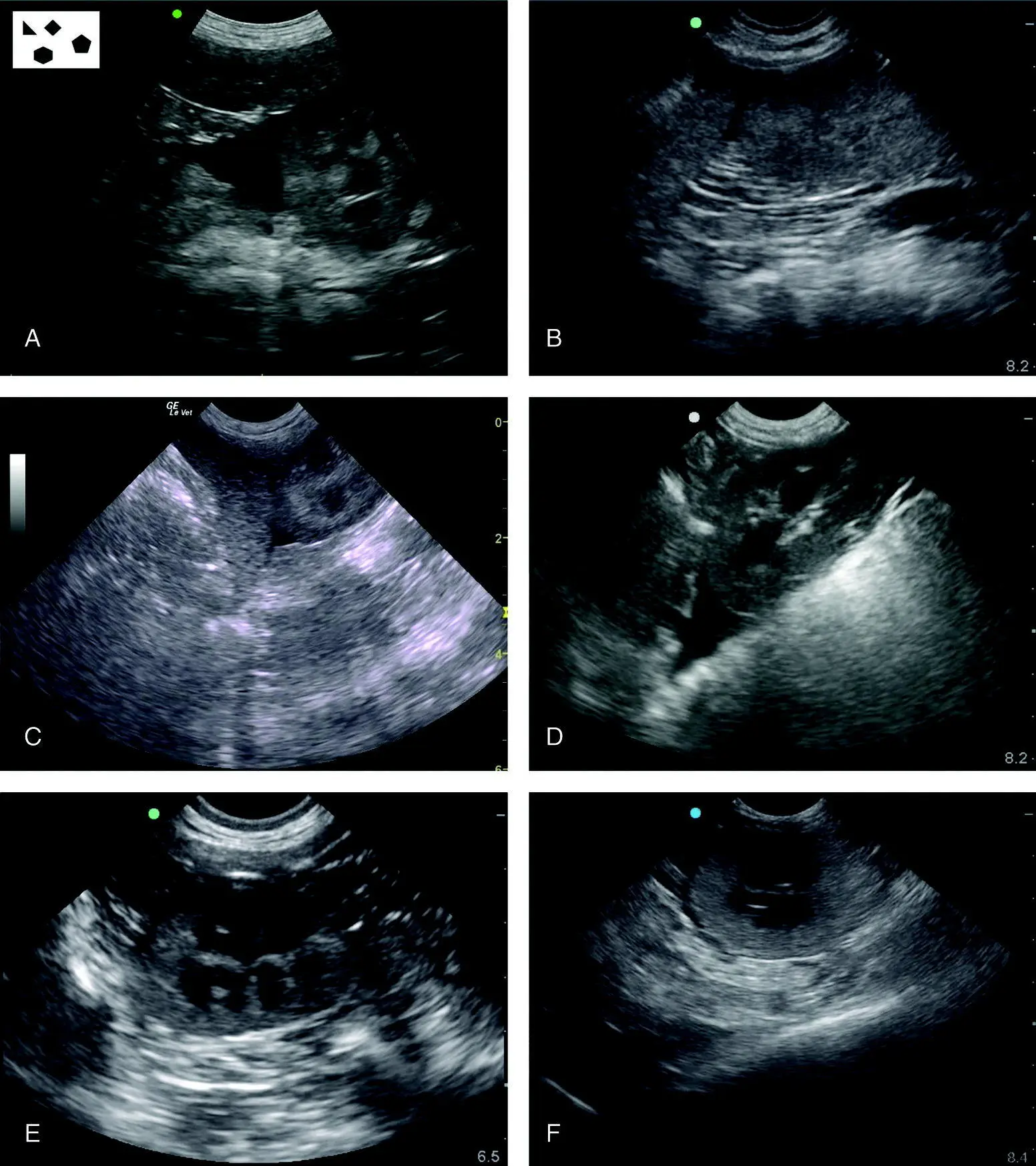
Figure 6.19. Examples of typical positive studies at the SR view. In (A) the spleen is in the near‐field identified by its characteristic location (importance of performing the SR view the same way every time), its hyperechoic capsule, and vessels splitting its capsule. Classic triangulation between the spleen and the left kidney. In (B) is another positive with a similar location of free fluid with the triangulation outlining the spleen from the stomach (screen left) and left kidney (screen right). With fanning through the region, the left kidney is easily identified to the right of the near‐field of the image. In (C) free fluid is located between the left kidney and spleen. In (D) is shown the classic triangulation off the cranial pole of the left kidney where it abuts the wall of the colon. In (E) there is some perirenal fluid to the left of the kidney that is similar to the image from another case in (F). Note differences in image quality and ultrasound machine settings making it easier or more difficult to see. Serial examinations are key when subtle findings are suspected. Moreover, the SR view is complemented by information from other AFAST views. Compare these images to the anatomy in Figures 6.17and 6.18. Note the consistency in the images with their proportionality and location of the relevant SR view structures of the left kidney, spleen, stomach and colon.
Source: Reproduced with permission of Dr Gregory Lisciandro, Hill Country Veterinary Specialists and FASTVet.com, Spicewood, TX.
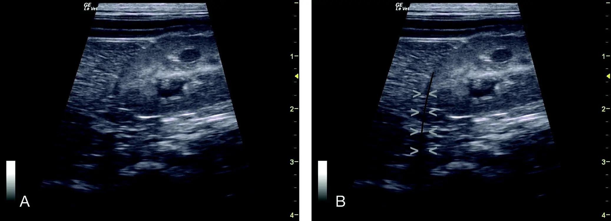
Figure 6.20. Edge shadowing artifact off the left kidney. (A) and (B) are the same image unlabeled and labeled for comparison. In (B) the hypoechoic linear edge shadowing artifact in (A) is outlined with (V) and with a thin black line. When free fluid is suspected off the margin of a curved surface, this artifact should always be considered as creating a false positive. Compare to Figure 6.19(E) and (F).
Source: Reproduced with permission of Dr Gregory Lisciandro, Hill Country Veterinary Specialists and FASTVet.com, Spicewood, TX.
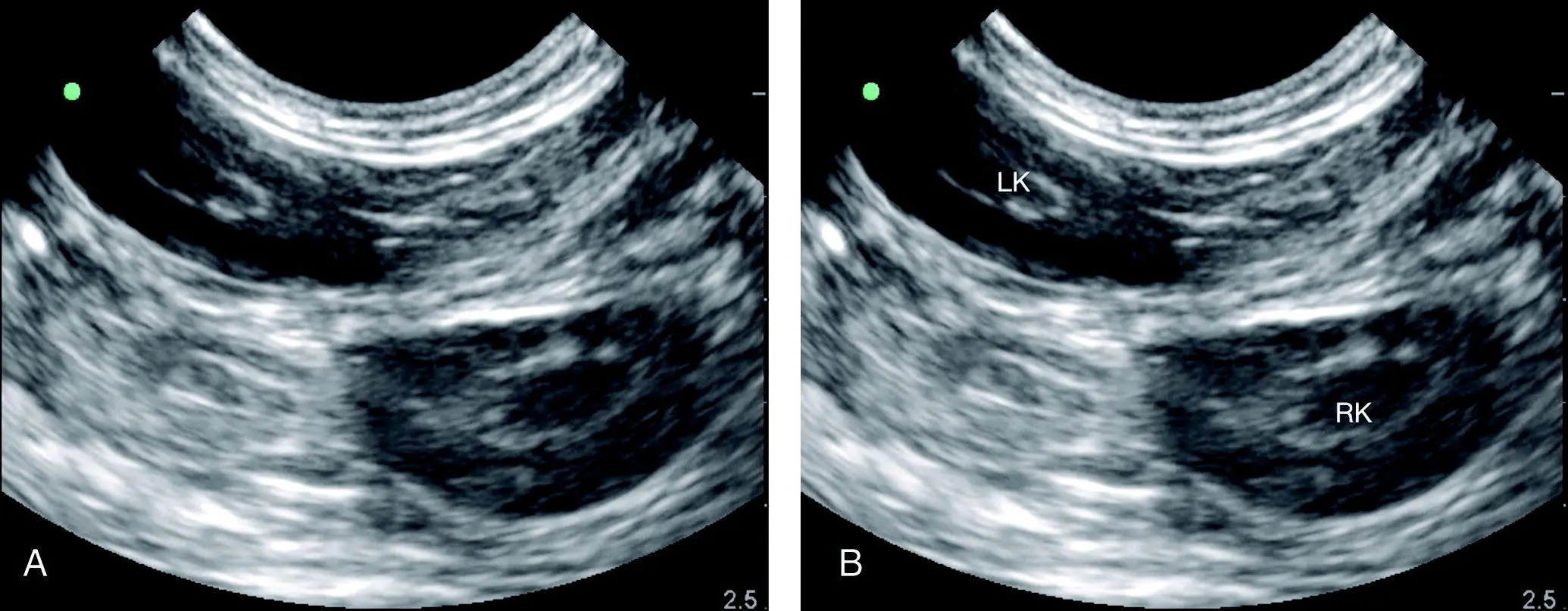
Figure 6.21. Both kidneys in view at the SR view. In the cat and the small dog, it is not uncommon to see both kidneys through the SR view. LK, left kidney; RK, right kidney.
Source: Reproduced with permission of Dr Gregory Lisciandro, Hill Country Veterinary Specialists and FASTVet.com, Spicewood, TX.
Air‐filled Stomach
Air causes interference (ultrasound cannot image through air) cranial or to the screen's left. Study the presence of the stomach and its dirty shadowing in Figures 6.17– 6.19.
Читать дальшеИнтервал:
Закладка:
Похожие книги на «Point-of-Care Ultrasound Techniques for the Small Animal Practitioner»
Представляем Вашему вниманию похожие книги на «Point-of-Care Ultrasound Techniques for the Small Animal Practitioner» списком для выбора. Мы отобрали схожую по названию и смыслу литературу в надежде предоставить читателям больше вариантов отыскать новые, интересные, ещё непрочитанные произведения.


Обсуждение, отзывы о книге «Point-of-Care Ultrasound Techniques for the Small Animal Practitioner» и просто собственные мнения читателей. Оставьте ваши комментарии, напишите, что Вы думаете о произведении, его смысле или главных героях. Укажите что конкретно понравилось, а что нет, и почему Вы так считаете.