Point-of-Care Ultrasound Techniques for the Small Animal Practitioner
Здесь есть возможность читать онлайн «Point-of-Care Ultrasound Techniques for the Small Animal Practitioner» — ознакомительный отрывок электронной книги совершенно бесплатно, а после прочтения отрывка купить полную версию. В некоторых случаях можно слушать аудио, скачать через торрент в формате fb2 и присутствует краткое содержание. Жанр: unrecognised, на английском языке. Описание произведения, (предисловие) а так же отзывы посетителей доступны на портале библиотеки ЛибКат.

- Название:Point-of-Care Ultrasound Techniques for the Small Animal Practitioner
- Автор:
- Жанр:
- Год:неизвестен
- ISBN:нет данных
- Рейтинг книги:5 / 5. Голосов: 1
-
Избранное:Добавить в избранное
- Отзывы:
-
Ваша оценка:
Point-of-Care Ultrasound Techniques for the Small Animal Practitioner: краткое содержание, описание и аннотация
Предлагаем к чтению аннотацию, описание, краткое содержание или предисловие (зависит от того, что написал сам автор книги «Point-of-Care Ultrasound Techniques for the Small Animal Practitioner»). Если вы не нашли необходимую информацию о книге — напишите в комментариях, мы постараемся отыскать её.
New chapters in
cover ultrasound-guided nerve blocks, musculoskeletal, brain imaging, and applications of focused ultrasound techniques in cats, exotics and marine mammals—making it an essential purchase for veterinarians wanting to incorporate point-of-care ultrasound techniques into their veterinary practices.
Presents a standardized approach to point-of-care ultrasound as an extension of the physical exam, including trauma, non-trauma, and monitoring applications Includes coverage of new techniques for focused ultrasound exams, including lung, anesthesia and ultrasound guided nerve blocks, transcranial brain imaging, musculoskeletal, volume status evaluation, and rapid assessment for treatable forms of shock Adds cats, exotic and wildlife mammals, and marine mammals to the existing canine coverage Emphasizes the integration of point-of-care ultrasound techniques for optimizing patient care and accurate patient assessment Offers access to a companion website with supplementary video clips showing many clinically relevant didactic examples The second edition of
is an excellent resource for veterinary practitioners, ranging from the general practitioner to nearly all clinical specialists, including internal medicine, oncology, cardiology, emergency and critical care, anesthesiology, ophthalmology, exotics, and zoo medicine specialists, and veterinary students.
Point-of-Care Ultrasound Techniques for the Small Animal Practitioner — читать онлайн ознакомительный отрывок
Ниже представлен текст книги, разбитый по страницам. Система сохранения места последней прочитанной страницы, позволяет с удобством читать онлайн бесплатно книгу «Point-of-Care Ultrasound Techniques for the Small Animal Practitioner», без необходимости каждый раз заново искать на чём Вы остановились. Поставьте закладку, и сможете в любой момент перейти на страницу, на которой закончили чтение.
Интервал:
Закладка:
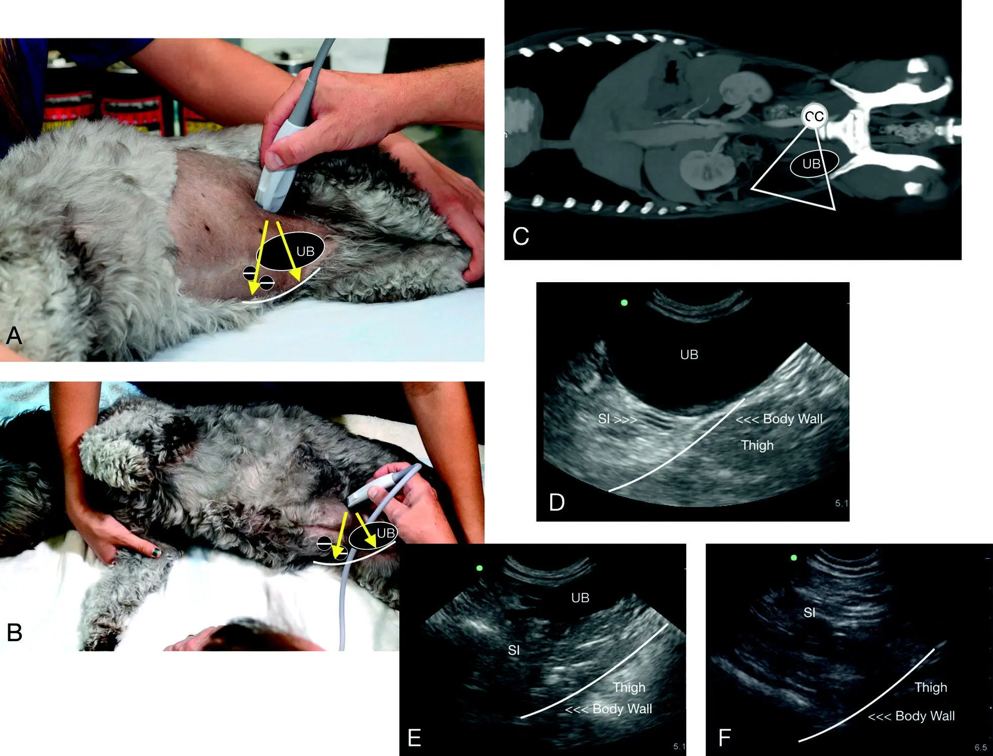
Figure 6.23. The CC view in a dog. In (A) the direction of the probe is shown externally dorsal to midline and directed toward the tabletop, or the most gravity‐dependent region of the view. In (B) is a similar image with anatomy overlay that correlates to the CT image. The urine‐filled urinary bladder serves as a recognizable landmark for the CC view and for identifying the “CC pouch” where the urinary bladder and body wall in its most gravity‐dependent region are next to one another ( curved line ) as this is where free fluid would pocket. The thigh is often in view through the far‐field and is screen right in (D–F). Computed tomography courtesy of Dr Daniel Rodriguez, VETTEM, and Dr Jesús Paredes, CVM, Mexico City, Mexico.
Source: Reproduced with permission of Dr Gregory Lisciandro, Hill Country Veterinary Specialists and FASTVet.com, Spicewood, TX.
The striated rounded soft tissue structure through the far‐field is often the patient's opposing thigh (“imaging ham”) and not a mass. The rounded anechoic circular structures in between the urinary bladder and the thigh are often appreciated and represent the femoral artery and vein ( Figure 6.25; see also Figures 6.23and 25.8). The compression technique can determine which is the vein and which the artery in addition to the use of color flow Doppler (see Chapter 25).
Pearl:In blunt trauma dogs, the probability of hemoabdomen to uroabdomen has been documented in separate veterinary studies to be ~98% to ~2–4%, respectively (Boysen et al. 2004; Lisciandro et al. 2009; Simpson et al. 2009; Hoffberg et al. 2016; Grimes et al. 2018). Thus, in most canine blunt trauma cases in which the urinary bladder is not visualized, the serial AFAST examination provides a second opportunity to evaluate for the integrity of an intact urinary bladder with its expected rounded contour (Lisciandro et al. 2009). In its absence, radiography, abdominal fluid sampling and analysis, such as comparative abdominal fluid to serum creatinine, or other more advanced imaging procedures should be considered.
Pearl:In cats with large‐volume effusion that survive blunt trauma, the probability of a uroabdomen is much higher than a hemoabdomen because cats generally don't survive large‐volume bleeds like dogs do.
Typical CC View Positives
The classic CC positive view with a small‐volume effusion has a small anechoic (black) triangle in the “CC pouch,” between the urinary bladder apex and the body wall in the most gravity‐dependent region (see Figure 6.25). Large‐volume positives are usually easy to see in real time with the wafting movement of small intestines and omentum. Urinary bladder pathology such as sediment and calculi is most commonly going to be in the same most gravity‐dependent view of the “CC pouch.” When a mass effect is found in the urinary bladder, the best way to differentiate a thrombus from a mass is by using color flow Doppler, with a thrombus (and artifact) with no flow versus a mass with pulsatile blood flow.
Pearl:When a urinary bladder mass is found, differentiate through the use of color flow Doppler, with a thrombus having no flow versus a mass that will have pulsatile blood flow.
Artifacts
The most relevant CC view artifacts are described below (see Figure 6.26). More detail is provided in Chapter 3.
Acoustic Enhancement Artifact
The fluid‐filled urinary bladder will make the soft tissue distal through it brighter (more echogenic), similar to what occurs with the gallbladder at the DH view. With the recommended probe positioning, the body wall will be brighter in the far‐field in the “CC pouch” because of this artifact and readily seen along the body wall in the far‐field deep to the urinary bladder.
Mirror Image Artifact
The mirror image artifact requires a strong soft tissue–air interface and is most commonly observed at the DH view, the CC view (bladder mirrored on the far side of the abdominal wall) and during echo views (heart mirrored on the far side of the pericardium).
Side‐lobe and Slice‐thickness Artifact
These artifacts mimic sediment and the author has even received images suggesting this artifact was a mass. Think about the artifact in terms of gravity dependency because sediment and calculi will settle into the bladder lumen and be best viewed within the “CC pouch.” Lowering the gain eliminates the artifact whereas true sediment persists, and color Doppler can assess for flow (no flow = either artifact or thrombus versus flow = a mass).
Probe Pressure Artifact
The sonographer should always be aware of the effects of probe pressure on the image. The urinary bladder is susceptible to probe pressure artifact creating misshapen appearances.
Reverberation and Dirty Shadowing Artifact
An overlying air‐filled colon will obscure the far‐field, making the bladder appear in odd shapes, and if colon or small bowel gets under the bladder, either may be mistaken for sediment, calculi, and masses (see Figure 6.27) (see Chapters 3and 11). It is a good habit to check where the probe and the beam (the scanning plane) are directed to help interpret your image (see Chapter 5).
Pearl:Change the position of the probe and alter the pressure of the probe on the patient (both a little more and a little less) to coax the colon out of your way or sweep toward midline while imaging in the “CC pouch” to get away from the air‐filled colon.
False Positives
False positives are generally not a problem at the CC view when directing the probe into the “CC pouch,” the most gravity‐dependent region between the urinary bladder and the body wall in the far‐field. The great vessels are in the opposite direction, located sublumbar, which is not an imaging plane for the CC view.
If you fan dorsally into the sublumbar area, generally unnecessary during AFAST, the great vessels, female reproductive tract, and lymph nodes may become confounders because they are generally anechoic fluid‐filled structures or hypoechoic structures, respectively.
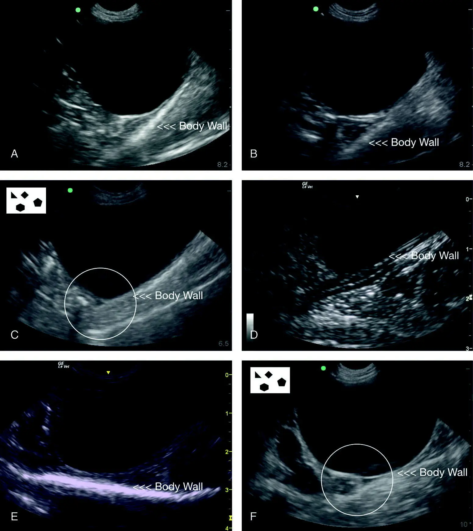
Figure 6.24. Examples of typical negative studies at the CC view. In all these images, note how readily recognizable is the filled urinary bladder as a landmark along with the body wall in the far‐field within the “CC pouch.” The “CC pouch” is very sensitive for detecting small‐volume effusions as noted in (C) and (F) and the small anechoic triangulation circled, which in fact may be considered a normal finding in juvenile puppies and kittens with abdominal fluid scores of no more than 1. In fact, adult dogs and cats may also have a small amount of free fluid (<3 mm) at the CC view as the only positive AFAST view. The only other typical abdominal structures in these images are small intestine. Because of its facial planes and acoustic enhancement through the fluid‐filled urinary bladder, the body wall is typically a hyperechoic (bright white) line. Images (A), (B), (D) and (E) are negative CC studies. Note the consistency in the images with their proportionality and location of the relevant CC view structures of the urinary bladder, “CC pouch”, and body wall.
Читать дальшеИнтервал:
Закладка:
Похожие книги на «Point-of-Care Ultrasound Techniques for the Small Animal Practitioner»
Представляем Вашему вниманию похожие книги на «Point-of-Care Ultrasound Techniques for the Small Animal Practitioner» списком для выбора. Мы отобрали схожую по названию и смыслу литературу в надежде предоставить читателям больше вариантов отыскать новые, интересные, ещё непрочитанные произведения.


Обсуждение, отзывы о книге «Point-of-Care Ultrasound Techniques for the Small Animal Practitioner» и просто собственные мнения читателей. Оставьте ваши комментарии, напишите, что Вы думаете о произведении, его смысле или главных героях. Укажите что конкретно понравилось, а что нет, и почему Вы так считаете.