Point-of-Care Ultrasound Techniques for the Small Animal Practitioner
Здесь есть возможность читать онлайн «Point-of-Care Ultrasound Techniques for the Small Animal Practitioner» — ознакомительный отрывок электронной книги совершенно бесплатно, а после прочтения отрывка купить полную версию. В некоторых случаях можно слушать аудио, скачать через торрент в формате fb2 и присутствует краткое содержание. Жанр: unrecognised, на английском языке. Описание произведения, (предисловие) а так же отзывы посетителей доступны на портале библиотеки ЛибКат.

- Название:Point-of-Care Ultrasound Techniques for the Small Animal Practitioner
- Автор:
- Жанр:
- Год:неизвестен
- ISBN:нет данных
- Рейтинг книги:5 / 5. Голосов: 1
-
Избранное:Добавить в избранное
- Отзывы:
-
Ваша оценка:
Point-of-Care Ultrasound Techniques for the Small Animal Practitioner: краткое содержание, описание и аннотация
Предлагаем к чтению аннотацию, описание, краткое содержание или предисловие (зависит от того, что написал сам автор книги «Point-of-Care Ultrasound Techniques for the Small Animal Practitioner»). Если вы не нашли необходимую информацию о книге — напишите в комментариях, мы постараемся отыскать её.
New chapters in
cover ultrasound-guided nerve blocks, musculoskeletal, brain imaging, and applications of focused ultrasound techniques in cats, exotics and marine mammals—making it an essential purchase for veterinarians wanting to incorporate point-of-care ultrasound techniques into their veterinary practices.
Presents a standardized approach to point-of-care ultrasound as an extension of the physical exam, including trauma, non-trauma, and monitoring applications Includes coverage of new techniques for focused ultrasound exams, including lung, anesthesia and ultrasound guided nerve blocks, transcranial brain imaging, musculoskeletal, volume status evaluation, and rapid assessment for treatable forms of shock Adds cats, exotic and wildlife mammals, and marine mammals to the existing canine coverage Emphasizes the integration of point-of-care ultrasound techniques for optimizing patient care and accurate patient assessment Offers access to a companion website with supplementary video clips showing many clinically relevant didactic examples The second edition of
is an excellent resource for veterinary practitioners, ranging from the general practitioner to nearly all clinical specialists, including internal medicine, oncology, cardiology, emergency and critical care, anesthesiology, ophthalmology, exotics, and zoo medicine specialists, and veterinary students.
Point-of-Care Ultrasound Techniques for the Small Animal Practitioner — читать онлайн ознакомительный отрывок
Ниже представлен текст книги, разбитый по страницам. Система сохранения места последней прочитанной страницы, позволяет с удобством читать онлайн бесплатно книгу «Point-of-Care Ultrasound Techniques for the Small Animal Practitioner», без необходимости каждый раз заново искать на чём Вы остановились. Поставьте закладку, и сможете в любой момент перейти на страницу, на которой закончили чтение.
Интервал:
Закладка:
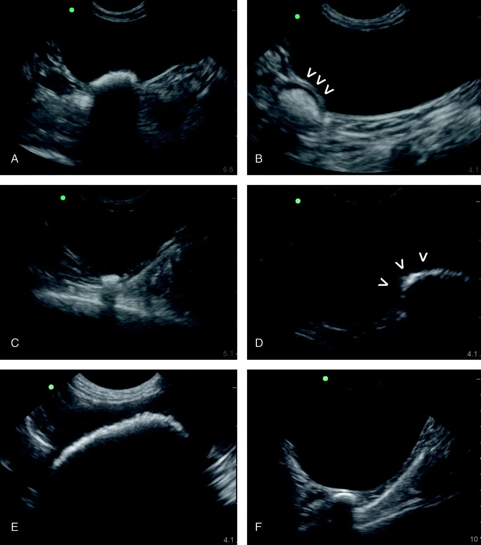
Figure 6.27. Calculi versus intestinal tract. The sonographer will readily appreciate deviations from the expected appearance of the urinary bladder at the CC view. In images (A), (C) and (E) bladder calculi are evident by clean shadowing through the far‐field and contained within the lumen of the urinary bladder. Moreover, they should settle at the “CC pouch” where free fluid would pocket being at the CC view's most gravity‐dependent region. In (B), (D) and (F) are examples of air‐filled intestinal tract that mimic urinary bladder calculi illustrating how care should be taken. By examining (and magnifying) more closely, the bladder lumen can be seen to be pushed into the lumen by the loop of intestinal tract as in (B) and (D) indicated by the cursors (V). Moreover, an abdominal radiograph may be added to the diagnostic work‐up.
Source: Reproduced with permission of Dr Gregory Lisciandro, Hill Country Veterinary Specialists and FASTVet.com, Spicewood, TX.
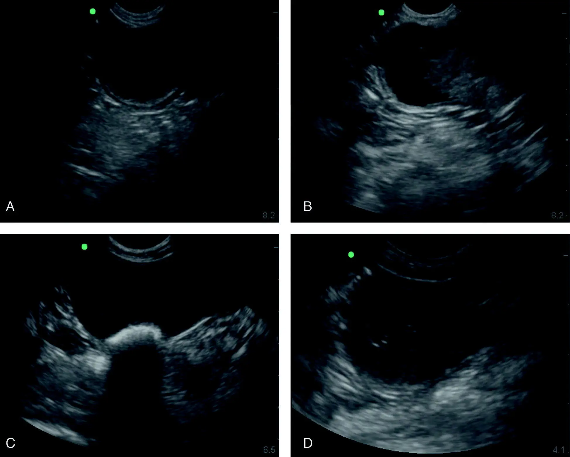
Figure 6.28. Various abnormal intraluminal urinary bladder findings. (A) An irregular urinary bladder wall in its apical region suggestive of chronic cystitis. (B) A soft tissue mass in the trigone region of the urinary bladder of an older female dog that was hit by a car (blunt trauma). The condition was captured and the dog diagnosed with a transitional cell carcinoma that lived with treatment for another 11 months. Color flow Doppler could be additionally applied to evaluate for blood flow (not shown). (C) Bladder calculi in a diabetic being admitted for hospitalized care. (D) A large thrombus in an older male bluntly traumatized dog. Color flow Doppler was helpful for documenting absent flow.
Source: Reproduced with permission of Dr Gregory Lisciandro, Hill Country Veterinary Specialists and FASTVet.com, Spicewood, TX.
Pitfall:Excessive probe pressure will cause small volumes of free fluid to move away to either side of the probe head and be potentially missed.
Pitfall:If the probe is not directed into the abdominal cavity or lacks adequate depth then you may be imaging through planes of body wall or “imaging through bacon,” as the author likes to say.
Pearl:The HRU view (SRU view in left lateral recumbency) is performed just ventral (right below) to the umbilicus to image the most gravity‐dependent area of the laterally recumbent dog or cat.
Pearl:Stay longitudinal (sagittal) at all sites (DH, SR, CC) except at the HRU view where it is helpful to perform both longitudinal (fanning) and transverse (rotating to the left or counterclockwise) imaging to discriminate free fluid (anechoic black triangles) from small intestine.
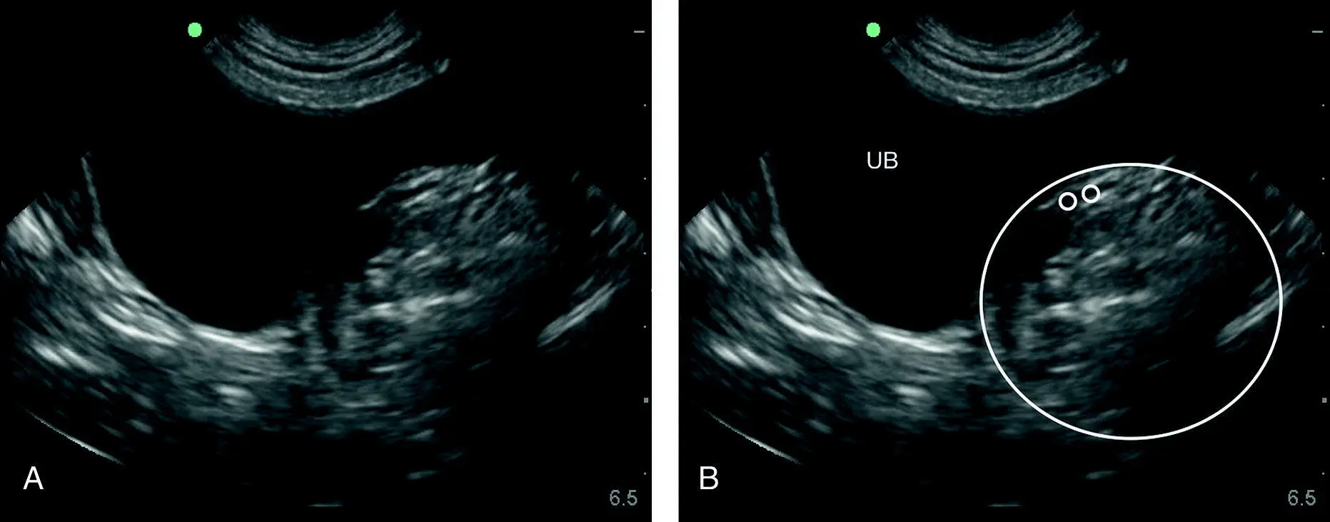
Figure 6.29. Pitfall of the thigh or a mass or other. In (A) and (B) are identical images unlabeled and labeled. The large circle encompasses the region of interest and its suspect origin of being the thigh muscle. The smaller circles are where typically the femoral artery and vein would be located with the patient in lateral recumbency and observing for pulsation or applying color Doppler would be helpful. The image shows how the thigh can push into the urinary bladder or stool in the colon or a caudal abdominal mass and make odd impressions when imaging the CC view with a patient in lateral recumbency. A digital rectal examination as part of a good physical examination with or without more advanced imaging is an ancillary evaluation to increase the probability of being correct. From the still B‐mode image, the exact origin of the circled structure may be unclear. UB, urinary bladder.
Source: Reproduced with permission of Dr Gregory Lisciandro, Hill Country Veterinary Specialists and FASTVet.com, Spicewood, TX.
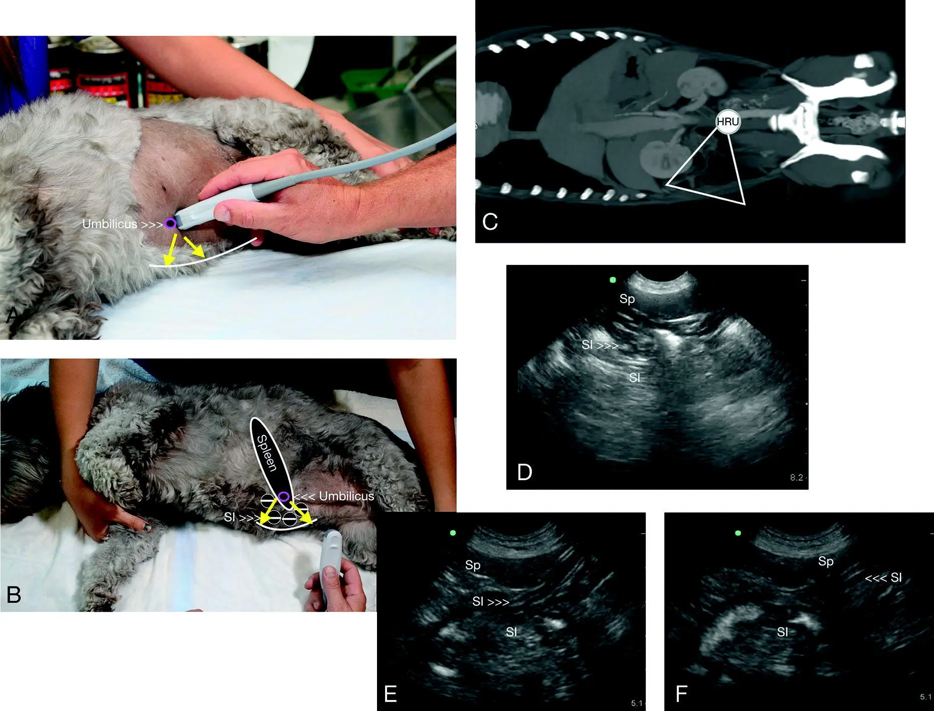
Figure 6.30. HRU view in a dog. In (A) and (B) the direction of the probe is shown externally ventral to midline at the level of the umbilicus with the objective to image and fan through the most gravity‐dependent “HRU pouch” illustrated by the white curved line. (B) A similar image with anatomy overlay that correlates with CT image in (C). The target organs for the HRU (and SRU) view are in reality the small intestine and spleen and either or both should be recognized to confirm that you are imaging the abdominal cavity. In (D), (E) and (F) the expected sonographic images are shown. Computed tomography courtesy of Dr Daniel Rodriguez, VETTEM, and Dr Jesús Paredes, CVM, Mexico City, Mexico.
Source: Reproduced with permission of Dr Gregory Lisciandro, Hill Country Veterinary Specialists and FASTVet.com, Spicewood, TX.
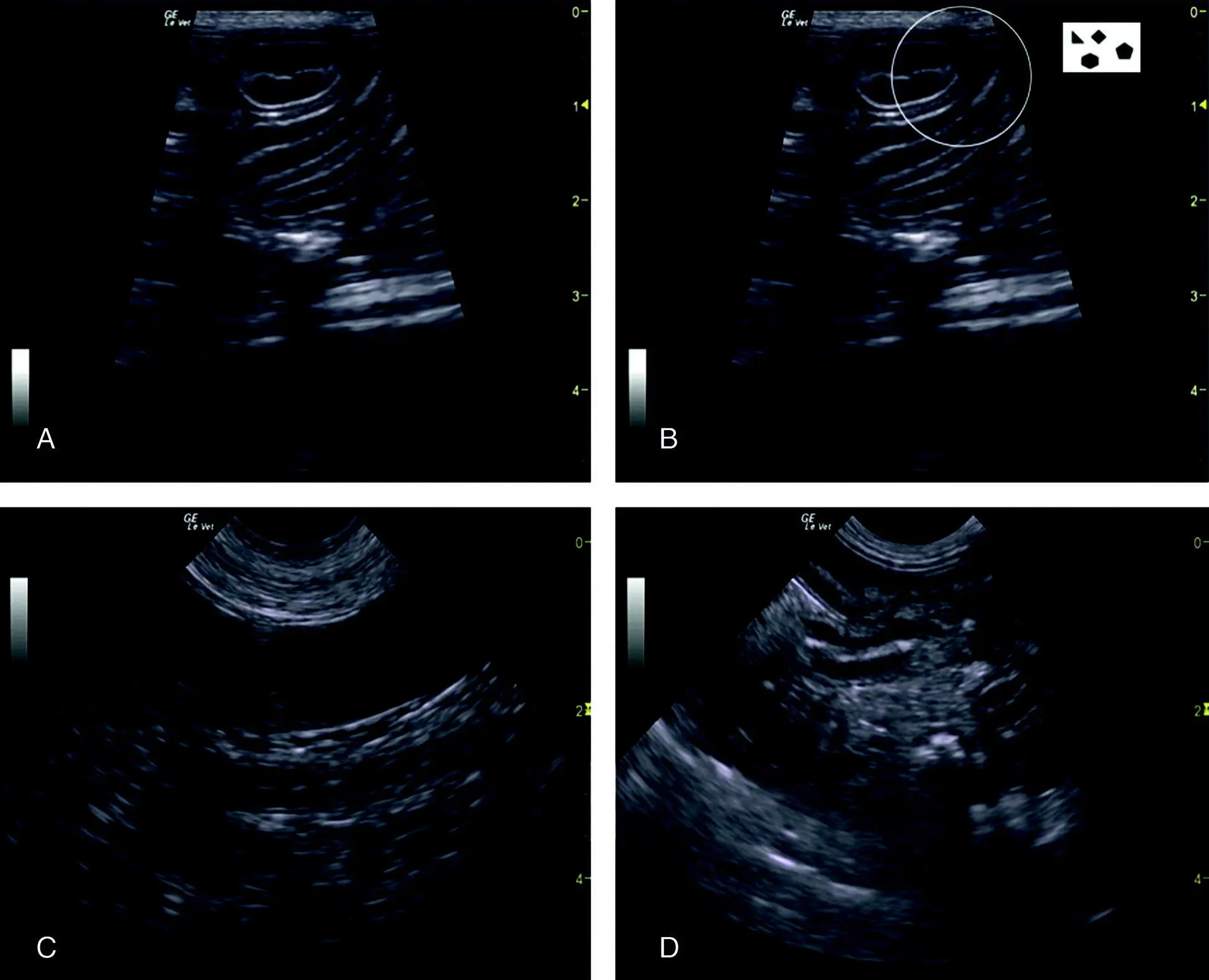
Figure 6.31. Examples of typical negative studies at the HRU (SRU) view. The target organs are the small intestine and spleen. By imaging either or both, the sonographer knows they are within the abdominal cavity. (A) and (B) are the same image labeled and unlabeled, showing how the small intestine in cross‐section, transverse orientation, appears like “hamburgers” and in longitudinal or sagittal appears like “highways.” In (B) the circle highlights an area that may or may not have a small triangulation of free fluid, illustrating the difficulty in seeing small pockets of fluids (milliliters) in between intestinal loops because of the anechoic layers of intestine. A better strategy is shown in (C) by using the spleen as an acoustic window and looking for free fluid on its far side. In (D) is another region in which soft tissue is in proximity to small intestine as a better strategy to detect small‐volume effusions at this view than in (A) and (B).
Source: Reproduced with permission of Dr Gregory Lisciandro, Hill Country Veterinary Specialists and FASTVet.com, Spicewood, TX.
If the right (left in left lateral recumbency) kidney needs to be imaged because of concern for retroperitoneal injury, or when hematuria exists, then the HR5th (SR5th in left lateral recumbency) bonus view should be performed. Once the four AFAST views used for the AFS are mastered, the HR5th/SR5th bonus views should be considered as an add‐on skills and incorporated for all subsequent patients.
Typical HRU View Positives
The HRU (SRU) view is the most gravity‐dependent view in right lateral recumbency (the SRU when in left lateral recumbency) and completes the four AFAST views and the calculation of its applied fluid scoring system and assigning the AFS (see Chapter 7). The “rabbit ear sign” is typical in large‐volume effusions created by small intestine and omentum wafting in the free fluid and typical positives are shown in Figure 6.32. Furthermore, if positives were previously seen at the other less gravity‐dependent AFAST views, the sonographer is already anticipating the likelihood of performing abdominocentesis at the HRU view (SRU view in left lateral recumbency).
Читать дальшеИнтервал:
Закладка:
Похожие книги на «Point-of-Care Ultrasound Techniques for the Small Animal Practitioner»
Представляем Вашему вниманию похожие книги на «Point-of-Care Ultrasound Techniques for the Small Animal Practitioner» списком для выбора. Мы отобрали схожую по названию и смыслу литературу в надежде предоставить читателям больше вариантов отыскать новые, интересные, ещё непрочитанные произведения.


Обсуждение, отзывы о книге «Point-of-Care Ultrasound Techniques for the Small Animal Practitioner» и просто собственные мнения читателей. Оставьте ваши комментарии, напишите, что Вы думаете о произведении, его смысле или главных героях. Укажите что конкретно понравилось, а что нет, и почему Вы так считаете.