Point-of-Care Ultrasound Techniques for the Small Animal Practitioner
Здесь есть возможность читать онлайн «Point-of-Care Ultrasound Techniques for the Small Animal Practitioner» — ознакомительный отрывок электронной книги совершенно бесплатно, а после прочтения отрывка купить полную версию. В некоторых случаях можно слушать аудио, скачать через торрент в формате fb2 и присутствует краткое содержание. Жанр: unrecognised, на английском языке. Описание произведения, (предисловие) а так же отзывы посетителей доступны на портале библиотеки ЛибКат.

- Название:Point-of-Care Ultrasound Techniques for the Small Animal Practitioner
- Автор:
- Жанр:
- Год:неизвестен
- ISBN:нет данных
- Рейтинг книги:5 / 5. Голосов: 1
-
Избранное:Добавить в избранное
- Отзывы:
-
Ваша оценка:
Point-of-Care Ultrasound Techniques for the Small Animal Practitioner: краткое содержание, описание и аннотация
Предлагаем к чтению аннотацию, описание, краткое содержание или предисловие (зависит от того, что написал сам автор книги «Point-of-Care Ultrasound Techniques for the Small Animal Practitioner»). Если вы не нашли необходимую информацию о книге — напишите в комментариях, мы постараемся отыскать её.
New chapters in
cover ultrasound-guided nerve blocks, musculoskeletal, brain imaging, and applications of focused ultrasound techniques in cats, exotics and marine mammals—making it an essential purchase for veterinarians wanting to incorporate point-of-care ultrasound techniques into their veterinary practices.
Presents a standardized approach to point-of-care ultrasound as an extension of the physical exam, including trauma, non-trauma, and monitoring applications Includes coverage of new techniques for focused ultrasound exams, including lung, anesthesia and ultrasound guided nerve blocks, transcranial brain imaging, musculoskeletal, volume status evaluation, and rapid assessment for treatable forms of shock Adds cats, exotic and wildlife mammals, and marine mammals to the existing canine coverage Emphasizes the integration of point-of-care ultrasound techniques for optimizing patient care and accurate patient assessment Offers access to a companion website with supplementary video clips showing many clinically relevant didactic examples The second edition of
is an excellent resource for veterinary practitioners, ranging from the general practitioner to nearly all clinical specialists, including internal medicine, oncology, cardiology, emergency and critical care, anesthesiology, ophthalmology, exotics, and zoo medicine specialists, and veterinary students.
Point-of-Care Ultrasound Techniques for the Small Animal Practitioner — читать онлайн ознакомительный отрывок
Ниже представлен текст книги, разбитый по страницам. Система сохранения места последней прочитанной страницы, позволяет с удобством читать онлайн бесплатно книгу «Point-of-Care Ultrasound Techniques for the Small Animal Practitioner», без необходимости каждый раз заново искать на чём Вы остановились. Поставьте закладку, и сможете в любой момент перейти на страницу, на которой закончили чтение.
Интервал:
Закладка:
Pearls and Pitfalls, The Final Say
By gaining a basic understanding of ultrasound physics and the common ultrasound artifacts, the nonradiologist veterinarian or veterinary sonographer can more clearly interpret the ultrasound image ( Table 3.1). Always keep in mind the basic assumptions used to generate the image when scanning or viewing the ultrasound image. Your interpretive and diagnostic skills, and hence your patient, will benefit greatly.
Knowing the basic assumptions used to generate the image leads to less misinterpretation of the ultrasound image.
Artifacts are one of the primary pitfalls of ultrasonographic imaging.
Table 3.1. Summary of common artifacts and examples.
| Name of artifact | Fluid‐associated | Air‐associated | Other | Common examples |
|---|---|---|---|---|
| Shadowing, clean | No | No | Bone/stone | Cystouroliths, ribs |
| Shadowing, dirty | No | Yes | Irregular/partial penetration into gas | Lung, stomach, colon, small intestine |
| Edge shadowing | No | No | Refraction off round structures | Stomach wall, gallbladder wall, urinary bladder wall |
| Acoustic enhancement | Yes | No | Decreased attenuation | Gallbladder, cysts, eye |
| Mirror image | No | Frequently | Reflection | Diaphragm/liverUrinary bladder/colon |
| Reverberation A‐lines | No | Yes | Typically used only in reference to lung ultrasound | Lung surface |
| Comet‐tail, ring‐down | No | No | Bone/stone/metal | Calculi, surgical clips, tissue mineralization |
| B‐lines, also called ultrasound lung rockets (ULRs) | Yes | Yes | Air–fluid interfaces | Lung |
| Pseudo B‐lines | Yes | Yes | Air–fluid interfaces (gastric luminal contents); strong soft tissue–air interfaces, i.e., lung nodules | Stomach contents, lung nodules |
| Side‐lobe | Yes | No | Multiple echoes | Urinary bladder lumen,gallbladder lumen |
| Slice‐thickness | Yes | No | Multiple echoes | False appearance of sludge in gallbladder, cysts |
References
1 Feldman MK, Katyal, S, Blackwood MS. 2009. Ultrasound artifacts. RadioGraphics 29:1179–1189.
2 Lichtenstein DA, Meziere GA. 2008. Relevance of lung ultrasound in the diagnosis of acute respiratory failure: the BLUE protocol. Chest 134(1):117–125.
3 Lichtenstein DA, Meziere GA, Lagoueyte J, et al. 2009. A‐lines and B‐lines. Lung ultrasound as a bedside tool for predicting pulmonary artery occlusion pressure in the critically ill. Chest 136(4):1014–1020.
4 Lisciandro GR. 2011. Abdominal and thoracic focused assessment with sonography for trauma, triage, and monitoring in small animals. J Vet Emerg Crit Care 21(2):104–122.
5 Nyland TG, Mattoon JS, Herrgesell EJ, et al. 2002. Physical principles, instrumentation, and safety of diagnostic ultrasound. In: Small Animal Diagnostic Ultrasound, 2nd edition, edited by Nyland TG, Mattoon JS. Philadelphia: WB Saunders, pp 1–18.
6 Penninck DG. 2002. Artifacts. In: Small Animal Diagnostic Ultrasound, 2nd edition, edited by Nyland TG, Mattoon JS. Philadelphia: WB Saunders, pp 19–29.
7 Reef V. 1998. Thoracic ultrasonography: noncardiac imaging. In: Equine Diagnostic Ultrasound, edited by Reef V. Philadelphia: WB Saunders, pp 187–214.
8 Volpicelli G, Elbarbary M, Blaivas M, et al. 2012. International evidence‐based recommendations for point‐of‐care lung ultrasound. Intensive Care Med 38:577–591.
Chapter Four POCUS: Basic Ultrasound Scanning
Robert M. Fulton
Introduction
Ultrasound is a user‐dependent imaging modality, with the quality of the image obtained directly dependent on the skill of the ultrasonographer. Many of the POCUS studies are not highly reliant on advanced skill or knowledge. Still, one must have a basic understanding of how to manipulate the probe and machine settings to obtain the prescribed images and optimize image quality.
What POCUS Basic Scanning Can Do
Provide a basic tutorial on ultrasound physics, image acquisition, and storage, and discuss basic ultrasound systematics.
What POCUS Basic Scanning Cannot Do
Cannot provide an in‐depth tutorial on how to learn ultrasound techniques.
Cannot replace experience and continued learning.
Indications
Provide a basic understanding of ultrasound principles and image acquisition, including probe type, probe manipulations and orientations, and ultrasound machine features to maximize accurate image interpretation.
Objectives
Provide a basic understanding of ultrasound image acquisition.
Provide a review of basic ultrasound techniques and systematics including:image planesimage orientationprobe maneuversimage optimizationimage documentationbasic machine selection and care.
Understanding Features of the Ultrasound Image
Imaging Planes
Understanding how the two‐dimensional image is formed from scanning through a three‐dimensional object is crucial not only to image interpretation but also communication of that information. The first piece of information is the imaging or scanning plane. The imaging plane may refer to the whole body, a part of the body (such as a leg or the head), or to a specific structure within the body, the left kidney, for instance ( Figures 4.1and 4.2).
The first of these, transverse, is any plane along the short axis of the structure being imaged.
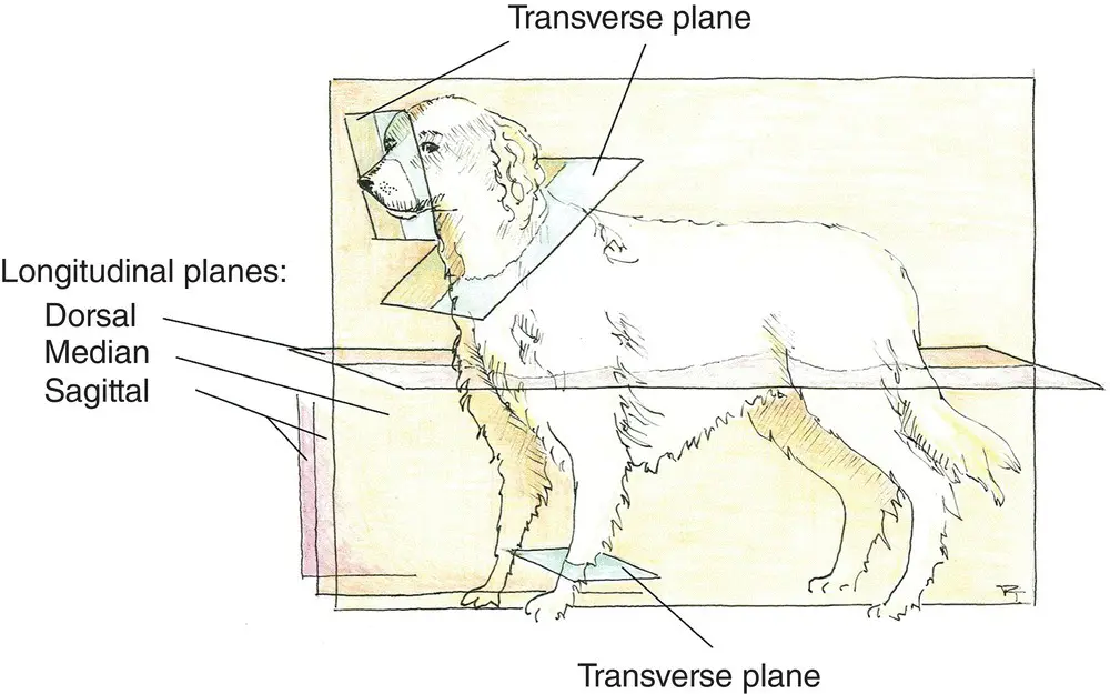
Figure 4.1. Longitudinal and transverse orientation shown on anatomical planes.
Source: Illustration courtesy of Randi Taggart, Richmond, VA.
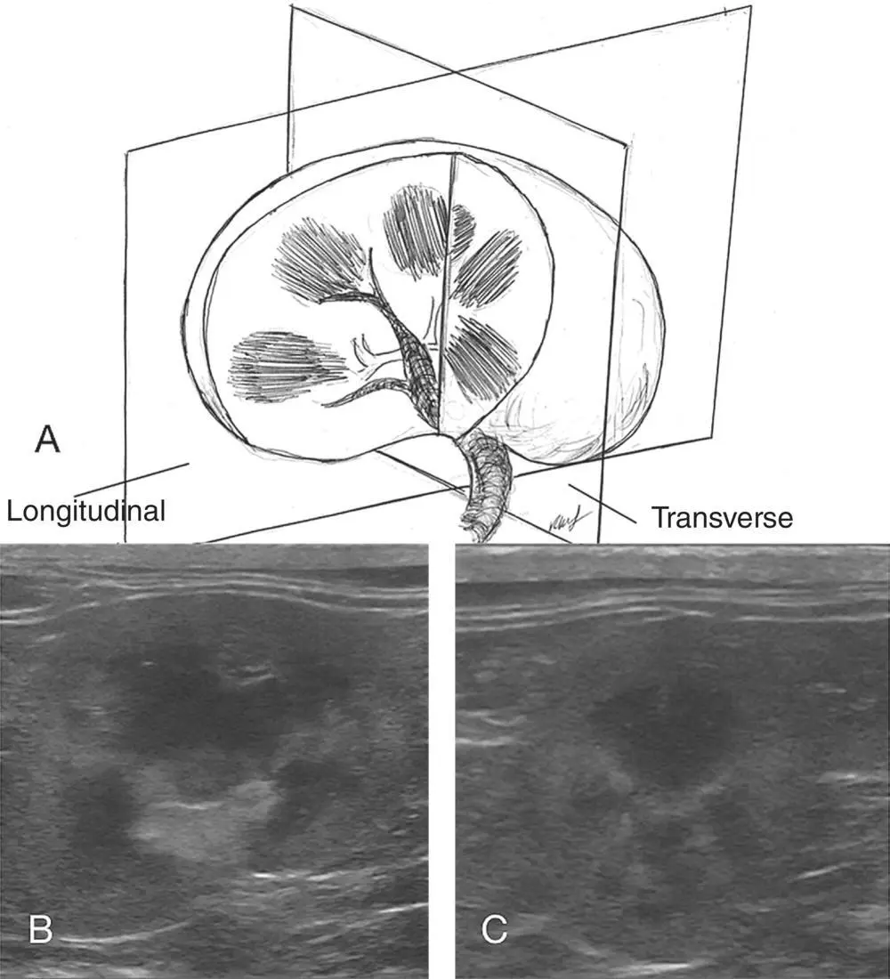
Figure 4.2. Examples of image planes for the kidney. In (A) the kidney is shown with longitudinal (sagittal) and transverse planes. In (B) the longitudinal (sagittal) plane is contrasted to (C) in transverse orientation on actual B‐mode ultrasound images. For most nonradiologist sonographers, the kidney is more readily recognized in the longitudinal (sagittal) orientation.
Source: Courtesy of Robert M. Fulton, DVM, Richmond, VA.
The second, longitudinal, is any plane that is perpendicular to the transverse plane or the long axis of the structure. Technically, the longitudinal plane has several specific types.
Median – this plane splits a symmetrical structure into equal left and right halves along the long axis.
Sagittal or paramedian – this plane is any plane that is parallel to the median plane. While technically incorrect, many will use sagittal interchangeably with longitudinal.
Dorsal – this plane splits a structure into dorsal and ventral segments and is analogous to the coronal plane in biped scanning terminology.
Commonly, veterinary sonographers tend to talk about only two planes, transverse and longitudinal, and this suffices for most communication and image understanding (see Figures 4.1and 4.2).
Читать дальшеИнтервал:
Закладка:
Похожие книги на «Point-of-Care Ultrasound Techniques for the Small Animal Practitioner»
Представляем Вашему вниманию похожие книги на «Point-of-Care Ultrasound Techniques for the Small Animal Practitioner» списком для выбора. Мы отобрали схожую по названию и смыслу литературу в надежде предоставить читателям больше вариантов отыскать новые, интересные, ещё непрочитанные произведения.


Обсуждение, отзывы о книге «Point-of-Care Ultrasound Techniques for the Small Animal Practitioner» и просто собственные мнения читателей. Оставьте ваши комментарии, напишите, что Вы думаете о произведении, его смысле или главных героях. Укажите что конкретно понравилось, а что нет, и почему Вы так считаете.