Point-of-Care Ultrasound Techniques for the Small Animal Practitioner
Здесь есть возможность читать онлайн «Point-of-Care Ultrasound Techniques for the Small Animal Practitioner» — ознакомительный отрывок электронной книги совершенно бесплатно, а после прочтения отрывка купить полную версию. В некоторых случаях можно слушать аудио, скачать через торрент в формате fb2 и присутствует краткое содержание. Жанр: unrecognised, на английском языке. Описание произведения, (предисловие) а так же отзывы посетителей доступны на портале библиотеки ЛибКат.

- Название:Point-of-Care Ultrasound Techniques for the Small Animal Practitioner
- Автор:
- Жанр:
- Год:неизвестен
- ISBN:нет данных
- Рейтинг книги:5 / 5. Голосов: 1
-
Избранное:Добавить в избранное
- Отзывы:
-
Ваша оценка:
Point-of-Care Ultrasound Techniques for the Small Animal Practitioner: краткое содержание, описание и аннотация
Предлагаем к чтению аннотацию, описание, краткое содержание или предисловие (зависит от того, что написал сам автор книги «Point-of-Care Ultrasound Techniques for the Small Animal Practitioner»). Если вы не нашли необходимую информацию о книге — напишите в комментариях, мы постараемся отыскать её.
New chapters in
cover ultrasound-guided nerve blocks, musculoskeletal, brain imaging, and applications of focused ultrasound techniques in cats, exotics and marine mammals—making it an essential purchase for veterinarians wanting to incorporate point-of-care ultrasound techniques into their veterinary practices.
Presents a standardized approach to point-of-care ultrasound as an extension of the physical exam, including trauma, non-trauma, and monitoring applications Includes coverage of new techniques for focused ultrasound exams, including lung, anesthesia and ultrasound guided nerve blocks, transcranial brain imaging, musculoskeletal, volume status evaluation, and rapid assessment for treatable forms of shock Adds cats, exotic and wildlife mammals, and marine mammals to the existing canine coverage Emphasizes the integration of point-of-care ultrasound techniques for optimizing patient care and accurate patient assessment Offers access to a companion website with supplementary video clips showing many clinically relevant didactic examples The second edition of
is an excellent resource for veterinary practitioners, ranging from the general practitioner to nearly all clinical specialists, including internal medicine, oncology, cardiology, emergency and critical care, anesthesiology, ophthalmology, exotics, and zoo medicine specialists, and veterinary students.
Point-of-Care Ultrasound Techniques for the Small Animal Practitioner — читать онлайн ознакомительный отрывок
Ниже представлен текст книги, разбитый по страницам. Система сохранения места последней прочитанной страницы, позволяет с удобством читать онлайн бесплатно книгу «Point-of-Care Ultrasound Techniques for the Small Animal Practitioner», без необходимости каждый раз заново искать на чём Вы остановились. Поставьте закладку, и сможете в любой момент перейти на страницу, на которой закончили чтение.
Интервал:
Закладка:
The amplitude of a reflected sound wave is proportional to the difference in acoustic impedance between two different tissues. Air has a low impedance and bone has a high impedance when compared to soft tissue (Reef 1998) ( Figure 2.2). Therefore, when a sound wave comes across a soft tissue–bone or a soft tissue–air interface (large difference in acoustic impedance), nearly all of the sound waves are strongly reflected (and a bright white echogenic line is formed at either interface). Reflection is why the sonographer cannot image through bone (solid) or lung (air) and this points up one of the most common misnomers in clinical ultrasonography: when imaging through the liver into the thorax, we term the bright, curved cranial border as the diaphragm. In reality, the diaphragm is uncommonly imaged except in bicavitary effusions. The bright white (hyperechoic), curved line is actually the strongly reflective surface of the lung (air) at the soft tissue–air boundary or interface serving as a strong reflector.
By comparing the acoustic impedance of most tissues in the body other than bone (solid) and lung (air), we see that they are very similar (there is little difference in acoustic impedance among them). This similarity makes ultrasound a great imaging tool for examining into and through soft tissues (their parenchyma). On the other hand, due to the large difference in acoustic impedance between soft tissue–air and soft tissue–bone interfaces, ultrasound is not an effective tool for examination beyond the surfaces of either aerated lung, gas‐containing hollow viscus or bone (Reef 1998).
Absorption, Scatter, and Reflection
Other ultrasound principles that affect our image include absorption, scatter, and angle of reflection. As the sound waves enter the body, some of them are absorbed by the tissues and are never reflected back to the probe. These waves are lost and do not contribute to the image. Furthermore, many of the waves are scattered by the tissues and their surface irregularities and either return to the probe (receiver) in a distorted path or do not return at all. As a result, the ultrasound waves are “misinterpreted” by the processor, and the image and its resolution are affected.
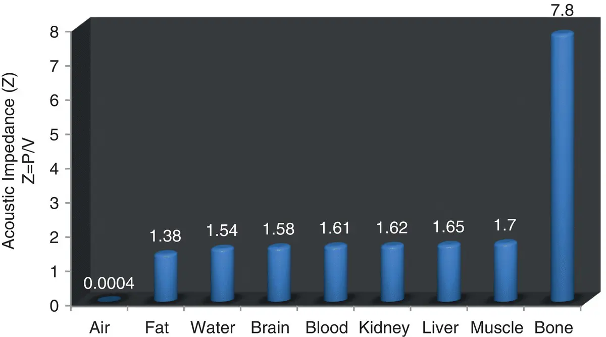
Figure 2.2. Acoustic impedance (106 kg/m2sec) of common body tissues or substances. This figure illustrates the degree of difference in acoustic impedance between substances that helps determine sound wave transmission. The greater the difference, the greater is reflection or loss of transmission. One can see how ultrasound is ideally suited for most soft tissue and why it is not suited for imaging bone or air‐filled structures (Reef 1998).
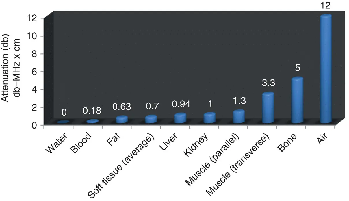
Figure 2.3. Attenuation (db/cm/MHz) in common tissues. Attenuation of sound energy within tissues varies with the frequency of the sound and is affected by reflection, scattering, and absorbance. Note how bone and air have the greatest attenuation values (Reef 1998).
Attenuation
All sound beams lose energy, or become attenuated, during transmission through tissues; therefore, the returning sound wave is weaker than when it started. Different frequencies (MHz) are attenuated to different degrees. Because higher frequency probes send more waves per second, there is greater tissue interaction with the sound waves. This provides improved resolution but undergoes more attenuation. Low‐frequency sound waves are less attenuated because of less tissue interaction, and therefore allow deeper tissue penetration. Strategies that include lowering the MHz for better penetration (depth) come at the expense of detail. Conversely, using higher frequency for more detail comes at the cost of less penetration (depth). Furthermore, high‐density tissues attenuate the sound waves more than low‐density tissues ( Figure 2.3).
Pearl:The analogy of hearing a boom box from a distance can help you remember which MHz penetrates more. The bass dominates (low MHz) over higher frequencies (high MHz); thus, low MHz penetrates deeply at the expense of detail, and high MHz give better detail at the expense of penetration.
The Final Say
By understanding the basic physical principles governing sound transmission and the limitations of the ultrasound processor, the ultrasonographer can better understand the image on the screen. Furthermore, this same knowledge is fundamental in understanding ultrasound artifacts covered in the next chapter.
References
1 Bushberg JT, Seibert JA, Leidholdt EM, Boone JM. 2002. The Essential Physics of Medical Imaging, 2nd edition. Philadelphia: Lippincott Williams and Wilkins.
2 Coltera M. 2010. Ultrasound physics in a nutshell. Otolaryngol Clin North Am 43(6):1149–1159.
3 Filly RA. 1988. Ultrasound: the stethoscope of the future, alas. Radiology 167:400.
4 Nyland TG, Mattoon JS, Herrgesell EJ, et al. 2002. Physical principles, instrumentation, and safety of diagnostic ultrasound. In: Small Animal Diagnostic Ultrasound, 2nd edition, edited by Nyland TG, Mattoon JS. Philadelphia: WB Saunders, pp 1–18.
5 Penninck DG. Artifacts. In: Small Animal Diagnostic Ultrasound, 2nd edition, edited by Nyland TG, Mattoon JS. Philadelphia: WB Saunders, pp 19–29.
6 Reef V. 1998. Thoracic ultrasonography: noncardiac imaging. In: Equine Diagnostic Ultrasound, edited by Reef V. Philadelphia: WB Saunders, pp 187–214.
7 Rozycki GS, Pennington SD, Feliciano DV, et al. 2001. Surgeon‐performed ultrasound in the critical care setting: its use as an extension of the physical examination to detect pleural effusion. J Trauma 50:636–642.
Further Reading
1 Volpicelli G, Elbarbary M, Blaivas M, et al. 2012. International evidence‐based recommendations for point‐of‐care lung ultrasound. Intensive Care Med 38:577–591.
Chapter Three POCUS: Basic Ultrasound Artifacts
Robert M. Fulton
Introduction
In this chapter, we'll look at the fundamental laws governing wave dynamics and see how ultrasound artifacts are created. Ultrasound machines rely on several physical assumptions to assign the location and intensity of each received echo. These assumptions are that the received echoes have originated from within the main ultrasound beam, an echo returns to the transducer after a single reflection, the depth of an object is directly related to the amount of time for an ultrasound pulse to return to the transducer, the speed of sound in tissue is constant (1540 m/sec), the sound beam and its echo travel in a straight path, and the acoustic energy in an ultrasound field is uniformly attenuated (Feldman et al. 2009). Artifacts may be grouped by the most important principles leading to their formation, including attenuation, velocity or propagation, and artifacts associated with multiple echoes. Many artifacts can be grouped by their association with air or fluid, making learning them easier.
What POCUS Basic Ultrasound Artifacts Can Do
Provide a basic understanding of how artifacts are formed to allow better interpretation of the ultrasound image.
What POCUS Basic Ultrasound Artifacts Cannot Do
Cannot provide an in‐depth discussion of ultrasound artifacts.
Cannot account for what artifacts your ultrasound machine's software is most biased towards.
Читать дальшеИнтервал:
Закладка:
Похожие книги на «Point-of-Care Ultrasound Techniques for the Small Animal Practitioner»
Представляем Вашему вниманию похожие книги на «Point-of-Care Ultrasound Techniques for the Small Animal Practitioner» списком для выбора. Мы отобрали схожую по названию и смыслу литературу в надежде предоставить читателям больше вариантов отыскать новые, интересные, ещё непрочитанные произведения.


Обсуждение, отзывы о книге «Point-of-Care Ultrasound Techniques for the Small Animal Practitioner» и просто собственные мнения читателей. Оставьте ваши комментарии, напишите, что Вы думаете о произведении, его смысле или главных героях. Укажите что конкретно понравилось, а что нет, и почему Вы так считаете.