Point-of-Care Ultrasound Techniques for the Small Animal Practitioner
Здесь есть возможность читать онлайн «Point-of-Care Ultrasound Techniques for the Small Animal Practitioner» — ознакомительный отрывок электронной книги совершенно бесплатно, а после прочтения отрывка купить полную версию. В некоторых случаях можно слушать аудио, скачать через торрент в формате fb2 и присутствует краткое содержание. Жанр: unrecognised, на английском языке. Описание произведения, (предисловие) а так же отзывы посетителей доступны на портале библиотеки ЛибКат.

- Название:Point-of-Care Ultrasound Techniques for the Small Animal Practitioner
- Автор:
- Жанр:
- Год:неизвестен
- ISBN:нет данных
- Рейтинг книги:5 / 5. Голосов: 1
-
Избранное:Добавить в избранное
- Отзывы:
-
Ваша оценка:
Point-of-Care Ultrasound Techniques for the Small Animal Practitioner: краткое содержание, описание и аннотация
Предлагаем к чтению аннотацию, описание, краткое содержание или предисловие (зависит от того, что написал сам автор книги «Point-of-Care Ultrasound Techniques for the Small Animal Practitioner»). Если вы не нашли необходимую информацию о книге — напишите в комментариях, мы постараемся отыскать её.
New chapters in
cover ultrasound-guided nerve blocks, musculoskeletal, brain imaging, and applications of focused ultrasound techniques in cats, exotics and marine mammals—making it an essential purchase for veterinarians wanting to incorporate point-of-care ultrasound techniques into their veterinary practices.
Presents a standardized approach to point-of-care ultrasound as an extension of the physical exam, including trauma, non-trauma, and monitoring applications Includes coverage of new techniques for focused ultrasound exams, including lung, anesthesia and ultrasound guided nerve blocks, transcranial brain imaging, musculoskeletal, volume status evaluation, and rapid assessment for treatable forms of shock Adds cats, exotic and wildlife mammals, and marine mammals to the existing canine coverage Emphasizes the integration of point-of-care ultrasound techniques for optimizing patient care and accurate patient assessment Offers access to a companion website with supplementary video clips showing many clinically relevant didactic examples The second edition of
is an excellent resource for veterinary practitioners, ranging from the general practitioner to nearly all clinical specialists, including internal medicine, oncology, cardiology, emergency and critical care, anesthesiology, ophthalmology, exotics, and zoo medicine specialists, and veterinary students.
Point-of-Care Ultrasound Techniques for the Small Animal Practitioner — читать онлайн ознакомительный отрывок
Ниже представлен текст книги, разбитый по страницам. Система сохранения места последней прочитанной страницы, позволяет с удобством читать онлайн бесплатно книгу «Point-of-Care Ultrasound Techniques for the Small Animal Practitioner», без необходимости каждый раз заново искать на чём Вы остановились. Поставьте закладку, и сможете в любой момент перейти на страницу, на которой закончили чтение.
Интервал:
Закладка:
Other Terms
We have done our best to make this second edition uniform in terminology and up to date with the current consensus in both human and veterinary medicine. Some examples in which multiple terms relating to similar things are used are listed here.
The preferred term is listed first but each may be considered synonymously.
POCUS exam and Focused exam
sonographer and ultrasonographer
ultrasonographically and sonographically
color Doppler and color flow Doppler
beam and scanning plane
acoustic window and view
fanning and tilting
longitudinal and sagittal (and long axis)
transverse (and short axis)
orientation and plane
probe and transducer
curvilinear probe and microconvex probe
B‐lines and ultrasound lung rockets
lung sliding and glide sign
gallbladder halo sign, gallbladder halo effect, and gallbladder double rim effect
FAST diaphragmatico‐hepatic view and subxiphoid view
acoustic coupling and probe–skin contact
We have provided a comprehensive list of abbreviations, terms, and definitions in the Appendices.
Recording Your Findings on Goal‐directed Templates
Recording data on goal‐directed templates with clear objectives for answering defined clinical questions is imperative to gain the respect of our colleagues, to stay disciplined during the respective POCUS or FAST examination, to allow post hoc evaluation of studies, and to detect training strengths and deficiencies. Without recording your data, you cannot measure. If you cannot measure, you cannot critically study with the potential to improve. We have made great strides in Global FAST training by reevaluating our results recorded on goal‐directed templates and saved video clips. A prime example is the establishment of clear tenets for the accurate TFAST diagnosis of pericardial effusion (Lisciandro 2016).
We have presented POCUS and FAST goal‐directed templates and methods to efficiently save video clips for you to adapt and modify for your practice according to the types of cases you see, your practice type, and your skill level in the Appendices and Chapter 45.
Echogenicity – Whites, Grays, and Blacks
The jargon of ultrasound can be intimidating to the novice sonographer. Clarity may be accomplished through acknowledging that ultrasound is generally the opposite of how free fluid, air, and soft tissue appear on radiographic studies (our brain needs to reformat itself). For example and very simplistically, air is white on ultrasound and black on radiographs. Fluid is black on ultrasound and white on radiographs. However, bone is black (shadows) on ultrasound? And white on radiographs? Or white along its proximal surface with acoustic shadowing through the far field. Now if we are losing you, hang in there. The figures help ( Figures 1.2, 1.3, 1.4, 1.5). Study them now .
The ultrasound terms describing whites, grays, and blacks are anechoic (black), degrees of echogenicity (hypoechoic, shades of gray), and hyperechoic (white). The terms may be used relatively between structures like “X relative to Y” and “Y relative to Z.” For example, the spleen is hyper echoic (brighter than) to the left kidney. The liver is hypo echoic (darker than) to the falciform fat. The feline cortex of the kidney is isoechoic (same as) to the spleen (see Figure 1.3).
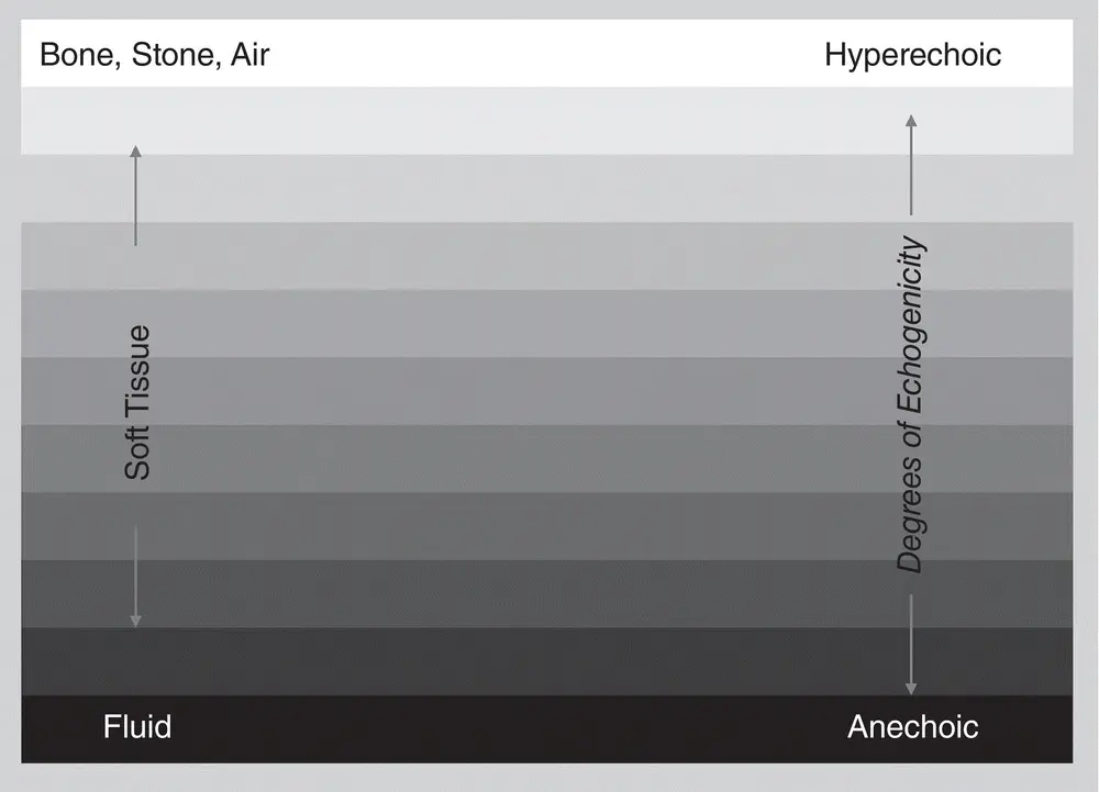
Figure 1.2. The figure shows the ultrasound differences in B‐mode gray scale of various tissues, with bone, stone and air being most reflective of the ultrasound beam and thus portrayed as white along its surface, to soft tissues being shades of gray, to fluid which is black. Ultrasound terminology corresponds to these gray‐scale colors as hyperechoic, degrees of echogenicity (hypoechoic), and anechoic. Study this chart to get the visual of what these gray‐scale terms mean. Courtesy of Dr Gregory Lisciandro, Hill Country Veterinary Specialists and FASTVet.com, Spicewood, TX.
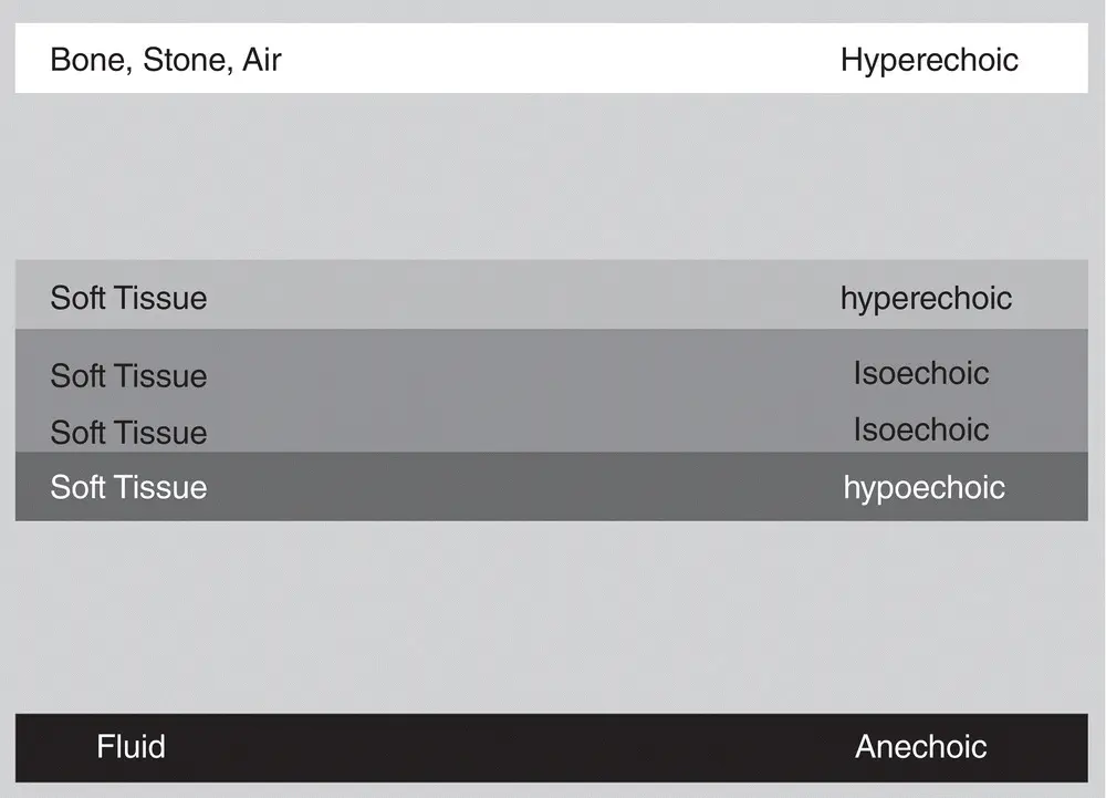
Figure 1.3. This figure is similar to Figure 1.2but with some of the gray scale removed to illustrate how descriptive ultrasound terminology is used for different tissues. The soft tissue is isoechoic (same as), hyperechoic (brighter than), and hypoechoic (darker than) relative to one another. Correlate with Table 1.1Courtesy of Dr Gregory Lisciandro, Hill Country Veterinary Specialists and FASTVet.com, Spicewood, TX.
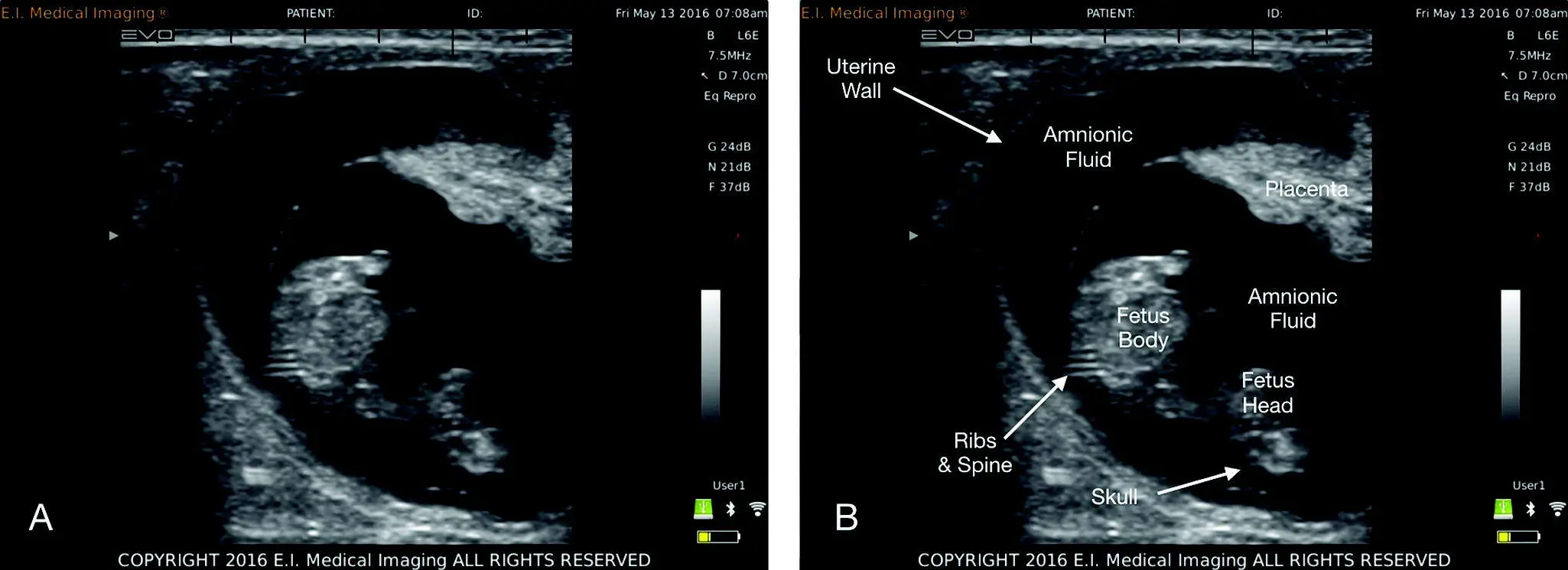
Figure 1.4. This figure is unlabeled and labeled for contrast of what tissue looks like in a gravid female, illustrating the different tissues and structures that correlate with concepts in Figures 1.1and 1.2. Image used with permission from E.I. Medical, Loveland, CO, USA. www.eimedical.com
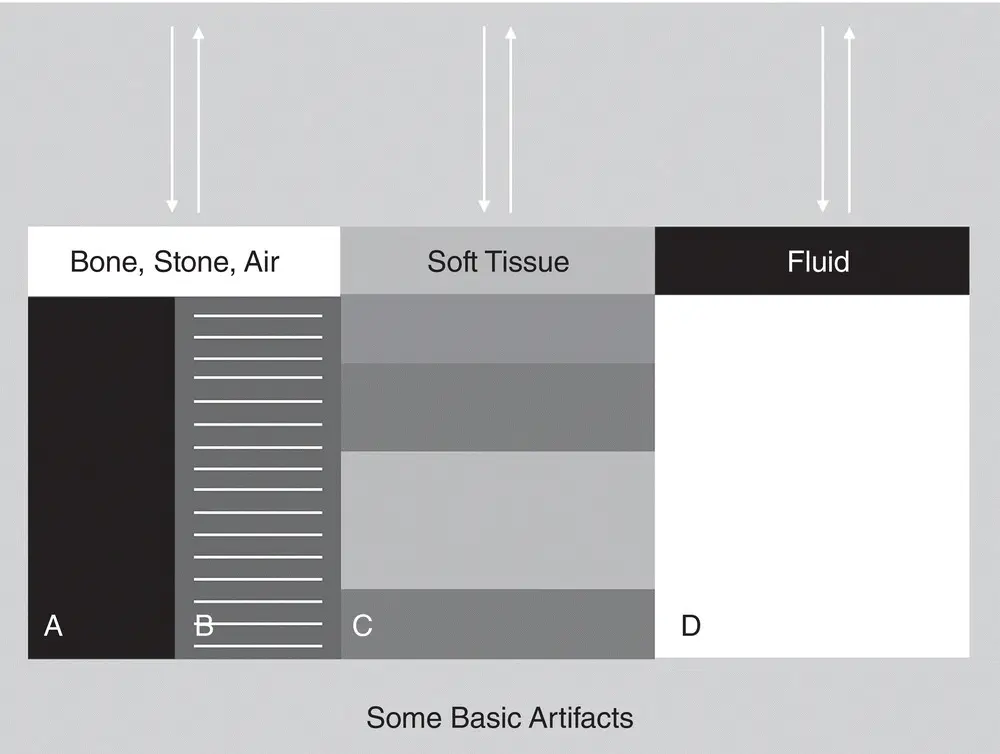
Figure 1.5. Ultrasound and different tissues and elements. Schematic that ties into the previous images showing various elements and the general behavior of ultrasound and its associated artifacts. The arrows represent the ultrasound beam being transmitted and returned to the transducer (probe). In (A) and (B), bone, stone, and air reflect because ultrasound (its echoes) does not transmit through them. As a result, they create shadowing of the beam, resulting in (A) “clean shadowing” (anechoic or black past the surface of the structure or element) or (B) “dirty shadowing,” including air reverberation called A‐lines (hyperechoic horizontal bars past the air). Soft tissues will absorb the ultrasound in different degrees depending on the soft tissue, thus (C) shows different degrees of echogenicity. Lastly, fluid will not absorb much of the ultrasound (echoes pass through the fluid), resulting in (D) acoustic enhancement through the far‐field beyond the distal boundary of fluid, making tissues more hyperechoic (brighter) than their adjacent counterparts outside the fluid‐related beam. Courtesy of Dr Gregory Lisciandro, Hill Country Veterinary Specialists and FASTVet.com, Spicewood, TX.
Anechoic (homogeneous black): occurs when no ultrasound waves are reflected back to the receiver. Thus, normal urine, normal bile, transudates, and blood all are purely anechoic (black).
Hypoechoic (shades of gray): occurs when variable degrees of ultrasound waves are reflected back to the transducer. Thus, the more echoes reflected back, the brighter gray the structure, and the fewer echoes, the darker the gray. Thus, all soft tissues that are not fully aerated are described relative to other distinct tissues, such that the liver is hypoechoic (darker than) relative to the spleen. Viscus organs (i.e., stomach) cannot be described this way, only their walls (i.e., stomach wall).
Hyperechoic (whites, bright whites): occurs when all or nearly 100% of ultrasound waves are reflected back to the transducer. Thus, bone, stone (metals), and air are strong reflectors resulting in hyperechoic (bright white) interfaces with shadowing, comet tail artifact, ring‐down artifact, ultrasound lung rockets, or reverberation artifact projected distal to the reflective surface.
Читать дальшеИнтервал:
Закладка:
Похожие книги на «Point-of-Care Ultrasound Techniques for the Small Animal Practitioner»
Представляем Вашему вниманию похожие книги на «Point-of-Care Ultrasound Techniques for the Small Animal Practitioner» списком для выбора. Мы отобрали схожую по названию и смыслу литературу в надежде предоставить читателям больше вариантов отыскать новые, интересные, ещё непрочитанные произведения.


Обсуждение, отзывы о книге «Point-of-Care Ultrasound Techniques for the Small Animal Practitioner» и просто собственные мнения читателей. Оставьте ваши комментарии, напишите, что Вы думаете о произведении, его смысле или главных героях. Укажите что конкретно понравилось, а что нет, и почему Вы так считаете.