Point-of-Care Ultrasound Techniques for the Small Animal Practitioner
Здесь есть возможность читать онлайн «Point-of-Care Ultrasound Techniques for the Small Animal Practitioner» — ознакомительный отрывок электронной книги совершенно бесплатно, а после прочтения отрывка купить полную версию. В некоторых случаях можно слушать аудио, скачать через торрент в формате fb2 и присутствует краткое содержание. Жанр: unrecognised, на английском языке. Описание произведения, (предисловие) а так же отзывы посетителей доступны на портале библиотеки ЛибКат.

- Название:Point-of-Care Ultrasound Techniques for the Small Animal Practitioner
- Автор:
- Жанр:
- Год:неизвестен
- ISBN:нет данных
- Рейтинг книги:5 / 5. Голосов: 1
-
Избранное:Добавить в избранное
- Отзывы:
-
Ваша оценка:
Point-of-Care Ultrasound Techniques for the Small Animal Practitioner: краткое содержание, описание и аннотация
Предлагаем к чтению аннотацию, описание, краткое содержание или предисловие (зависит от того, что написал сам автор книги «Point-of-Care Ultrasound Techniques for the Small Animal Practitioner»). Если вы не нашли необходимую информацию о книге — напишите в комментариях, мы постараемся отыскать её.
New chapters in
cover ultrasound-guided nerve blocks, musculoskeletal, brain imaging, and applications of focused ultrasound techniques in cats, exotics and marine mammals—making it an essential purchase for veterinarians wanting to incorporate point-of-care ultrasound techniques into their veterinary practices.
Presents a standardized approach to point-of-care ultrasound as an extension of the physical exam, including trauma, non-trauma, and monitoring applications Includes coverage of new techniques for focused ultrasound exams, including lung, anesthesia and ultrasound guided nerve blocks, transcranial brain imaging, musculoskeletal, volume status evaluation, and rapid assessment for treatable forms of shock Adds cats, exotic and wildlife mammals, and marine mammals to the existing canine coverage Emphasizes the integration of point-of-care ultrasound techniques for optimizing patient care and accurate patient assessment Offers access to a companion website with supplementary video clips showing many clinically relevant didactic examples The second edition of
is an excellent resource for veterinary practitioners, ranging from the general practitioner to nearly all clinical specialists, including internal medicine, oncology, cardiology, emergency and critical care, anesthesiology, ophthalmology, exotics, and zoo medicine specialists, and veterinary students.
Point-of-Care Ultrasound Techniques for the Small Animal Practitioner — читать онлайн ознакомительный отрывок
Ниже представлен текст книги, разбитый по страницам. Система сохранения места последней прочитанной страницы, позволяет с удобством читать онлайн бесплатно книгу «Point-of-Care Ultrasound Techniques for the Small Animal Practitioner», без необходимости каждый раз заново искать на чём Вы остановились. Поставьте закладку, и сможете в любой момент перейти на страницу, на которой закончили чтение.
Интервал:
Закладка:
Image Orientation
Any part of a medical record must contain the essentials of basic medical communication to have value. As veterinarians, we are taught how to communicate with each other in such a way regardless of our individual personality and training. One veterinarian can describe a lesion to another half a world away and pass along vital information. POCUS exams likewise need to have standard image orientation and recording of findings to give the study meaning.
Ultrasonography Compared to Radiography
For standard flat film radiography, the lateral film is oriented with the patient's head to the left, and the spine or dorsum is at the top of the viewer. This is the same for either a right or left lateral image. For the ventrodorsal or dorsoventral view, the radiograph is positioned with the head pointed up, and the patient's right side toward the left‐hand side of the view box.
Ultrasound follows similar conventions. When we scan from the ventral aspect, as when the patient is in dorsal recumbency, the following orientations apply.
Longitudinal image: the ventrum is on the top of the screen, dorsum on the bottom. Cranial (head) is to the left and caudal (tail) is to the right, similar to lateral radiography ( Figure 4.3).
Transverse image: ventral and dorsal remain top and bottom, respectively, and the patient's right side is represented on the left side of the screen, and the patient's left side is represented on the right side of the screen similar to ventrodorsal radiography (see Figure 4.3).
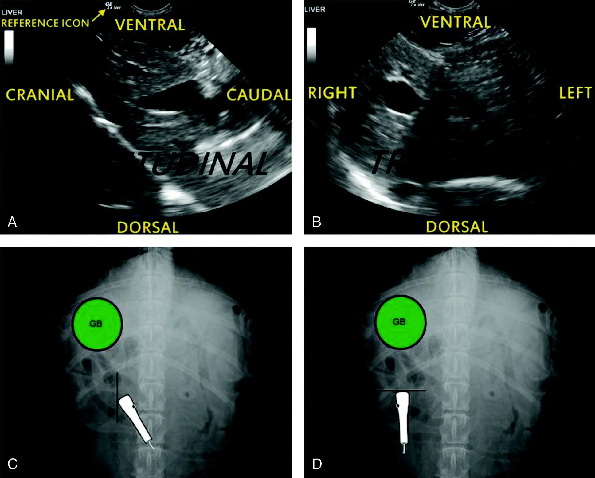
Figure 4.3. Standard ultrasound screen orientation, longitudinal (sagittal) and transverse. The radiograph for each orientation is located below the respective ultrasound image. Figures (A) and (C) illustrate longitudinal (or sagittal) and (B) and (D) transverse orientation with the corresponding probe position during interrogation of the liver and gallbladder via the subxiphoid region of a dog. Note that the reference icon (GELe) corresponds with the probe reference marker (dot on the probe) (labeled with arrow in (A) to the left on the ultrasound image). The best way to make standard ultrasound imaging a habit is to have the probe marker toward the head for longitudinal (or sagittal) orientation (black dot on the probe in (C) and turn (the probe head) left or counterclockwise for transverse orientation (black dot on the probe in (D) with the reference icon to the screen's left, shown at the top of the ultrasound image in (A) and (B)). If your reference icon is to the right of the ultrasound image, most ultrasound machines have a "reverse" button on their keyboard to flip the reference icon back to the standard left side (with the exception of echocardiography orientation; see Chapters 19– 21).
Pearl:Ultrasound orientation is the same as radiography: in lateral radiography, the head is to the left and tail to the right when scanning the long axis of the patient; and in ventrodorsal radiography, the left side of the patient is on the right side of the screen when scanning the transverse axis.
This ultrasound image orientation convention is the most intuitive if the patient is positioned in dorsal recumbency with its head facing the same way the sonographer is facing with both toward the machine and its screen. Many emergent patients are not stable enough to be placed in dorsal recumbency and all FAST scans actually prescribe lateral recumbency or sternal or standing positioning because the positioning is safer. So the sonographer may need to do some mental gymnastics at times to orient the image on the screen with the patient. What helps is thinking in terms of gravity dependent (fluid falls) and gravity nondependent (air rises) and each in terms of where the transducer is placed.
Pearl:When imaging, picture the gravity‐dependent and gravity‐nondependent regions of your patient. Keep in mind that fluid falls (gravity dependent) and air rises (gravity nondependent).
When scanning from the lateral aspect of the patient, that is, in a dorsal plane, the following conventions apply.
Longitudinal image: nonrecumbent side is on top of the screen, recumbent side is on the bottom. Cranial (head) is to the left side of the screen, and caudal (tail) is to the right (see Figure 4.3).
Transverse image: nonrecumbent side is still on top of the screen, recumbent side still on screen's bottom. Ventral is on the left, dorsal is on the right (see Figure 4.3).
Pearl:Develop the habit of having the marker toward the patient's head (longitudinal imaging) and turning left for transverse imaging to maintain proper orientation etiquette, similar to radiography.
Probe Orientation and Reference Markers
All ultrasound probes have a reference marker to allow for proper orientation. The reference marker may be a raised dot or line molded into the plastic, or possibly a small LED light. On the image screen, there will be a symbol, often the company's logo, that corresponds with the probe's reference marker. The marker on the screen is commonly referred to as the “reference icon” (see Figure 4.3).
Sonographers should familiarize themselves with the various types of ultrasound probes (also called transducers): phased‐array (also called sector), linear, and curvilinear. Furthermore, they should know that by looking at the shape of the ultrasound image, the probe is readily apparent as follows: pie‐shaped pointed near‐field (phased‐array or sector), rectangular (linear), and pie‐shaped with curved concave near‐field (curvilinear) ( Figure 4.4).
Pearl:You can tell what probe was used for the published image by looking at the image's near‐field as follows: pie‐shaped pointed near‐field (phased‐array or sector), rectangular (linear), and pie‐shaped with curved concave near‐field (curvilinear).
Most veterinarians are taught that when scanning the abdomen in long axis, the probe's reference marker is pointed toward the patient's head. Therefore, to maintain convention, the reference icon on the screen will also be positioned on the left‐hand side of the screen (screen left = cranial, screen right = caudal). When the probe is turned into the transverse orientation, the reference marker is pointed toward the patient's right, making a counterclockwise motion (“turning left”) if one views the probe from its tail or cable end (screen left = right side of patient, screen right = left side of patient). Again, the orientation is consistent with lateral and ventrodorsal radiographic orientation.
Some veterinarians are trained to orient the reference marker of the probe to be pointed caudally. Therefore, to maintain convention, the reference icon on the screen will be positioned on the right‐hand side of the screen. When switching into the transverse orientation, the probe is still rotated counterclockwise. The reference marker will be pointed towards the patient’s left, and the reference icon on the screen will still be on the right‐hand side of the screen.
No matter which training a veterinarian has received, the image on the screen should follow proper orientation convention with the patient's head to the left (screen left is cranial) and the patient's tail to the right (screen right is caudal) as with a lateral radiograph, and the patient's right is to the left of the screen as with a ventrodorsal radiograph.
Probe Maneuvers
There is one basic concept and six basic probe movements used in acquiring the ultrasound image. Beginning sonographers often miss their mark because they lack structured movement of the probe when scanning . Being able to make one type of movement at a time is essential to obtain a good image. To the casual observer, it may appear that the experienced sonographer makes free‐flowing and complex moves with the probe; however, the reality is that they are simply integrating the six foundational movements one movement at a time .
Читать дальшеИнтервал:
Закладка:
Похожие книги на «Point-of-Care Ultrasound Techniques for the Small Animal Practitioner»
Представляем Вашему вниманию похожие книги на «Point-of-Care Ultrasound Techniques for the Small Animal Practitioner» списком для выбора. Мы отобрали схожую по названию и смыслу литературу в надежде предоставить читателям больше вариантов отыскать новые, интересные, ещё непрочитанные произведения.


Обсуждение, отзывы о книге «Point-of-Care Ultrasound Techniques for the Small Animal Practitioner» и просто собственные мнения читателей. Оставьте ваши комментарии, напишите, что Вы думаете о произведении, его смысле или главных героях. Укажите что конкретно понравилось, а что нет, и почему Вы так считаете.