James C. Kessler - Fundamentals of Fixed Prosthodontics
Здесь есть возможность читать онлайн «James C. Kessler - Fundamentals of Fixed Prosthodontics» — ознакомительный отрывок электронной книги совершенно бесплатно, а после прочтения отрывка купить полную версию. В некоторых случаях можно слушать аудио, скачать через торрент в формате fb2 и присутствует краткое содержание. Жанр: unrecognised, на английском языке. Описание произведения, (предисловие) а так же отзывы посетителей доступны на портале библиотеки ЛибКат.
- Название:Fundamentals of Fixed Prosthodontics
- Автор:
- Жанр:
- Год:неизвестен
- ISBN:нет данных
- Рейтинг книги:3 / 5. Голосов: 1
-
Избранное:Добавить в избранное
- Отзывы:
-
Ваша оценка:
Fundamentals of Fixed Prosthodontics: краткое содержание, описание и аннотация
Предлагаем к чтению аннотацию, описание, краткое содержание или предисловие (зависит от того, что написал сам автор книги «Fundamentals of Fixed Prosthodontics»). Если вы не нашли необходимую информацию о книге — напишите в комментариях, мы постараемся отыскать её.
Fundamentals of Fixed Prosthodontics — читать онлайн ознакомительный отрывок
Ниже представлен текст книги, разбитый по страницам. Система сохранения места последней прочитанной страницы, позволяет с удобством читать онлайн бесплатно книгу «Fundamentals of Fixed Prosthodontics», без необходимости каждый раз заново искать на чём Вы остановились. Поставьте закладку, и сможете в любой момент перейти на страницу, на которой закончили чтение.
Интервал:
Закладка:

Fig 4-12Registration material is removed from the lingual aspect of the registration.
Fig 4-13Registration material is removed from the facial aspect of the registration by cutting along a line through the facial cusp tips.
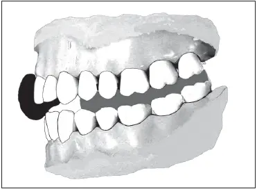
Fig 4-14The registration is checked to determine if the teeth seat fully in the registration.
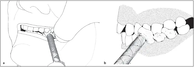
Fig 4-15 (a and b) Registration material is injected between the prepared teeth and the opposing arch.
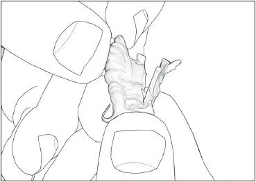
Fig 4-16The excess material is removed from the facial and lingual aspects of the registration.
Maximal Intercuspation Record
Although diagnostic mountings are done with the condyles in a centric relation position, casts that are to be used for the fabrication of restorations for a small portion of the occlusion are attached to the articulator in a position of maximal intercuspation. Mounting them in a centric relation position could result in a restoration with a built-in interference.
Armamentarium
Polyvinyl siloxane registration material
Impression material dispenser
Laboratory knife with no. 25 blade
Technique
The technique employed to index the intercuspal position for restoration fabrication produces an interocclusal record with the maxillary and mandibular teeth in full contact.
An impression gun is assembled in the same manner as when obtaining a centric relation record with registration material (see Fig 4-7). With the patient’s mouth slightly open, material is injected between the prepared teeth and the opposing arch ( Fig 4-15). The patient is instructed to close firmly until all posterior teeth are contacting normally. The dentist parts the lips and verifies that the patient has not closed in a protrusive or working relationship. The patient is instructed to keep the teeth together until asked to open. The record is left in place until the material has hardened. The bite registration is removed from the mouth and rinsed under running tap water. It should be inspected to ensure that all of the necessary teeth have been captured. A laboratory knife with a no. 25 blade is used to cut off all excess material on the facial and lingual sides of the prepared teeth ( Fig 4-16). Any material that extends over the unprepared teeth adjacent to the preparations should be removed.
Excess thickness is removed from the upper and lower surfaces of the record ( Fig 4-17). On the unprepared teeth opposing the preparation(s), enough material should be removed so that little more than the cusp tip indentations remain. Any material that reproduces edentulous ridges, gingival crevices, or the central fossae of the opposing occlusal surfaces is likely to produce incomplete seating of a cast with imperfections in those areas, so it is important to eliminate it ( Fig 4-18). The overall thickness of the record should be approximately 4.0 mm, with an equal amount having been removed from its upper and lower aspects.
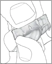
Fig 4-17The excess thickness from the upper and lower surfaces of the record are removed.
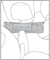
Fig 4-18All material contacting soft tissue or the central fossae of the opposing occlusal surfaces is removed.
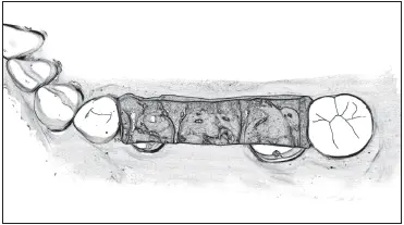
Fig 4-19The registration is trimmed along the facial cusps to verify complete seating of the record.
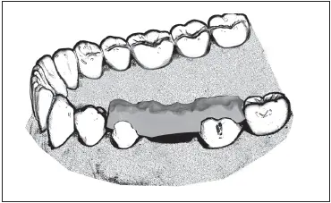
Fig 4-20The record is placed on the mandibular cast, and complete seating is confirmed.
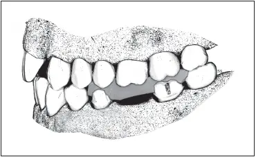
Fig 4-21The maxillary cast is placed in the completed record and articulated with the mandibular cast.
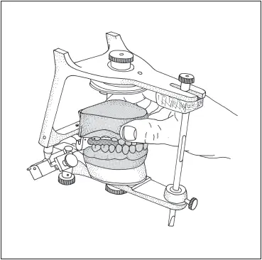
Fig 4-22The mandibular cast is mounted on the articulator using the record.
To verify seating of the casts into the record, its thickness along the facial cusps of the mandibular teeth is cut through completely using a laboratory knife with a no. 25 blade ( Fig 4-19). The registration is then rinsed with a hospital-grade disinfectant before proceeding.
The record is set on the mandibular cast, and its complete seating is verified ( Fig 4-20). The teeth of the maxillary cast are placed completely into the index while the teeth on the opposite side of the arch and those near the preparation(s) are articulated ( Fig 4-21). The record is used to articulate the casts, and the mandibular cast is mounted on the articulator ( Fig 4-22).
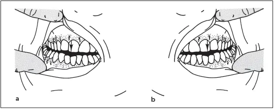
Fig 4-23The patient is guided into working excursions on the right (a) and left (b) sides.
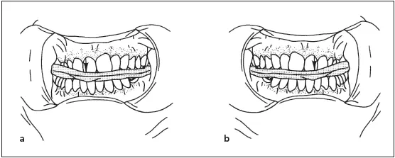
Fig 4-24Right (a) and left (b) lateral interocclusal records are made in wax wafers.
Lateral Interocclusal Record
Lateral interocclusal records are made in the mouth for the purpose of capturing the position of the condyles in their respective fossae. These records are then used to set the condylar guides to approximate the anatomical limits of the temporomandibular joints (TMJs). This allows the maximum benefit from using an articulator, facilitating the fabrication of accurate restorations with minimal time required for intraoral adjustment when the restoration is cemented.
Because the configuration of the TMJs has a strong determining influence on the movements of the mandible, the occlusal morphology of any restoration placed in the mouth must be in harmony with the movements of the mandible to prevent the initiation of occlusal disharmony and trauma. Cusp placement, cusp height, groove direction, and groove depth are all features ultimately affected by TMJ configuration. 9
Читать дальшеИнтервал:
Закладка:
Похожие книги на «Fundamentals of Fixed Prosthodontics»
Представляем Вашему вниманию похожие книги на «Fundamentals of Fixed Prosthodontics» списком для выбора. Мы отобрали схожую по названию и смыслу литературу в надежде предоставить читателям больше вариантов отыскать новые, интересные, ещё непрочитанные произведения.


Обсуждение, отзывы о книге «Fundamentals of Fixed Prosthodontics» и просто собственные мнения читателей. Оставьте ваши комментарии, напишите, что Вы думаете о произведении, его смысле или главных героях. Укажите что конкретно понравилось, а что нет, и почему Вы так считаете.