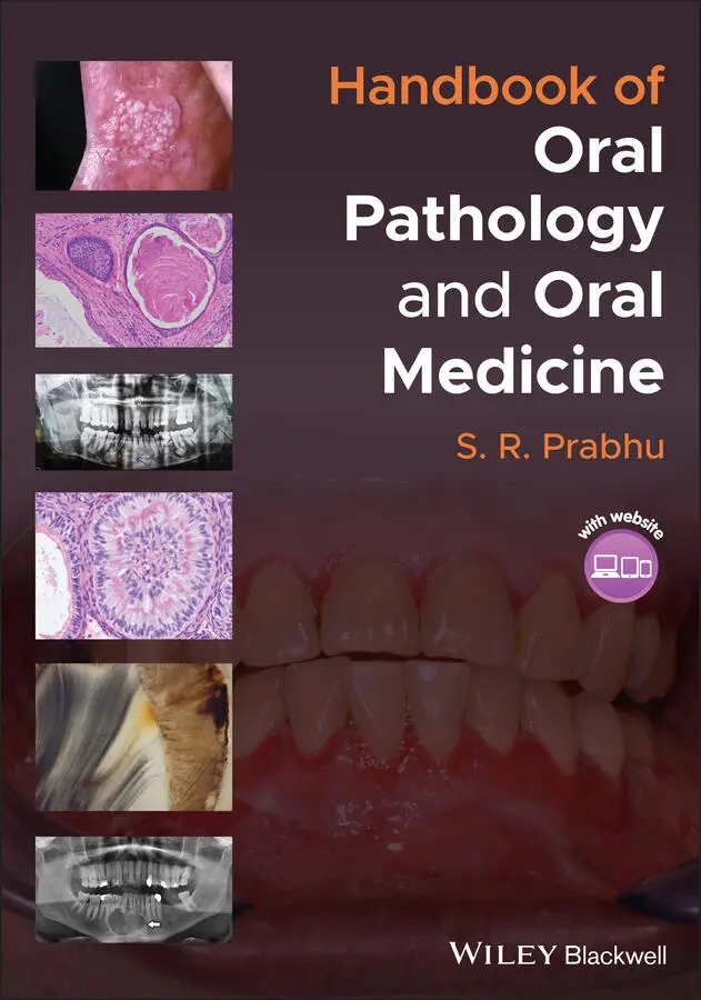
S. R. Prabhu - Handbook of Oral Pathology and Oral Medicine
Здесь есть возможность читать онлайн «S. R. Prabhu - Handbook of Oral Pathology and Oral Medicine» — ознакомительный отрывок электронной книги совершенно бесплатно, а после прочтения отрывка купить полную версию. В некоторых случаях можно слушать аудио, скачать через торрент в формате fb2 и присутствует краткое содержание. Жанр: unrecognised, на английском языке. Описание произведения, (предисловие) а так же отзывы посетителей доступны на портале библиотеки ЛибКат.
- Название:Handbook of Oral Pathology and Oral Medicine
- Автор:
- Жанр:
- Год:неизвестен
- ISBN:нет данных
- Рейтинг книги:5 / 5. Голосов: 1
-
Избранное:Добавить в избранное
- Отзывы:
-
Ваша оценка:
Handbook of Oral Pathology and Oral Medicine: краткое содержание, описание и аннотация
Предлагаем к чтению аннотацию, описание, краткое содержание или предисловие (зависит от того, что написал сам автор книги «Handbook of Oral Pathology and Oral Medicine»). Если вы не нашли необходимую информацию о книге — напишите в комментариях, мы постараемся отыскать её.
Discover a concise overview of the most common oral diseases in a reader-friendly book Handbook of Oral Pathology and Oral Medicine
Handbook of Oral Pathology and Oral Medicine
Handbook of Oral Pathology and Oral Medicine — читать онлайн ознакомительный отрывок
Ниже представлен текст книги, разбитый по страницам. Система сохранения места последней прочитанной страницы, позволяет с удобством читать онлайн бесплатно книгу «Handbook of Oral Pathology and Oral Medicine», без необходимости каждый раз заново искать на чём Вы остановились. Поставьте закладку, и сможете в любой момент перейти на страницу, на которой закончили чтение.
Интервал:
Закладка:
Apical periodontitis:Identify and eliminate the cause: endodontic therapy is effectiveExtraction of the tooth if exudate is to be drained (apical abscess)Usually, antibiotics are not required for simple (non‐suppurative) cases of periodontitisPain management with analgesics
Periapical granuloma:Endodontic treatmentExtraction of the tooth if tooth cannot be restored
3.4 Apical Abscess (Dentoalveolar Abscess)
3.4.1 Definition/Description
An odontogenic infection characterized by localization of pus in the structures that surround the teeth
3.4.2 Frequency
Shows a wide range of variation
Common in children: accounts for 47% of all dental‐related attendances at paediatric emergency rooms in the United States
3.4.3 Aetiology/Risk Factors
Secondary to dental caries, trauma, or failed root canal treatment
Bacteria and their toxic products enter the periapical tissues via the apical foramen and induce acute inflammation and pus formation
Polymicrobial odontogenic infection:Anaerobic cocci, Prevotella speciesFusobacterium speciesViridans group streptococci
3.4.4 Clinical and Radiographical Features
Non‐vital tooth in most cases
Involved tooth is tender to percussion
Lower molars commonly involved
In the initial stages there is no swelling, only intense, throbbing pain
Gingiva related to the tooth is red and tender
In established abscess buccal or labial painful gingival swellings ( Figure 3.3a)
Palatal swelling for maxillary molars is common
Trismus due to pain and swelling
Cervical lymphadenopathy is common
Early dental abscesses, within the first 10 days, may not show any radiographical features
Mild thickening of apical PDL, loss of lamina dura and loss of trabecular bone become evident as abscess advances ( Figure 3.3b)
Discharge of pus may occur in established abscess; pain subsides after the discharge of pus
Complications include cellulitis/Ludwig's angina or osteomyelitis
Life‐threatening complications include thrombophlebitis and septicaemia
Complete blood count for leucocytosis
3.4.5 Differential Diagnosis
Acute periodontitis
Infected radicular cyst
Focal sclerosing osteomyelitis
Periapical granuloma Figure 3.3 Apical abscess. (a) Presenting as a fluctuant gingival swelling; a decayed, broken down tooth with pulpal necrosis has caused this apical (periapical) abscess. Note draining pus via an intraoral sinus(source: Damdent, https://commons.wikimedia.org/wiki/File:Abces_parulique.jpg. Licensed Under CC BY‐SA 3.0.(b) Radiographical appearance of an established periapical abscess. Note the loss of periodontal ligament, lamina dura, and trabecular bone.
3.4.6 Diagnosis
History
Clinical examination
Radiography (periapical and panoramic views)
Needle aspirate for aerobic and anaerobic cultures
Blood culture studies
3.4.7 Management
Most dental abscesses respond to incision and drainage, root canal, or extraction. These procedures eliminate the source of infection
Antibiotics are usually not recommended for localized abscesses
If drainage is not possible or if the patient shows signs of systemic involvement, or is immunocompromised, antibiotics are required for a period of seven days
3.5 Condensing Osteitis
3.5.1 Definition/Description
Condensing osteitis refers to focal areas of bone sclerosis associated with apices of teeth with pulpitis or pulpal necrosis
Also known as focal sclerosing osteitis and focal sclerosing osteomyelitis
3.5.2 Frequency
Occurs in 4–7% of population
3.5.3 Aetiology
Low‐grade inflammatory stimulus from an inflamed dental pulp
3.5.4 Clinical Features
Most frequently in young adults
Asymptomatic
Most lesions are discovered on routine radiography
3.5.5 Radiographical Features
Localized increased radiodensity adjacent to the apex of the tooth ( Figure 3.4)
Widened periodontal ligament space
3.5.6 Microscopic Features
Normally bone biopsy is not indicated unless significant pathology (other than condensing osteitis) is suspected
The following microscopic features confirm the diagnosis of condensing osteitis:Replacement of marrow spaces and cancellous bone by dense, sclerotic compact boneBone may show prominent incremental linesMay see fibrosis replacing fatty marrow or scant connective tissueInflammatory changes are absent or minimal
3.5.7 Differential Diagnosis
Differential diagnoses on radiography include:Periapical cemental dysplasiaOsteomaComplex odontomaCementoblastomaOsteoblastomaHypercementosis Figure 3.4 Condensing osteitis. Note the increased radiodensity around the roots of a carious molar (black arrow).(Source: by kind permission of Professor Charles Dunlap, Kansas City, USA.)
3.5.8 Diagnosis
Clinical history is non‐contributory since condensing osteitis is asymptomatic
Most cases of condensing osteitis are incidentally detected on routine radiography
3.5.9 Management
No treatment is required for asymptomatic bony lesions
Tooth that caused the condensing osteitis needs to be identified and treated
Recommended Reading
1 Abbott, P.V. (2004). Classification, diagnosis, and clinical manifestations of apical periodontitis. Endodontic Topics 8: 36–54.
2 Abbott, P.V. and Yu, C. (2007). A clinical classification of the status of the pulp and the root canal system. Australian Dental Journal 52 (1 Suppl): S17–S31.
3 Braz‐Silva, P.H., Bergamini, M.L., Mardegan, A.P. et al. (2019). Inflammatory profile of chronic apical periodontitis: a literature review. Acta Odontologica Scandinavica 77: 173–180.
4 Dabuleanu, M. (2013). Pulpitis (reversible/irreversible). Journal of the Canadian Dental Association 79: d90.
5 Neville, B.W., Damm, D.D., Allen, C.M., and Chi, C.A. (2016). Pulpal and periapical disease. In: Oral and Maxillofacial Pathology, 4ee, 111–136. St Louis, MO: Elsevier.
6 Odell, E.W. (2017). Pulpitis and apical periodontitis. In: Cawson's Essentials of Oral Pathology and Oral Medicine, 9ee, 73–81. Edinburgh: Elsevier.
4 Tooth Wear, Pathological Resorption of Teeth, Hypercementosis and Cracked Tooth Syndrome
CHAPTER MENU
1 4.1 Tooth Wear: Attrition, Abrasion, Erosion, and Abfraction
2 4.2 Pathological Resorption of Teeth
3 4.3 Hypercementosis
4 4.4 Cracked Tooth Syndrome
4.1 Tooth wear: Attrition, Abrasion, Erosion, and Abfraction
4.1.1 Definition/Description
Attrition is a normal physiological process characterized by tooth wear caused by tooth‐to‐tooth contact. Usually this affects the tips of cusps of canines and posterior teeth and incisal edges of incisor teeth
Abrasion is tooth wear caused by an abrasive external agent. It affects both enamel and dentin
Erosion is a process characterized by progressive solubilization of tooth substance, usually by exposure to external or gastric acids
Abfraction is the pathological loss of hard tooth substance caused by biomechanical loading forces
4.1.2 Frequency
Prevalence of tooth wear varies from region to region
A universal agreement on prevalence of attrition, abrasion, and erosion does not exist
Читать дальшеИнтервал:
Закладка:
Похожие книги на «Handbook of Oral Pathology and Oral Medicine»
Представляем Вашему вниманию похожие книги на «Handbook of Oral Pathology and Oral Medicine» списком для выбора. Мы отобрали схожую по названию и смыслу литературу в надежде предоставить читателям больше вариантов отыскать новые, интересные, ещё непрочитанные произведения.


Обсуждение, отзывы о книге «Handbook of Oral Pathology and Oral Medicine» и просто собственные мнения читателей. Оставьте ваши комментарии, напишите, что Вы думаете о произведении, его смысле или главных героях. Укажите что конкретно понравилось, а что нет, и почему Вы так считаете.