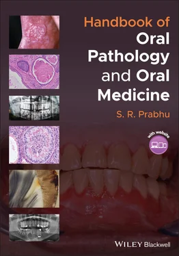
S. R. Prabhu - Handbook of Oral Pathology and Oral Medicine
Здесь есть возможность читать онлайн «S. R. Prabhu - Handbook of Oral Pathology and Oral Medicine» — ознакомительный отрывок электронной книги совершенно бесплатно, а после прочтения отрывка купить полную версию. В некоторых случаях можно слушать аудио, скачать через торрент в формате fb2 и присутствует краткое содержание. Жанр: unrecognised, на английском языке. Описание произведения, (предисловие) а так же отзывы посетителей доступны на портале библиотеки ЛибКат.
- Название:Handbook of Oral Pathology and Oral Medicine
- Автор:
- Жанр:
- Год:неизвестен
- ISBN:нет данных
- Рейтинг книги:5 / 5. Голосов: 1
-
Избранное:Добавить в избранное
- Отзывы:
-
Ваша оценка:
- 100
- 1
- 2
- 3
- 4
- 5
Handbook of Oral Pathology and Oral Medicine: краткое содержание, описание и аннотация
Предлагаем к чтению аннотацию, описание, краткое содержание или предисловие (зависит от того, что написал сам автор книги «Handbook of Oral Pathology and Oral Medicine»). Если вы не нашли необходимую информацию о книге — напишите в комментариях, мы постараемся отыскать её.
Discover a concise overview of the most common oral diseases in a reader-friendly book Handbook of Oral Pathology and Oral Medicine
Handbook of Oral Pathology and Oral Medicine
Handbook of Oral Pathology and Oral Medicine — читать онлайн ознакомительный отрывок
Ниже представлен текст книги, разбитый по страницам. Система сохранения места последней прочитанной страницы, позволяет с удобством читать онлайн бесплатно книгу «Handbook of Oral Pathology and Oral Medicine», без необходимости каждый раз заново искать на чём Вы остановились. Поставьте закладку, и сможете в любой момент перейти на страницу, на которой закончили чтение.
Интервал:
Закладка:
Occasionally, supernumerary roots may be detected in mandibular third molars, mandibular canines and premolars
No treatment is required
Detection is critical for endodontic treatment
Recommended Reading
1 Consolaro, A., Hadaya, O., Oliveira Miranda, D.A., and Colsolaro, R.B. (2020). Concrescence: can the teeth involved be moved or separated? Dental Press Journal of Orthodontics 25 (1): 20–25.
2 Miletich, I. and Sharpe, P.T. (2003). Normal and abnormal dental development. Human Molecular Genetics 12 (Suppl 1): R69–R73.
3 Neville, B.W., Damm, D.D., Allen, C.M., and Chi, C.A. (2016). Developmental alterations of teeth. In: Oral and Maxillofacial Pathology, 4ee, 49–92. St. Louis, MO: Elsevier.
4 Odell, E.W. (2017). Disorders of tooth development. In: Cawson's Essentials of Oral Pathology and Oral Medicine, 9ee, 23–42. Edinburgh: Elsevier.
5 Parsa, A. and Rapala, H. (2016). Endodontic treatment of a mandibular 6 years molar with three roots: a pedodontist perspective. International Journal of Pedodontic Rehabilitation 1: 64–67.
6 Rhoads, S.G., Hendricks, H.M., and Frazier‐Bowers, S.A. (2013). Establishing the diagnostic criteria for eruption disorders based on genetic and clinical data. American Journal of Orthodontics and Dentofacial Orthopaedics 144 (2): 194–202.
7 Rosa, D.C.L., Simukawa, E.R., Capelozza, A.L.A. et al. (2019). Alveolodental ankylosis: biological bases and diagnostic criteria. RGO Revista Gaúcha de Odontologia 67: e2019003.
8 Seow, W.K. (2014). Developmental defects of enamel and dentine: challenges for basic science research and clinical management. Australian Dental Journal 59 (1 Suppl): 143–154.
2 Dental caries
CHAPTER MENU
1 2.1 Definition/Description
2 2.2 Frequency
3 2.3 Aetiology/Risk Factors/Pathogenesis
4 2.4 Classification of Caries
5 2.5 Clinical Features
6 2.6 Differential Diagnosis
7 2.7 Diagnosis
8 2.8 Microscopic Features
9 2.9 Management
10 2.10 Prevention
2.1 Definition/Description
Dental caries is an infectious, transmissible disease resulting in destruction of tooth structure by acid‐forming bacteria found in dental plaque (an intraoral biofilm) in the presence of fermentable carbohydrates
The infection results in the loss of tooth minerals that begins with the outer surface of the tooth and can progress through the dentin to the pulp, ultimately compromising the vitality of the tooth
2.2 Frequency
60–90% of schoolchildren worldwide; the disease is most prevalent in Asian and Latin American countries
2.3 Aetiology/Risk Factors/Pathogenesis
Biofilm acid‐producing bacteria metabolize sugars and produce acids that lower the biofilm pH creating conditions that demineralize tooth enamel and dentin
Acid‐producing bacteria are Mutans streptococci and Lactobacillus species
Acids produced include lactic, acetic, formic and propionic acids. These acids are capable of demineralizing enamel and dentin
Cycles of demineralization and remineralization continue in the mouth in the presence of cariogenic bacteria, fermentable carbohydrates and saliva
Plaque microorganisms, substrate, susceptible tooth and time are essential factors for the development of caries
2.4 Classification of Caries
Classification is based on:Rate of progression: acute and chronic cariesAffected dental hard tissues: enamel, dentin and cemental (root surface) cariesLocation on the tooth surface involved: pit and fissure caries, approximal/smooth surface caries and root surface caries ( Table 2.1) Table 2.1 American Dental Association caries classification system: Site Definitions‐OriginSource: Based on Ismail, A.I., Tellez, M., Pitts, N.B. et al. (2013). Caries management pathways preserve dental tissues and promote oral health. Community Dentistry and Oral Epidemiology 41 (1): e12–e40.SiteDefinitionPit and fissureThe anatomical pits or fissures (clefts or valleys in the tooth surface) of the teeth at the occlusal, facial or lingual surfaces of posterior teeth OR the lingual surfaces of the maxillary incisors or caninesApproximal surfaceThe contact point(s) between adjacent teethCervical and smooth surfaceThe cervical area or any other smooth enamel surface of the anatomic crown adjacent to an edentulous space (toothless space); may exist anywhere around the full circumference of the toothRootThe root surface apical to the anatomic crown
2.5 Clinical Features
Asymptomatic in the initial stages
Mild pain, tooth sensitivity when carious lesion gets larger
Visible cavity in the tooth
Brown, black or white chalky discoloration
Oral malodour
2.5.1 Primary Caries
Decay at a location that has not previously experienced decay
2.5.2 Secondary Caries (Recurrent Caries)
Appears at a location with a previous history of caries
Frequently found on the margins of fillings and other dental restorations
2.5.3 Arrested Caries
A lesion on a tooth that was previously demineralized but was remineralized before causing a cavitation
2.5.4 Rampant Caries
Severe decay on multiple surfaces of many teeth ( Figure 2.1)
Those at risk: individuals with xerostomia, poor oral hygiene, drug‐induced dry mouth, large sugar intake and radiation to the head and neck region
Treatment options include therapeutic and preventive strategies, including diet modifications Figure 2.1 Rampant caries(source: From Mary A. Aubertin. 2014. Common Benign Dental and Periodontal Lesions. In: Diagnosis and Management of Oral Lesions and Conditions: A Resource Handbook for the Clinician, ed. Cesar A. Migliorati and Fotinos S. Panagakos, IntechOpen, doi: 10.5772/57597).
2.5.5 Early Childhood Caries
Rampant dental caries in infants and toddlers; also known as baby‐bottle caries
Most likely affected teeth: maxillary anterior deciduous teeth ( Figure 2.2)
Cause: allowing children to fall asleep with sweetened liquids in their bottles or feeding children sweetened liquids multiple times during the day
Education of parents/carers to follow healthy dietary and feeding habits to prevent the development of early childhood caries is important
2.5.6 Methamphetamine‐Induced Caries
Rampant caries often found in methamphetamine users and is often called ‘meth mouth’
The dental symptoms of methamphetamine users include poor oral hygiene, gingival inflammation, xerostomia, rampant caries and excessive tooth wear Figure 2.2 Early childhood caries(source: by kind permission of Dr Sadashivmurthy Prashanth, JSS Dental College, Mysuru, India). Figure 2.3 Caries in a methamphetamine user(source: From Mary A. Aubertin. 2014. Common Benign Dental and Periodontal Lesions. In: Diagnosis and Management of Oral Lesions and Conditions: A Resource Handbook for the Clinician, ed. Cesar A. Migliorati and Fotinos S. Panagakos, IntechOpen, doi: 10.5772/57597).
The pattern of caries is distinctive: it tends to start near the gums and involves the buccal smooth surface of the posterior teeth and the interproximal space of the anterior teeth, and progresses to complete destruction of the coronal portion of the tooth ( Figure 2.3)
The key to successful dental treatment is cessation of methamphetamine use.
2.5.7 Radiation Caries
Radiation caries is a complication of head and neck cancer radiotherapy
Typical radiation caries is characterized by enamel erosion and dentin exposure ( Figure 2.4)
Читать дальшеИнтервал:
Закладка:
Похожие книги на «Handbook of Oral Pathology and Oral Medicine»
Представляем Вашему вниманию похожие книги на «Handbook of Oral Pathology and Oral Medicine» списком для выбора. Мы отобрали схожую по названию и смыслу литературу в надежде предоставить читателям больше вариантов отыскать новые, интересные, ещё непрочитанные произведения.
Обсуждение, отзывы о книге «Handbook of Oral Pathology and Oral Medicine» и просто собственные мнения читателей. Оставьте ваши комментарии, напишите, что Вы думаете о произведении, его смысле или главных героях. Укажите что конкретно понравилось, а что нет, и почему Вы так считаете.
