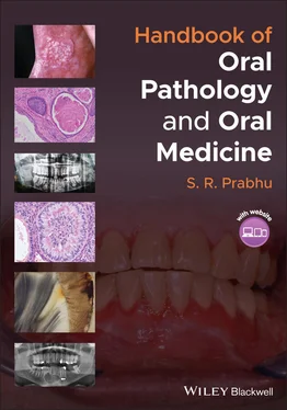
S. R. Prabhu - Handbook of Oral Pathology and Oral Medicine
Здесь есть возможность читать онлайн «S. R. Prabhu - Handbook of Oral Pathology and Oral Medicine» — ознакомительный отрывок электронной книги совершенно бесплатно, а после прочтения отрывка купить полную версию. В некоторых случаях можно слушать аудио, скачать через торрент в формате fb2 и присутствует краткое содержание. Жанр: unrecognised, на английском языке. Описание произведения, (предисловие) а так же отзывы посетителей доступны на портале библиотеки ЛибКат.
- Название:Handbook of Oral Pathology and Oral Medicine
- Автор:
- Жанр:
- Год:неизвестен
- ISBN:нет данных
- Рейтинг книги:5 / 5. Голосов: 1
-
Избранное:Добавить в избранное
- Отзывы:
-
Ваша оценка:
- 100
- 1
- 2
- 3
- 4
- 5
Handbook of Oral Pathology and Oral Medicine: краткое содержание, описание и аннотация
Предлагаем к чтению аннотацию, описание, краткое содержание или предисловие (зависит от того, что написал сам автор книги «Handbook of Oral Pathology and Oral Medicine»). Если вы не нашли необходимую информацию о книге — напишите в комментариях, мы постараемся отыскать её.
Discover a concise overview of the most common oral diseases in a reader-friendly book Handbook of Oral Pathology and Oral Medicine
Handbook of Oral Pathology and Oral Medicine
Handbook of Oral Pathology and Oral Medicine — читать онлайн ознакомительный отрывок
Ниже представлен текст книги, разбитый по страницам. Система сохранения места последней прочитанной страницы, позволяет с удобством читать онлайн бесплатно книгу «Handbook of Oral Pathology and Oral Medicine», без необходимости каждый раз заново искать на чём Вы остановились. Поставьте закладку, и сможете в любой момент перейти на страницу, на которой закончили чтение.
Интервал:
Закладка:
Oculodentodigital dysplasia
Segmental odontomaxillary dysplasia
Odonto‐onychodermal dysplasia
Odontochondrodysplasia
1.9.7 Diagnosis
History
Clinical examination
Radiography
1.9.8 Management
Unerupted teeth to remain without any interference
Erupted teeth: steel crowns
Non‐salvageable teeth to be extracted
1.10 Delayed Tooth Eruption
1.10.1 Definition/Description
Delayed tooth eruption is the emergence of a tooth into the oral cavity at a time that deviates significantly from norms established for different races, ethnic groups and sexes
1.10.2 Frequency
Delayed eruption is relatively common; racial and gender variations exist
Failure of eruption is less common
Agenesis of teeth cause failure of eruption
1.10.3 Aetiology/Risk Factors
Local causes associated with delayed tooth eruption:Supernumerary teethMucosal barrier scar tissue due to trauma/surgery/gingival hyperplasiaTumours: odontogenic or non‐odontogenic tumoursAnkylosis of deciduous teethEnamel pearlsInjuries to primary teethRegional odontodysplasiaEctopic eruptionImpacted permanent teethEmbedded primary teethOral cleftsRadiation damage
Systemic causes associated with delayed tooth eruption:Nutritional deficienciesVitamin D‐resistant ricketsHypoparathyroidismHypopituitarismLong‐term chemotherapyCerebral palsyPrematurity or low birth weightPhenytoin useGenetic disorders
1.10.4 Clinical and Radiographical Features
Local factors causing delayed tooth eruption are frequently detected by radiography
Systemic factors causing delayed tooth eruption are detected by systemic clinical features and laboratory findings
Failure of tooth eruption: congenital absence of teeth (third molars, mandibular second premolars and maxillary lateral incisors) results in failure of tooth eruption
Radiographical evidence of absence of teeth is diagnostic
1.10.5 Diagnosis
History
Clinical examination
Radiography (panoramic view is ideal)
Laboratory tests if systemic factors are suspected
1.10.6 Management
Patient with eruption delay of more than 12 months (delayed eruption) of the normal age range should be referred to a paediatric dentist for further evaluation
Identification of the causes and their elimination is important
Surgical exposure followed by orthodontic treatment may be required for some patients with delayed eruption
1.11 Tooth Impaction (Impacted Teeth)
1.11.1 Definition/Description
Teeth that are completely or partially retained in the jaws beyond their normal date of eruption
1.11.2 Frequency
Common; variations in incidence and prevalence exist
The mandibular third molars are the most common impacted teeth, with their prevalence ranging from 27% to 68.8% in various parts of the world
The reported prevalence of impacted teeth of canines and second premolars ranges from 2.9% to 13.7%
1.11.3 Aetiology
Lack of space for tooth eruption due to inadequate arch length
Crowding of teeth
Dense overlying bone
Excessive soft tissue in the path of eruption
Genetic abnormalities
Long tortuous path of eruption (for canines)
1.11.4 Clinical Features
Frequently impacted teeth: mandibular third molars followed by the maxillary third molars, maxillary canines and mandibular premolars
Young adults are commonly affected; often detected on routine radiography
Impacted deciduous teeth are extremely rare
Impacted permanent first and second molars are rare
Often supernumerary teeth are impacted (detected on radiography)
Impacted teeth may or may not be symptomatic
With no history of extraction, clinically the number of teeth present in the dentition is less than normal
Impaction can be full or partial
Symptomatic patients with lower third molar may complain of earache or paraesthesia of the lip
Pericoronitis may occur (pain, inability to open the mouth, swelling of the pericoronal soft tissue)
Often, all four third molars may be impacted
Occasionally impactions are associated with syndromes or odontogenic cysts and tumours
1.11.5 Radiographical Features
Types of impaction: mesioangular, distoangular, vertical or horizontal impaction for third molars ( Figure 1.9a‐d). Canine impaction may be bilateral ( Figure 9 e) or inverted
Proximity of the impacted tooth to the inferior dental nerve for lower third molar impactions may cause paraesthesia
Impacted teeth may be associated with cysts or odontogenic tumours
1.11.6 Diagnosis
History
Clinical examination
Radiography (panoramic view)
1.11.7 Management
No treatment is required for asymptomatic impactions
Surgical removal for symptomatic impacted teeth
Surgery for impacted teeth associated with cysts or tumours Figure 1.9(a) Mesioangular impaction of the mandibular third molar. (b) Distoangular impaction of the mandibular third molar (c) Vertical impaction of the mandibular third molar. (d) Horizontal impaction of the mandibular third molar. (e) Bilateral impaction of maxillary canines.
1.12 Dens Invaginatus and Dens Evaginatus
1.12.1 Definition/Description
Dens invaginatus refers to an exaggeration of the process of formation of lingual pit causing invagination (also called dens in dente or dilated odontome)
Dens evaginatus refers to an enamel and dentin covered spur extending outward from the occlusal surfaces of molars or premolars and rarely lingual surfaces of lower anterior teeth. This is the opposite of dens evaginatus (also called evaginated odontome)
1.12.2 Frequency
Dens invaginatus: prevalence: 0.3–10%, affecting more males than females
Dens evaginatus: more common in people of Asian descent; prevalence: 0.06–7.7%; 15% in Inuit and Native American populations
1.12.3 Aetiology/Risk Factors
Dens invaginatus:Deepening or invagination of the enamel organ into the dental papilla prior to calcification of the dental tissuesGenetics may play a role
Dens evaginatus:A result of an unusual growth and folding of the inner enamel epithelium and ectomesenchymal cells of dental papilla into the stellate reticulum of the enamel organ
1.12.4 Clinical Features
Dens invaginatus:The permanent maxillary lateral incisors appear to be the most frequently affected tooth (90% of all cases)Maxillary posterior teeth: 6.5% of all casesMandibular teeth are very rarely affected
May be associated with taurodontism, microdontia, gemination, supernumerary tooth and dentinogenesis imperfecta Figure 1.10(a) Dens invaginatus; radiograph showing dens invaginatus in a peg lateral incisor(source: by kind permission of Professor Charles Dunlap, Kansas City, Kansas, USA).(b) Dens evaginatus; radiograph showing dens evaginatus. Note a tubercle extending outward from the occlusal surface of the premolar.
Causes food debris deposits and renders tooth vulnerable to caries
Dens evaginatus:More common in mandibular premolar teethMay be bilateral and symmetrical tubercles on the occlusal surfaces of posterior teeth or on lingual surfaces of lower anteriorSlight female sex predilectionCan cause malocclusion with opposing teethAbnormal wear and fracture of the tubercle may occur
1.12.5 Radiographical features
Интервал:
Закладка:
Похожие книги на «Handbook of Oral Pathology and Oral Medicine»
Представляем Вашему вниманию похожие книги на «Handbook of Oral Pathology and Oral Medicine» списком для выбора. Мы отобрали схожую по названию и смыслу литературу в надежде предоставить читателям больше вариантов отыскать новые, интересные, ещё непрочитанные произведения.
Обсуждение, отзывы о книге «Handbook of Oral Pathology and Oral Medicine» и просто собственные мнения читателей. Оставьте ваши комментарии, напишите, что Вы думаете о произведении, его смысле или главных героях. Укажите что конкретно понравилось, а что нет, и почему Вы так считаете.
