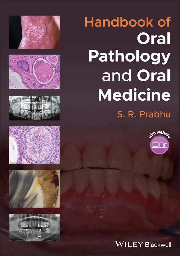
S. R. Prabhu - Handbook of Oral Pathology and Oral Medicine
Здесь есть возможность читать онлайн «S. R. Prabhu - Handbook of Oral Pathology and Oral Medicine» — ознакомительный отрывок электронной книги совершенно бесплатно, а после прочтения отрывка купить полную версию. В некоторых случаях можно слушать аудио, скачать через торрент в формате fb2 и присутствует краткое содержание. Жанр: unrecognised, на английском языке. Описание произведения, (предисловие) а так же отзывы посетителей доступны на портале библиотеки ЛибКат.
- Название:Handbook of Oral Pathology and Oral Medicine
- Автор:
- Жанр:
- Год:неизвестен
- ISBN:нет данных
- Рейтинг книги:5 / 5. Голосов: 1
-
Избранное:Добавить в избранное
- Отзывы:
-
Ваша оценка:
Handbook of Oral Pathology and Oral Medicine: краткое содержание, описание и аннотация
Предлагаем к чтению аннотацию, описание, краткое содержание или предисловие (зависит от того, что написал сам автор книги «Handbook of Oral Pathology and Oral Medicine»). Если вы не нашли необходимую информацию о книге — напишите в комментариях, мы постараемся отыскать её.
Discover a concise overview of the most common oral diseases in a reader-friendly book Handbook of Oral Pathology and Oral Medicine
Handbook of Oral Pathology and Oral Medicine
Handbook of Oral Pathology and Oral Medicine — читать онлайн ознакомительный отрывок
Ниже представлен текст книги, разбитый по страницам. Система сохранения места последней прочитанной страницы, позволяет с удобством читать онлайн бесплатно книгу «Handbook of Oral Pathology and Oral Medicine», без необходимости каждый раз заново искать на чём Вы остановились. Поставьте закладку, и сможете в любой момент перейти на страницу, на которой закончили чтение.
Интервал:
Закладка:
Dilaceration:No treatment for mild dilacerationIf symptomatic due to gross dilaceration, tooth requires surgical extraction
1.6 Amelogenesis Imperfecta
1.6.1 Definition/Description
A group of inherited disorders caused by defects in the genes that encode enamel matrix proteins, resulting in defective structure of the enamel involving both dentitions
1.6.2 Incidence/Prevalence
Global prevalence: 0.5%
1.6.3 Aetiology/Risk Factors
Caused by mutations or altered expression in five genes:AMEL (amelogenin)ENAM (enamelin)MMP20 (matrix metalloproteinase‐20)KLK4 (kallikrein‐4)FAM83H. 6–16
Inheritance can be autosomal dominant, recessive or x‐linked
1.6.4 Clinical/Radiographical Features
Three types of amelogenesis imperfecta have been identified – hypoplastic, hypocalcified and hypomaturation:Hypoplastic type:Enamel is of reduced thickness due to a defect in the formation of normal matrixEnamel is pitted, grooved, stained and thinEnamel is normally mineralized; hard and translucentRadiographically, the enamel contrasts normally from dentineHypocalcified type:Enamel matrix is normal in quantityEnamel calcification is defectiveEnamel is weak in structure and vulnerable to attritionTeeth become opaque, stained and rapidly wear down ( Figure 1.6)Radiographically, enamel is less radio‐opaque than dentineHypomaturation type:Enamel is normal in thickness, shows opaque brownish‐yellow patchesEnamel mimics fluorotic mottled enamel in appearanceEnamel is soft and vulnerable to attrition
Other features that may occur in any of the above types of amelogenesis imperfecta include:Delay in dental eruptionMicrodontiaDeviant crown and morphologyRoot resorptionShort rootsEnlarged pulp chamberPulp stonesDens in dente (dens invaginatus)Tooth agenesisCrowding of teeth Figure 1.6 Amelogenesis imperfecta (hypocalcified type); the enamel is stained and vulnerable to attrition(source: by kind permission of Professor Charles Dunlap, Kansas City, USA).
1.6.5 Differential diagnosis
Dental fluorosis
Dentinogenesis imperfecta
Enamel hypoplasia
Trauma
Molar incisor hypomineralization
1.6.6 Diagnosis
History including a detailed family history
Pedigree plotting (family health history tree)
Clinical examination
Radiography
1.6.7 Management
Aesthetic treatment
Treatment for symptoms if present (e.g. tooth sensitivity)
1.7 Dentinogenesis Imperfecta
1.7.1 Definition/Description
A group of autosomal dominant genetic conditions characterized by abnormal dentin structure affecting both the primary and secondary dentitions
1.7.2 Frequency
Incidence of 1 in 6000 to 1 in 8000
1.7.3 Aetiology/Risk Factors
Mutations in dentin sialoprotein genes
1.7.4 Clinical Features
Primary and permanent teeth are affected
Teeth appear amber, brown/blue, or opalescent brown ( Figure 1.7a)
Syndromic form, osteogenesis imperfecta: opalescent dentine, blue sclera and short stature
1.7.5 Radiographical features
The crowns may appear bulbous ( Figure 1.7b)
Pulp chambers are often small or obliterated
The roots are often narrow with small or with obliterated root canals Figure 1.7 Dentinogenesis imperfecta. (a) Note tooth wear and opalescent crowns. (b) Radiograph shows bulbous crowns and cervical constriction of molars.(source: by kind permission of Professor Charles Dunlap, Kansas City, USA.)
1.7.6 Differential Diagnosis
Hypocalcified forms of amelogenesis imperfecta
Osteogenesis imperfecta
Congenital erythropoietic porphyria
Conditions leading to early tooth loss
Permanent teeth discolouration due to tetracyclines
Vitamin D‐dependent and vitamin D‐resistant rickets
1.7.7 Diagnosis
Family history
Pedigree construction
Detailed clinical examination
Radiography
1.7.8 Management
The aims of treatment are to remove sources of infection or pain, restore aesthetics and protect posterior teeth from wear
Preservation of occlusal face height, maintenance of function and aesthetic needs are priorities
For the primary dentition, stainless steel crowns are recommended
1.8 Dentinal Dysplasia (Dentin Dysplasia)
1.8.1 Definition/Description
A rare inherited disorder of dentin formation characterized by either absent or short conical roots
Two types occur: radicular dentin dysplasia (type 1) and coronal dentin dysplasia (type 2)
Coronal dentin dysplasia is a severe form of dentinogenesis imperfecta
1.8.2 Frequency
Type 1: 1 in 100 000
Type 2: 1 in 6000 to 1 in 8000
1.8.3 Aetiology/Risk Factors
Defective dentin sialoprotein gene for both types
1.8.4 Clinical Features
Radicular type (type 1):Diffusely affects both dentitions: severe manifestations in deciduous teethCoronal enamel and dentin are normalRadicular dentin is defective: short roots result in tooth mobility and premature tooth lossStrength of roots is reducedHypersensitive dentinLoss of pulpal vitalityAbsent root canalsBifurcation is close to the apex in molarsPeriapical pathology is common (seen as periapical radiolucency)
Coronal type (type 2):Primary and permanent teeth are affectedTeeth appear amber, brown/blue or opalescent brown
1.8.5 Radiographical features
Coronal type:The crowns may appear bulbousPulp chambers are often small or obliteratedThe roots are often narrow with small or obliterated root canalsRoots may be absent ( Figure 1.8) Figure 1.8 Dentinal dysplasia radiograph showing absence of roots(source: by kind permission of Professor Charles Dunlap, Kansas City, USA).
1.8.6 Differential Diagnosis
Dentinogenesis imperfecta
Osteogenesis imperfecta
Conditions that cause premature loss of teeth
1.8.7 Diagnosis
Family history
Clinical examination
Radiography
1.8.8 Management
Symptomatic and preventive care and meticulous oral hygiene
1.9 Regional Odontodysplasia (Ghost Teeth)
1.9.1 Definition/Description
A rare non‐hereditary dental anomaly involving enamel, dentin and cementum of both dentitions, but mostly the teeth of one quadrant
1.9.2 Frequency
A rare disorder
1.9.3 Aetiology/Risk Factors
Unknown
Probably alteration in vascular supply in the jaws around developing teeth
1.9.4 Clinical Features
Female predilection (female to male ratio 1.7 : 1)
Both dentitions are involved
Mostly one but rarely two quadrants are involved
Age at diagnosis: 2–4 years for deciduous teeth and 7–11 years for permanent teeth
Maxillary predominance (ratio of maxillary to mandibular width 1.6 : 1)
Failure of tooth eruption is common
Erupted teeth exhibit small brown crowns
Pulp necrosis is common
Early tooth exfoliation
1.9.5 Radiographical features
Thin enamel and dentin appear surrounding enlarged radiolucent pulp chamber (hence the name ghost tooth)
Pulp stones are occasionally detected on radiography
1.9.6 Differential Diagnosis
Интервал:
Закладка:
Похожие книги на «Handbook of Oral Pathology and Oral Medicine»
Представляем Вашему вниманию похожие книги на «Handbook of Oral Pathology and Oral Medicine» списком для выбора. Мы отобрали схожую по названию и смыслу литературу в надежде предоставить читателям больше вариантов отыскать новые, интересные, ещё непрочитанные произведения.


Обсуждение, отзывы о книге «Handbook of Oral Pathology and Oral Medicine» и просто собственные мнения читателей. Оставьте ваши комментарии, напишите, что Вы думаете о произведении, его смысле или главных героях. Укажите что конкретно понравилось, а что нет, и почему Вы так считаете.