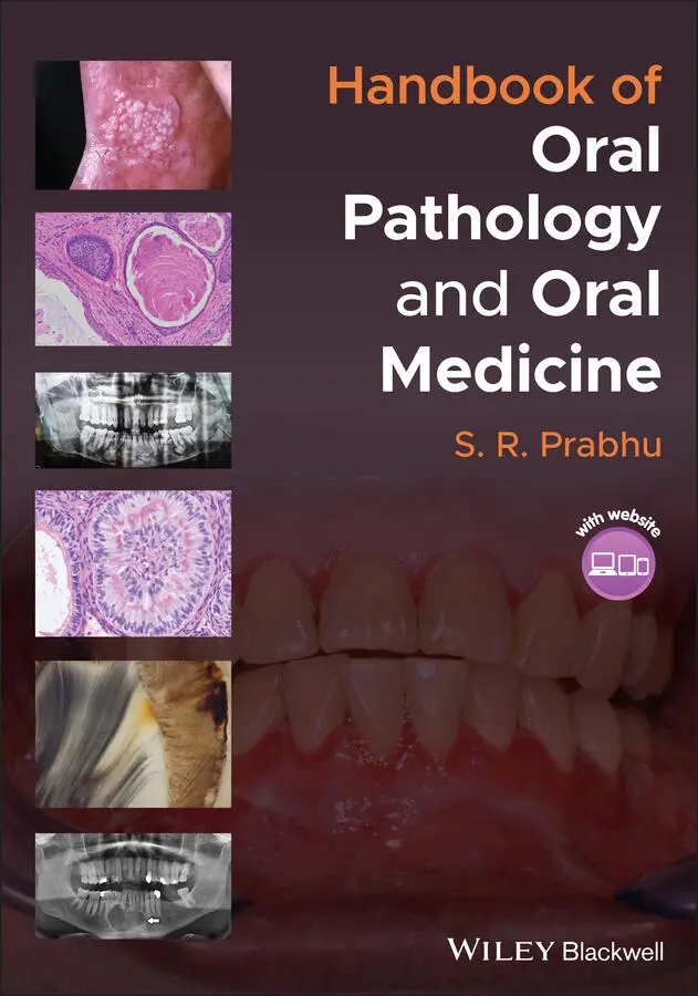
S. R. Prabhu - Handbook of Oral Pathology and Oral Medicine
Здесь есть возможность читать онлайн «S. R. Prabhu - Handbook of Oral Pathology and Oral Medicine» — ознакомительный отрывок электронной книги совершенно бесплатно, а после прочтения отрывка купить полную версию. В некоторых случаях можно слушать аудио, скачать через торрент в формате fb2 и присутствует краткое содержание. Жанр: unrecognised, на английском языке. Описание произведения, (предисловие) а так же отзывы посетителей доступны на портале библиотеки ЛибКат.
- Название:Handbook of Oral Pathology and Oral Medicine
- Автор:
- Жанр:
- Год:неизвестен
- ISBN:нет данных
- Рейтинг книги:5 / 5. Голосов: 1
-
Избранное:Добавить в избранное
- Отзывы:
-
Ваша оценка:
Handbook of Oral Pathology and Oral Medicine: краткое содержание, описание и аннотация
Предлагаем к чтению аннотацию, описание, краткое содержание или предисловие (зависит от того, что написал сам автор книги «Handbook of Oral Pathology and Oral Medicine»). Если вы не нашли необходимую информацию о книге — напишите в комментариях, мы постараемся отыскать её.
Discover a concise overview of the most common oral diseases in a reader-friendly book Handbook of Oral Pathology and Oral Medicine
Handbook of Oral Pathology and Oral Medicine
Handbook of Oral Pathology and Oral Medicine — читать онлайн ознакомительный отрывок
Ниже представлен текст книги, разбитый по страницам. Система сохранения места последней прочитанной страницы, позволяет с удобством читать онлайн бесплатно книгу «Handbook of Oral Pathology and Oral Medicine», без необходимости каждый раз заново искать на чём Вы остановились. Поставьте закладку, и сможете в любой момент перейти на страницу, на которой закончили чтение.
Интервал:
Закладка:
Three types of dens invaginatus occur which can be detected on radiography:Type I: invagination ends in a blind sac, limited to the tooth crown ( Figure 1.10a)Type II: invagination extends to the cementoenamel junction extending in a blind sac. It may or may not extend into the root pulpType III: invagination extends to the interior of the root providing an opening to the periodontium, sometimes this presents another foramen in the apical region of toothDens evaginatus shows a tubercle on the occlusal surface ( Figure 1.10b)
1.12.6 Diagnosis
History
Clinical examination (tooth morphology)
Radiography (intraoral periapical views)
1.12.7 Management
Dens invaginatus: placement of sealants and endodontic treatment for severe cases
Dens evaginatus: removal of the tubercle and application of fluorides
1.13 Fluorosis (Mottled Enamel)
1.13.1 Definition/Description
Dental fluorosis (mottled enamel) is a qualitative defect of enamel resulting from an increase in fluoride concentration during enamel formation
1.13.2 Frequency
Fluorosis is extremely common
Global variations exist. The global prevalence of fluorosis is reported to be about 32%
1.13.3 Aetiology/Risk Factors
A higher than normal amount of fluoride ingestion while teeth are forming
When the level of fluoride is above 1.5 mg/l (1.5 ppm) in drinking water, dental fluorosis occurs
The severity of fluorosis is dependent on the dose and time of exposure to fluoride levels
Other sources of fluoride: toothpastes, mouth rinses, fluoride supplements, beverages (brick tea, tea and butter tea) and food (infant formula, fish, beans, potatoes and wheat)
1.13.4 Clinical Features
Severity of fluorosis is dose dependent
Mild fluorosis: opaque lines following the perikymata
Moderate fluorosis: the opaque lines merge and more irregular cloudy areas become visible
Severe fluorosis: enamel is grossly defective with opaque chalky appearance and punched out pits. Extrinsic brown staining in the pits is frequent ( Figure 1.11)
Moderate to severe enamel fluorosis is called mottled enamel
Teeth with fluorosis are weak but resistant to caries
1.13.5 Differential Diagnosis
Turner's hypoplasia
Hypoplastic teeth in systemic disorders
Amelogenesis imperfecta
Early carious lesions
Tetracycline staining of teeth
1.13.6 Diagnosis
History (residence/ migration, water fluoridation, other sources of fluorides in the diet)
Clinical examination
1.13.7 Management
Aesthetic procedures as required Figure 1.11 Yellow‐brown discoloration of maxillary incisors due to fluorosis(source: From Mary A. Aubertin. 2014. Common Benign Dental and Periodontal Lesions. In: Diagnosis and Management of Oral Lesions and Conditions: A Resource Handbook for the Clinician. ed. Cesar A. Migliorati and Fotinos S. Panagakos. IntechOpen. doi: 10.5772/57597).
Close monitoring of sources of fluoride during the first three years of age
Water fluoridation: 0.7–1 ppm recommended
Use of fluoride tooth paste after 12 months of age
Infant formula with fluoridated water to be avoided
Fluoride supplements only in non‐fluoridated areas
1.14 Tetracycline‐Induced Discoloration of Teeth: Key Features
Tetracycline is a broad‐spectrum antibiotic commonly used for infections
Tetracycline has several different analogues such as doxycycline, oxytetracycline, minocycline, chlortetracycline, demeclocycline
Tetracycline can stain teeth if ingested by the mother in the third trimester or by the child during the years of tooth formation of deciduous and permanent dentition
The discoloration, which is permanent, varies from yellow or grey to brown ( Figure 1.12)
Administration of tetracycline to pregnant women must be avoided during the second or third trimester of gestation and to children up to eight years of age.
Tetracycline‐stained teeth must be differentiated from dentinogenesis imperfecta Figure 1.12 Tetracycline‐induced grey/brown discolouration of deciduous teeth in a child(source: by kind permission of Professor Charles Dunlap, Kansas City, Kansas, USA).
1.15 Enamel Pearl: Key Features
The enamel pearl is a globule of enamel formation located on the root surface ( Figure 1.13)
It is characterized by a core of dentin covered by enamel and may contain a pulp chamber
Enamel pearl may cause periodontal pockets and periodontitis Figure 1.13 Enamel pearl on the cementum(source: by kind permission of Professor Charles Dunlap, Kansas City, Kansas, USA).
1.16 Talon Cusp: Key Features
Talon cusp is a rare developmental anomaly presenting as a wisp‐like structure arising from the cervical region of anterior teeth
Resembles an eagle's talon Figure 1.14 Periapical radiograph of talon cusp on a partially erupted upper left permanent maxillary incisor in an eight‐year‐old boy. Note V‐shaped radiopaque structure overlapping the affected crown with its apex directed incisally(source: Matthew Fergusson, https://en.wikipedia.org/wiki/File:Talon_cusp.png. Licensed under CC BY‐SA 4.0).
In canines and incisors, it originates usually in the palatal cingulum as a tubercle projecting from the palatal surface
Its prevalence varies from less than 1% to approximately 8%
Radiographically, talon cusp appears as appears as a V‐shaped radiopaque structure overlapping the affected crown with its apex directed incisally ( Figure 1.14)
Symptoms include interference with occlusion, irritation of soft tissues, accidental cusp fracture and susceptible to dental caries
1.17 Hutchinson's Incisors and Mulberry Molars: Key Features
‘Hutchinson's incisors’ and ‘Mulberry molars’ are dental developmental defects seen in children with congenital syphilis; they are rare conditions
In Hutchinson's incisors, the incisal edge is either notched or screwdriver shaped. The bulbous crown is short and narrow (‘barrel shaped’). In the centre of the incisal edge a deep vertical central notch may be present ( Figure 1.15a)
Mulberry molars are characterized by multiple rounded rudimentary enamel cusps on the permanent first molars ( Figure 1.15b)
Hutchinson's incisors and mulberry molars are caused by direct invasion of tooth germs by Treponema organisms during tooth development Figure 1.15 (a) Hutchinson's incisors of congenital syphilis. Note screwdriver shape and central notch on the crowns of upper and lower permanent incisors. (b) Mulberry molars of congenital syphilis. Note multiple poorly formed globular cusps on the occlusal surfaces of mandibular permanent first molars.(source: images by kind permission of Professor Charles Dunlap, Kansas City, Kansas, USA.)
1.18 Tooth Ankylosis: Key Features
Anatomical fusion of tooth cementum with the alveolar bone:Mandibular primary first molars are frequently ankylosedAnkylosis of permanent teeth is uncommonA sharp solid note on percussion is noted, suggesting ankylosisPeriodontal ligament space is absent on radiographyIn many examples, the permanent successor is missing Figure 1.16 An extracted mandibular molar with three roots.
1.19 Supernumerary Roots: Key Features
Normally, the permanent mandibular first molar has two roots, one mesial and one distal root
Rarely, an additional third root is seen, which is found distolingually, called the radix entomolaris ( Figure 1.16)
Читать дальшеИнтервал:
Закладка:
Похожие книги на «Handbook of Oral Pathology and Oral Medicine»
Представляем Вашему вниманию похожие книги на «Handbook of Oral Pathology and Oral Medicine» списком для выбора. Мы отобрали схожую по названию и смыслу литературу в надежде предоставить читателям больше вариантов отыскать новые, интересные, ещё непрочитанные произведения.


Обсуждение, отзывы о книге «Handbook of Oral Pathology and Oral Medicine» и просто собственные мнения читателей. Оставьте ваши комментарии, напишите, что Вы думаете о произведении, его смысле или главных героях. Укажите что конкретно понравилось, а что нет, и почему Вы так считаете.