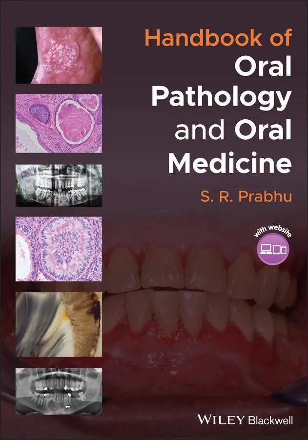
S. R. Prabhu - Handbook of Oral Pathology and Oral Medicine
Здесь есть возможность читать онлайн «S. R. Prabhu - Handbook of Oral Pathology and Oral Medicine» — ознакомительный отрывок электронной книги совершенно бесплатно, а после прочтения отрывка купить полную версию. В некоторых случаях можно слушать аудио, скачать через торрент в формате fb2 и присутствует краткое содержание. Жанр: unrecognised, на английском языке. Описание произведения, (предисловие) а так же отзывы посетителей доступны на портале библиотеки ЛибКат.
- Название:Handbook of Oral Pathology and Oral Medicine
- Автор:
- Жанр:
- Год:неизвестен
- ISBN:нет данных
- Рейтинг книги:5 / 5. Голосов: 1
-
Избранное:Добавить в избранное
- Отзывы:
-
Ваша оценка:
Handbook of Oral Pathology and Oral Medicine: краткое содержание, описание и аннотация
Предлагаем к чтению аннотацию, описание, краткое содержание или предисловие (зависит от того, что написал сам автор книги «Handbook of Oral Pathology and Oral Medicine»). Если вы не нашли необходимую информацию о книге — напишите в комментариях, мы постараемся отыскать её.
Discover a concise overview of the most common oral diseases in a reader-friendly book Handbook of Oral Pathology and Oral Medicine
Handbook of Oral Pathology and Oral Medicine
Handbook of Oral Pathology and Oral Medicine — читать онлайн ознакомительный отрывок
Ниже представлен текст книги, разбитый по страницам. Система сохранения места последней прочитанной страницы, позволяет с удобством читать онлайн бесплатно книгу «Handbook of Oral Pathology and Oral Medicine», без необходимости каждый раз заново искать на чём Вы остановились. Поставьте закладку, и сможете в любой момент перейти на страницу, на которой закончили чтение.
Интервал:
Закладка:
Indirect effects of radiotherapy include changes in salivary quantity and composition, together with alteration of the oral flora. These changes are widely regarded as the major causes of radiation caries Figure 2.4 Radiation caries(source: by kind permission from Dr Vlaho Brailo, School of Dental Medicine, University of Zagreb, Zagreb, Croatia).
Management of radiation caries includes management of xerostomia and restorative treatment radiation‐induced dental caries. Glass ionomer cements have proved to be a better alternative to composite resins in irradiated patients.
2.6 Differential Diagnosis
Hypoplastic enamel
Hypocalcified enamel
Fluorosis
Stains
2.7 Diagnosis
History
Clinical examination: initially a chalky white spot lesion
Blowing air across the suspected tooth surface is useful
Later stages cavitation
Radiography/laser detection
2.8 Microsopic Features
2.8.1 Enamel Caries
Early enamel lesion shows:Conical lesion with its apex towards dentinLesion shows four distinct zones of differing translucency ( Figure 2.5a and b)Translucent zone (deepest zone) Figure 2.5 (a) Early approximal enamel caries. Undecalcified section of a precavitation stage of enamel caries showing a cone‐shaped carious lesion on the proximal surface of the tooth with its apex towards dentine. The intact surface layer and the body of the lesion are visible. Evidence of early demineralization of dentine is seen beneath the amelodentinal junction deep to the carious enamel lesion. This is due to the diffusion of acids from the enamel lesion into the dentine. The dentine also shows numerous dead tracts. (b) Early pit and fissure enamel caries. Undecalcified section showing precavitation stage of enamel caries surrounding an occlusal pit. The dense surface zone, main body of the lesion, dark zone and peripheral translucent zones are visible.(Source: by kind permission of David Wilson, Adelaide, Australia.)Dark zone (superficial to the translucent zone)Body of the lesion (extends from beneath the surface zone to the dark zone)Surface zoneCaries reaches enamel–dentin junction and spreads laterally, undermining the enamel
Characteristics of enamel preceding cavitation:Translucent zone:1% mineral lossDark zone:2‐4% mineral loss overall. A zone of remineralization behind the advancing front becomes evidentBody of the lesion:5‐25% mineral lossSurface zone:1% mineral loss
2.8.2 Dentinal Caries
Dentin caries shows a conical lesion with broad base at the enamel–dentin junction and apex towards pulp
Bacterial colonies infiltrate dentinal tubules ( Figure 2.6a and b)
Three zones of dentinal caries seen:zone of demineralizationzone of bacterial penetrationzone of dentine destruction Figure 2.6 Dentine caries. (a) Carious tooth with clinical crown lost to decay. Note bacterial colonies infiltrating dentinal tubules(source: by kind permission of Associate Professor Kelly Magliocca, Department of Pathology and Laboratory Medicine, Winship Cancer Institute at Emory University, Atlanta, GA, USA).(b) Decalcified section showing softened dentinal tubules filled with colonies of bacteria. Multiple clefts caused by spreading infection of dentine are visible(source: by kind permission of David Wilson, Adelaide, Australia).
2.9 Management
Goal: preserve tooth structure and prevent further spread
Non‐cavitated lesions: arrest of caries by remineralization (optimum oral hygiene and topical fluoride application) and reduction of frequency of refined sugar intake (non‐operative treatment)
Cavitated lesions: dental restorations with dental amalgam, composite resin, porcelain etc.
Tooth extraction: non‐restorable carious teeth
Dental sealants
2.10 Prevention
Oral hygiene maintenance
Dietary modification
Use of fluoridated water (0.7–1.0 ppm) during tooth development periods
Topical fluoride applications (fluoride toothpaste, varnish, and mouth wash)
Recommended Reading
1 Featherstone, J.D.B. (2008). Dental caries: a dynamic disease process. Australian Dental Journal 53: 286–293.
2 Machiulskiene, V., Campus, G., Carvalho, J.C. et al. (2020). Terminology of dental caries and dental caries management: consensus report of a workshop organized by ORCA and Cariology research group of IADR. Caries Research 54: 7–14.
3 Major, I. (2005). Clinical diagnosis of recurrent caries. Journal of the American Dental Association 136 (10): 1426–1433.
4 Odell, E.W. (2017). Dental caries. In: Cawson's Essentials of Oral Pthology and Oral Medicine, 9ee (ed. E.W. Odell), 53–70. Edinburgh: Elsevier.
5 Petersen, P.E., Bourgeois, D., Ogawa, H. et al. (2005). The global burden of oral diseases and risks to oral health. Bull World Health Organ 83: 661–669.
6 Philip, N., Suneja, B., and Walsh, L.J. (2018). Ecological approaches to dental caries prevention: paradigm shift or shibboleth? Caries Research 52: 153–165.
7 Pitts, N., Zero, D., and Partnership, C.P. (2012). White Paper on Dental Caries Prevention and Management. A summary of the current evidence and the key issues in controlling this preventable disease. Geneva: FDI World Dental Federation.
3 Diseases of the Pulp and Apical Periodontal Tissues
CHAPTER MENU
1 3.1 Classification of Diseases of the Pulp and Apical Periodontal Tissues
2 3.2 Pulpitis
3 3.3 Apical Periodontitis and Periapical Granuloma
4 3.4 Apical Abscess (Dentoalveolar Abscess)
5 3.5 Condensing Osteitis
3.1 Classification of Diseases of the Pulp and Apical Periodontal Tissues
| Disease | Description |
| Reversible pulpitis | A clinical diagnosis based upon subjective and objective findings indicating that the inflammation should resolve and the pulp return to normal |
| Symptomatic Irreversible pulpitis | A clinical diagnosis based on subjective and objective findings indicating that the vital inflamed pulp is incapable of healing. Additional descriptors: lingering thermal pain, spontaneous pain, referred pain |
| Asymptomatic irreversible pulpitis | A clinical diagnosis based on subjective and objective findings indicating that the vital inflamed pulp is incapable of healing. Additional descriptors: no clinical symptoms but inflammation produced by caries, caries excavation, trauma, etc. |
| Pulp necrosis | A clinical diagnostic category indicating death of the dental pulp. The pulp is usually nonresponsive to pulp testing |
| Symptomatic apical periodontitis | Inflammation, usually of the apical periodontium, producing clinical symptoms including a painful response to biting and/or percussion or palpation. It may or may not be associated with an apical radiolucent area |
| Asymptomatic apical periodontitis | Inflammation and destruction of apical periodontium that is of pulpal origin, appears as an apical radiolucent area, and does not produce clinical symptoms |
| Acute apical abscess | An inflammatory reaction to pulpal infection and necrosis characterized by rapid onset, spontaneous pain, tenderness of the tooth to pressure, pus formation and swelling of associated tissues |
| Chronic apical abscess | An inflammatory reaction to pulpal infection and necrosis characterized by gradual onset, little or no discomfort, and the intermittent discharge of pus through an associated sinus tract |
| Condensing osteitis | Diffuse radiopaque lesion representing a localized bony reaction to a low‐grade inflammatory stimulus, usually seen at apex of tooth |
Source: Based on ENDODONTICS: Colleagues for Excellence, Endodontic Diagnosis, Fall 2013. American Association of Endodontists (2012 update)
Читать дальшеИнтервал:
Закладка:
Похожие книги на «Handbook of Oral Pathology and Oral Medicine»
Представляем Вашему вниманию похожие книги на «Handbook of Oral Pathology and Oral Medicine» списком для выбора. Мы отобрали схожую по названию и смыслу литературу в надежде предоставить читателям больше вариантов отыскать новые, интересные, ещё непрочитанные произведения.


Обсуждение, отзывы о книге «Handbook of Oral Pathology and Oral Medicine» и просто собственные мнения читателей. Оставьте ваши комментарии, напишите, что Вы думаете о произведении, его смысле или главных героях. Укажите что конкретно понравилось, а что нет, и почему Вы так считаете.