Jan Bellows - Feline Dentistry
Здесь есть возможность читать онлайн «Jan Bellows - Feline Dentistry» — ознакомительный отрывок электронной книги совершенно бесплатно, а после прочтения отрывка купить полную версию. В некоторых случаях можно слушать аудио, скачать через торрент в формате fb2 и присутствует краткое содержание. Жанр: unrecognised, на английском языке. Описание произведения, (предисловие) а так же отзывы посетителей доступны на портале библиотеки ЛибКат.
- Название:Feline Dentistry
- Автор:
- Жанр:
- Год:неизвестен
- ISBN:нет данных
- Рейтинг книги:3 / 5. Голосов: 1
-
Избранное:Добавить в избранное
- Отзывы:
-
Ваша оценка:
Feline Dentistry: краткое содержание, описание и аннотация
Предлагаем к чтению аннотацию, описание, краткое содержание или предисловие (зависит от того, что написал сам автор книги «Feline Dentistry»). Если вы не нашли необходимую информацию о книге — напишите в комментариях, мы постараемся отыскать её.
delivers a comprehensive exploration of the specific considerations required to provide dental care to cats that emphasizes their unique needs.
The updated Second Edition includes brand-new material and approximately 300 new images illustrating diseases, conditions, and procedures discussed within the book. The new edition combines the pathology and treatment information to provide additional context which helps make it more clinically relevant. The book also offers:
A thorough introduction to feline oral assessment, including anatomy, oral examinations, radiology, and charting Comprehensive explorations of dental pathology and treatment in cats, including necessary equipment and materials and anesthesia and pain control Practical discussions of dental pathology prevention in felines, including plaque and tartar control Perfect for veterinary general practitioners and veterinary students,
will also be useful to veterinary technicians seeking a one-stop, visual resource on feline-specific dentistry.
Feline Dentistry — читать онлайн ознакомительный отрывок
Ниже представлен текст книги, разбитый по страницам. Система сохранения места последней прочитанной страницы, позволяет с удобством читать онлайн бесплатно книгу «Feline Dentistry», без необходимости каждый раз заново искать на чём Вы остановились. Поставьте закладку, и сможете в любой момент перейти на страницу, на которой закончили чтение.
Интервал:
Закладка:
Figure 1.37 Right maxillary and mandibular apical and coronal directions.
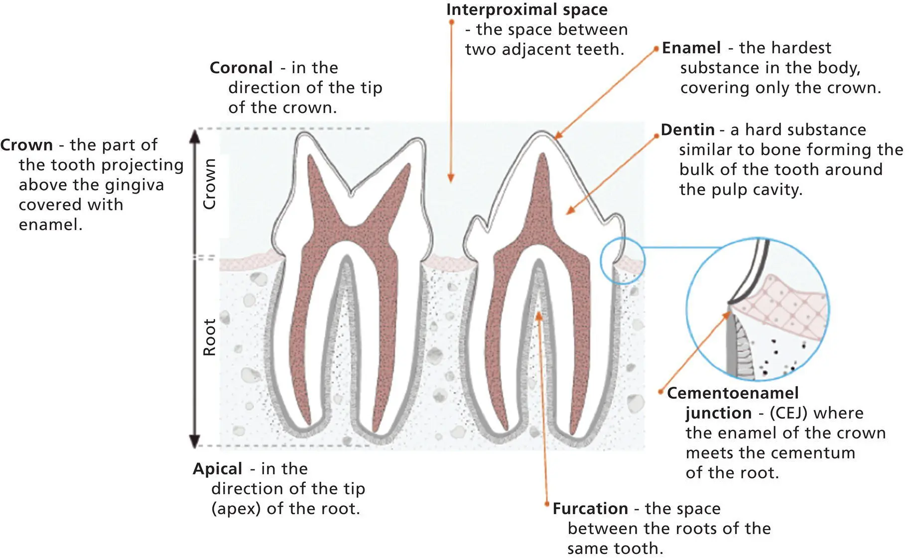
Illustration 1.2 Tooth anatomy and direction nomenclature.
Surfaces of Teeth and Directions in the Mouth
Vestibular/Buccal/Labial
Vestibularis the correct term referring to the surface of the tooth facing the vestibule or lips ; buccaland labialare acceptable alternatives. The term “facial” specifically refers to the surfaces of the rostral teeth visible from the front. According to Dr. A.J. Bezuidenhout, a veterinary anatomist at Cornell University, “facial” is a bit of a misnomer. Traditionally “facial” has been used in human dentistry for the aspect of teeth visible from the front, i.e. incisors and canines.
Lingual/Palatal
Lingual: The surface of a mandibular or maxillary tooth facing the tongue is the lingual surface. Palatal can also be used when referring to the lingual surface of maxillary teeth.
Mesial/Distal
Mesialand distalare terms applicable to tooth surfaces. The mesialsurface of the first incisor is next to the median plane; on other teeth it is directed toward the first incisor. The distalsurface is opposite from the mesial surface.
Rostral/Caudal
Rostraland caudalare the positional and directional anatomical terms applicable to the head in a sagittal plane in non‐human vertebrates. Rostralrefers to a structure closer to, or a direction toward, the most forward structure of the head. Caudalrefers to a structure closer to, or a direction toward, the tail ( Figures 1.36and 1.37) ( Illustration 1.2).
Further Reading
1 American Veterinary Dental College (n.d.). Veterinary dental nomenclature. https://avdc.org/avdc‐nomenclature/(accessed 27 August 2021).
2 Anderson, K.V. and Pearl, G. (1974). Transmedian innervation of canine tooth pulp in cats. Exp. Neurol. 44: 35–40.
3 Arredondo, J., Agut, A., Rodríguez, M.J. et al. (2013). Anatomy of the temporomandibular joint in the cat: a study by microdissection, cryosection and vascular injection. J. Feline Med. Surg. 15: 111–116.
4 Arzi, B. and Staszyk, C. (2019). The temporomandibular joint through the lens of comparative anatomy. In: Contemporary Management of Temporomandibular Disorders (eds. S.T. Connelly, G.M. Tartaglia and R.G. Silva), 41–50. Cham: Springer.
5 Barton‐Lamb, A.L., Martin‐Flores, M., Scrivani, P.V. et al. (2013). Evaluation of maxillary arterial blood flow in anesthetized cats with the mouth closed and open. Vet. J. 196: 325–331.
6 Berman, E. (1974). The time and pattern of eruption of the permanent teeth of the cat. Lab. Anim. Sci. 24 (6): 929–931.
7 Bishop, M.A. and Malhotra, M. (1990). An investigation of lymphatic vessels in the feline dental pulp. Am. J. Anat. 187: 247–253.
8 Buckland‐Wright (1975). Structure and function of cat skull bones in relation to the transmission of biting forces. PhD thesis. University of London.
9 Byers, M.R. and Matthews, B. (1981). Autoradiographic demonstration of ipsilateral and contralateral sensory nerve endings in cat dentin, pulp, and periodontium. Anat. Rec. 201 (2): 249–260.
10 Crossley, D.A. (1995). Tooth enamel thickness in the mature dentition of domestic dogs and cats: preliminary study. J. Vet. Dent. 12: 111–113.
11 Dummett, C.O. and Barens, G. (1981). Feline oral pigmentation. J. Periodontol. 41 (12): 696–701.
12 Floyd, M.R. (1991). The modified Triadan system: nomenclature for veterinary dentistry. J. Vet. Dent. 8 (4): 18–19.
13 Gavin, J.B. (1981). The ultrastructure of the crevicular epithelium of cat gingiva. Am. J. Anat. 123: 283–296.
14 Gioso, M.A. and Carvalho, V.G.G. (2005). Oral anatomy of the dog and cat in veterinary dentistry practice. Vet. Clin. North Am. Small Anim. Pract. 35: 763–780.
15 Gracis, M. (2007). Orodental anatomy and physiology. In: BSAVA Manual of Canine and Feline Dentistry, 3e (eds. C. Tutt, J. Deeprose and D. Crossley), 1–21. Gloucester: BSAVA.
16 Gracis, M. (1999). Radiographic study of the maxillary canine tooth of four mesaticephalic cats. J. Vet. Dent. 16: 115–128.
17 Grant, D. and Bernick, S. (1971). Morphodifferentiation and structure of Hertwig's root sheath in the cat. J. Dent. Res. 50 (6): 1580–1588.
18 Harvey, C.E. and Emily, P.P. (1993). Function, Formation, and Anatomy of Oral Structures in Carnivores, Small Animal Dentistry, 1–18. St Louis, MO: Mosby.
19 Harvey, C.E. (1985). Anatomy of the oral cavity in the dog and cat. In: Veterinary Dentistry (ed. C.E. Harvey), 5–22. Philadelphia, PA: WB Saunders.
20 Hayashi, K. and Kiba, H. (1989). Microhardness of enamel and dentine of cat premolar teeth. Nippon Juigaku Zasshi (Jpn. J. Vet. Sci.) 51: 1033–1035.
21 Hennet, P. (1995). Dental anatomy and physiology of small carnivores. In: BSAVA Manual of Small Animal Dentistry, 2e (eds. D.A. Crossley and S. Penman), 93–104. Cheltenham: BSAVA.
22 Hennet, P.R. and Harvey, C.E. (1991). Root canal anatomy in the cat. Proceedings of the Veterinary Dental Forum, New Orleans.
23 Hennet, P.R. and Harvey, C.E. (1996). Apical root canal anatomy of canine teeth in cats. Am. J. Vet. Res. 57: 1545–1548.
24 Hock, J. and Nuki, K. (1981). A vital microscopy study of the morphology of normal and inflamed gingiva. J. Periodontal Res. 6: 81–88.
25 Holland, G.R. (1975). The dentinal tubule and odontoblast process in the cat. J. Anat. 120 (1): 169–177.
26 Hudson, L.C. and Hamilton, W.P. (1993). Atlas of Feline Anatomy for Veterinarians. Philadelphia, PA: WB Saunders.
27 Izumi, H., Kuriwada, S., Karita, K. et al. (1990). The nervous control of gingival flow in cats. Microvasc. Res. 39 (1): 94–104.
28 Johnson, G.K., Squier, C.A., Johnson, W.T. et al. (1987). Blood flow and epithelial thickness in different regions of feline oral mucosa and skin. J. Oral Pathol. 16: 317–321.
29 Latorre, R., Arredondo, J., Rodríguez, M.J. et al. (2010). Clinical anatomy of the relation between the temporomandibular joint and the mandibular nerve in the cat. Clin. Anat. 23: 1005–1040.
30 Lombardero, M., Alonso‐Peñarando, D., and Yllera, M.D.M. (2021). The cat mandible (I): anatomical basis to avoid iatrogenic damage in veterinary clinical practice. Animals (Basel) 11 (2): 405.
31 Kishi, Y., Shimozato, N., and Takahashi, K. (1989). Vascular architecture of cat pulp using corrosive resin cast under scanning electron microscope. J. Endod. 15 (10): 478–483.
32 McClure, R.C., Pallman, M.S., and Garrett, P.G. (1973). Cat Anatomy: An Atlas, Text and Dissection Guide. Philadelphia, PA: Lea & Febiger.
33 Nalbandian, J. and Frank, R.M. (1980). Electron microscopic study of the regeneration of cementum and periodontal connective tissue attachment in the cat. J. Periodontal Res. 15: 71–89.
34 Nanci, A. (2003). Ten Cate's Oral Histology, Development, Structure, and Function, 6e. St. Louis, MO: Mosby.
35 Negro, V.B., Hernandez, S.Z., Maresca, B.M., and Lorenzo, C.E. (2004). Furcation canals of the maxillary fourth premolar and the mandibular first molar teeth in cats. J. Vet. Dent. 21: 10–14.
36 Okuda, A. and Harvey, C.E. (1992). Odontoclastic resorptive lesions in cats‐microscopic findings. Vet. Clin. North Am. Small Anim. Pract. 22: 1385.
37 Okuda, A., Inoue, E., and Asari, M. (1996). The membranous bulge lingual to the mandibular molar tooth of a cat contains a small salivary gland. J. Vet. Dent. 13: 61–64.
Читать дальшеИнтервал:
Закладка:
Похожие книги на «Feline Dentistry»
Представляем Вашему вниманию похожие книги на «Feline Dentistry» списком для выбора. Мы отобрали схожую по названию и смыслу литературу в надежде предоставить читателям больше вариантов отыскать новые, интересные, ещё непрочитанные произведения.


Обсуждение, отзывы о книге «Feline Dentistry» и просто собственные мнения читателей. Оставьте ваши комментарии, напишите, что Вы думаете о произведении, его смысле или главных героях. Укажите что конкретно понравилось, а что нет, и почему Вы так считаете.