Jan Bellows - Feline Dentistry
Здесь есть возможность читать онлайн «Jan Bellows - Feline Dentistry» — ознакомительный отрывок электронной книги совершенно бесплатно, а после прочтения отрывка купить полную версию. В некоторых случаях можно слушать аудио, скачать через торрент в формате fb2 и присутствует краткое содержание. Жанр: unrecognised, на английском языке. Описание произведения, (предисловие) а так же отзывы посетителей доступны на портале библиотеки ЛибКат.
- Название:Feline Dentistry
- Автор:
- Жанр:
- Год:неизвестен
- ISBN:нет данных
- Рейтинг книги:3 / 5. Голосов: 1
-
Избранное:Добавить в избранное
- Отзывы:
-
Ваша оценка:
Feline Dentistry: краткое содержание, описание и аннотация
Предлагаем к чтению аннотацию, описание, краткое содержание или предисловие (зависит от того, что написал сам автор книги «Feline Dentistry»). Если вы не нашли необходимую информацию о книге — напишите в комментариях, мы постараемся отыскать её.
delivers a comprehensive exploration of the specific considerations required to provide dental care to cats that emphasizes their unique needs.
The updated Second Edition includes brand-new material and approximately 300 new images illustrating diseases, conditions, and procedures discussed within the book. The new edition combines the pathology and treatment information to provide additional context which helps make it more clinically relevant. The book also offers:
A thorough introduction to feline oral assessment, including anatomy, oral examinations, radiology, and charting Comprehensive explorations of dental pathology and treatment in cats, including necessary equipment and materials and anesthesia and pain control Practical discussions of dental pathology prevention in felines, including plaque and tartar control Perfect for veterinary general practitioners and veterinary students,
will also be useful to veterinary technicians seeking a one-stop, visual resource on feline-specific dentistry.
Feline Dentistry — читать онлайн ознакомительный отрывок
Ниже представлен текст книги, разбитый по страницам. Система сохранения места последней прочитанной страницы, позволяет с удобством читать онлайн бесплатно книгу «Feline Dentistry», без необходимости каждый раз заново искать на чём Вы остановились. Поставьте закладку, и сможете в любой момент перейти на страницу, на которой закончили чтение.
Интервал:
Закладка:
1.11 Cementum
Cementum covers the healthy root and provides attachment for the periodontal ligament. Cementum is produced continuously by cementoblasts, slightly increasing in thickness throughout life. Acellular cementum is present at the coronal one‐third of the root, while cellular cementum appears at the root apex. Cementum is capable of formation, destruction, and repair, and is involved in both resorptive and reparative processes. It is nourished from vessels within the periodontal ligament. Cementocytes in cellular cementum communicate with underlying dentin with each other via canaliculi.
1.12 Alveolar Bone
Alveolar processes house the alveoli, which support the teeth by providing attachment for the fibers of the periodontal ligament. An alveolus can be divided into two parts:
1 Alveolar bone proper, which is a thin layer of bone surrounding the root that allows attachment to the periodontal ligament.
2 Supporting alveolar bone, which consists of compact, cortical, or cancellous bone on the vestibular and oral aspects of the alveolar process. The alveolar bone proper is also referred to as the cribriform plate. It covers the alveolar socket and is identified on radiographs as lamina dura ( Figure 1.10).
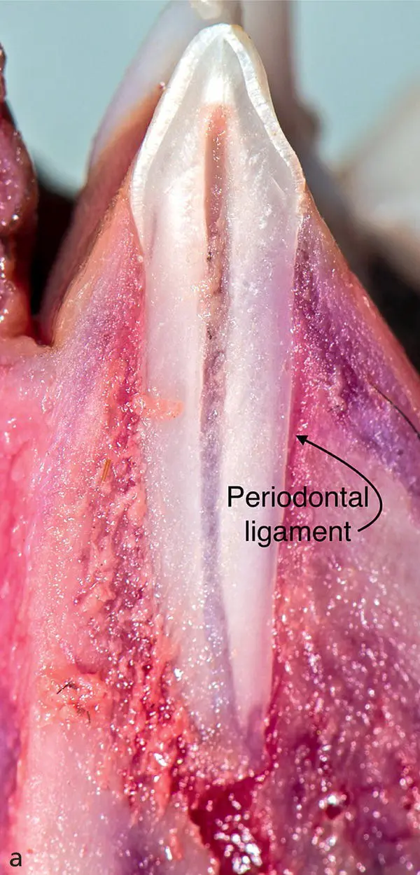
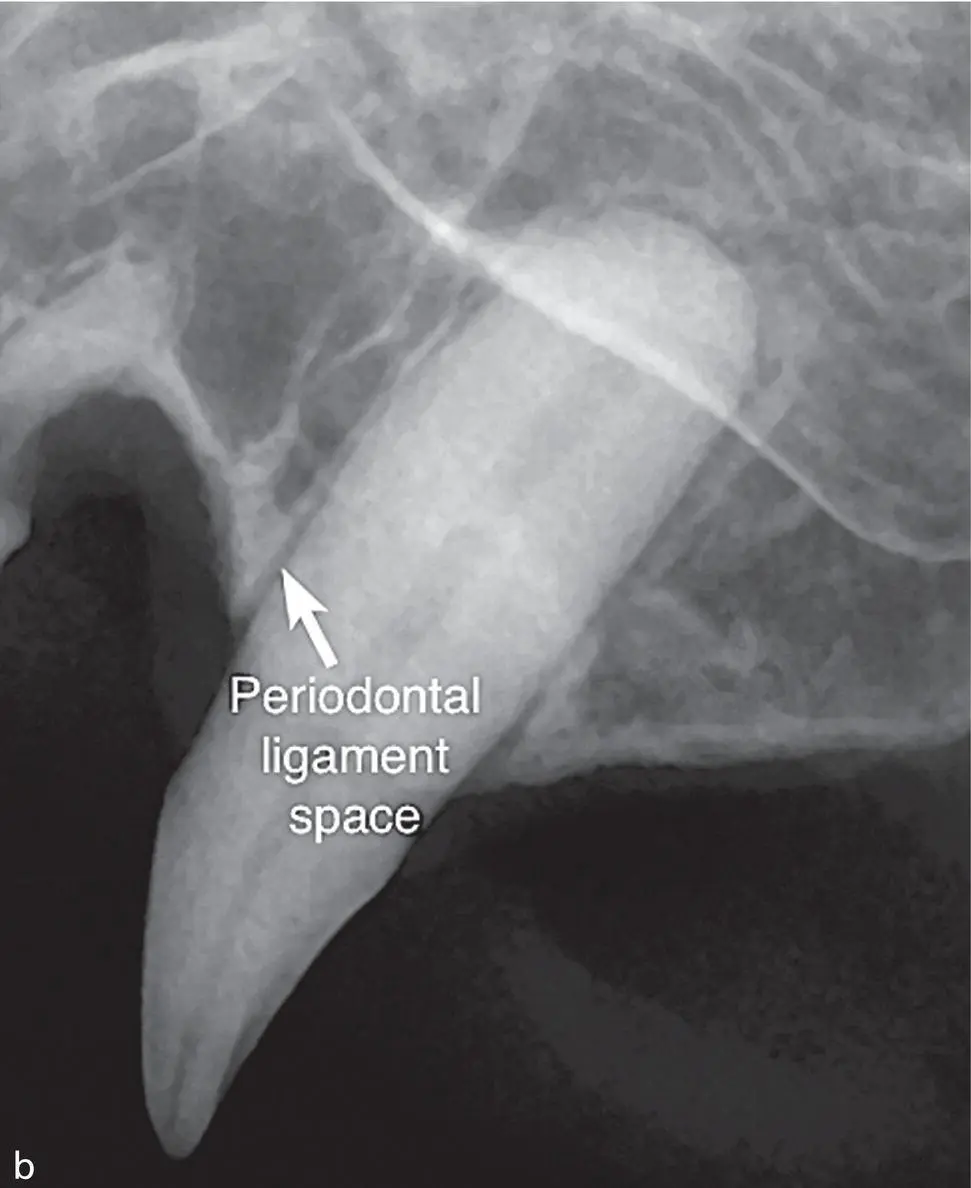
Figure 1.9 (a) Sagittal section revealing the periodontal ligament location (arrow). (b) Intraoral radiograph of the left maxillary canine demonstrating the periodontal ligament space.
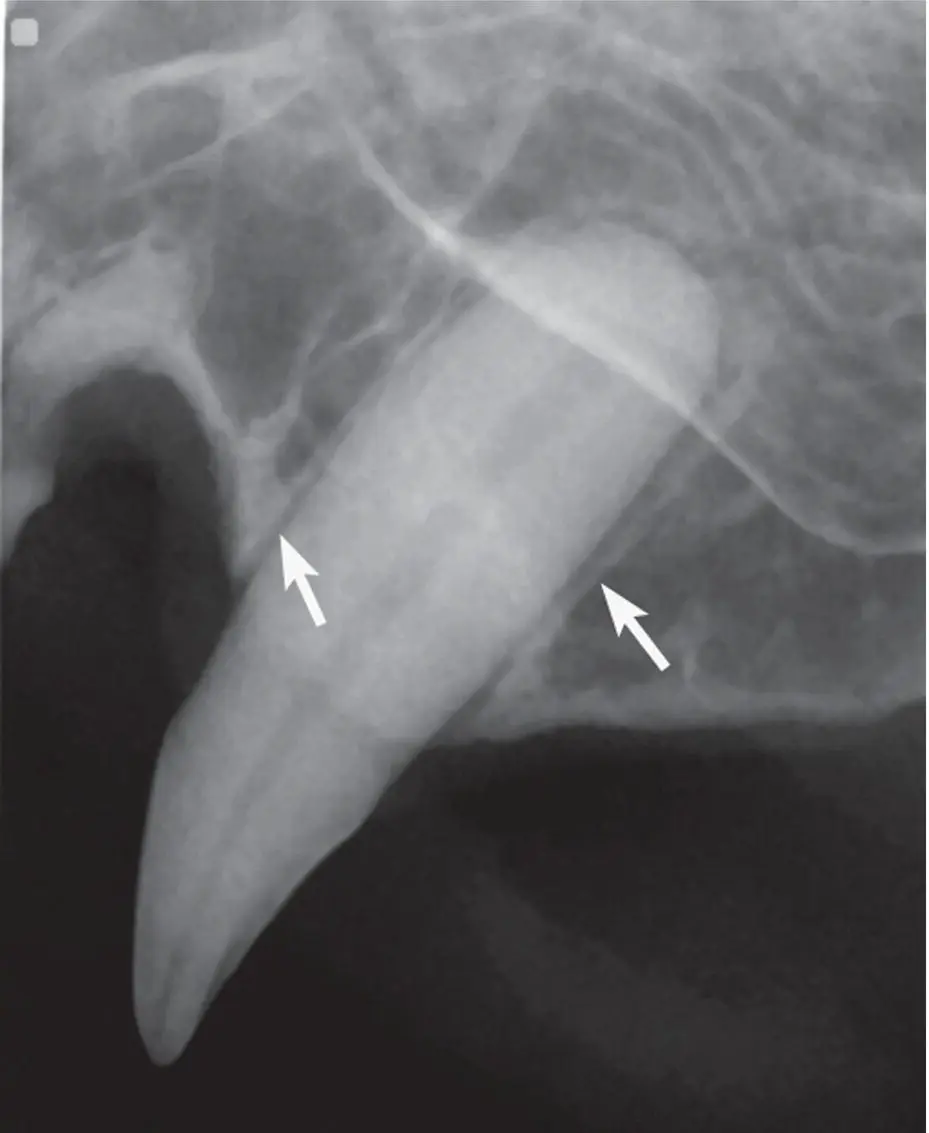
Figure 1.10 Lamina dura (arrows pointing to the white line surrounding the tooth root).
The alveolar bone and cortical plates are thickest in the mandible. The shape and structure of the trabeculae of spongy bone reflect the stress‐bearing requirements of a particular site. In some areas, alveolar bone is thin with no spongy bone.
The alveolar bone height is in equilibrium between bone formation and bone resorption. When bone resorption exceeds formation, the alveolar bone height is reduced ( Figure 1.11a,b).
1.13 Bones and Joints
1.13.1 Cranium
The skull can be divided into the fused bones of the calvaria, the upper jaw (maxillae), and lower jaw (mandibles). The dorsal aspect of the skull (cranium) is composed of the paired frontal and parietal bones. The occipital region of the cranium is the caudal aspect of the skull formed by the occipital bone. The temporal region is composed of the lateral walls of the cranium formed by the temporal bones. The rostral wall of the cranium is formed by the ethmoid bone ( Figure 1.12a,b).
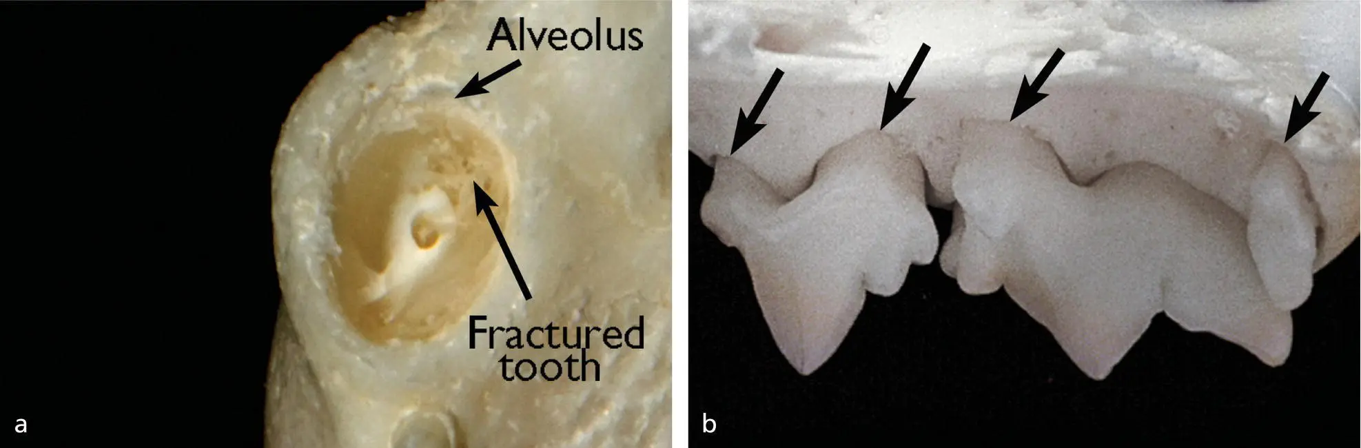
Figure 1.11 (a) Alveolus encasing a fractured maxillary canine tooth. (b) Decreased alveolar margin height (arrows) secondary to periodontal disease.
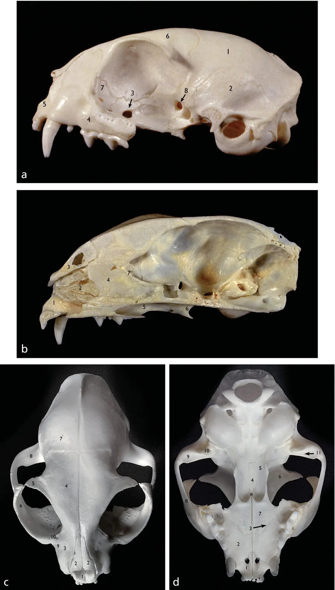
Figure 1.12 (a) Left lateral aspect of the skull with the zygomatic arch removed; 1. Parietal bone; 2. Squamous temporal bone; 3. Sphenopalatine foramen; 4. Maxilla; 5. Incisive bone; 6. Frontal bone; 7. Lacrimal bone; 8. Optic canal. (b) Medial aspect of a sagittal section of the left aspect of the skull: 1. Incisive bone; 2. Maxilloturbinates; 3. Nasal bone; 4. Nasal septum; 5. Palatine bone; 6. Pterygoid bone; 7. Ethmoid bone. (c) Dorsal aspect of the skull: 1. Incisive bone; 2. Nasal bone; 3. Maxilla; 4. Frontal bone; 5. Zygomatic process of frontal bone; 6. Zygomatic bone; 7. Parietal bone; 8. Zygomatic process of temporal bone; 9. Lacrimal foramen; 10. Infraorbital foramen. (d) Ventral aspect of the skull: 1. Incisive bone; 2. Palatine process of the maxilla; 3. Major palatine foramen; 4. Vomer bone; 5. Pterygoid bone; 6. Frontal bone; 7. Palatine bone; 8. Temporal process of the zygomatic bone; 9. Zygomatic process of the temporal bone; 10. Retroarticular process; 11. Mandibular fossa of the articular surface of the temporomandibular joint.
Source: Images reprinted with permission of Morton Publishing Company.
1.13.2 Facium
The facial part of the skull, which encloses the nasal and oral cavities, is divided into oral, nasal, and orbital regions. The oral region surrounding the oral cavity is composed of the incisive, maxillary, palatine, and mandibular bones.
The region surrounding the nasal cavity is composed of the nasal, maxillary, palatine, and incisive bones. The orbital region is formed by the frontal, lacrimal, palatine, sphenoid, and zygomatic bones surrounding the orbit ( Figure 1.12c,d).
1.13.3 Maxillae and Mandibles
Normal cats have two maxillas (or maxillae) and two mandibles. The adjective maxillary is often used in a wider sense, e.g. “maxillary fractures,” to include other facial bones, in addition to the maxillary bone itself.
1.14 Maxillary Region
The maxillary bones (maxillae) form the lateral parts of the face and the part of the hard palate that houses the canines and upper cheek teeth. The maxilla articulates with the incisive bone rostrally, the nasal bone dorsally, the vomer bone medially, and the lacrimal and zygomatic bones caudally ( Figure 1.13a,b). The maxillae extend to the caudal border of the hard palate laterally and are joined medially by the paired palatine bones to complete the hard palate. The infraorbital canal is located apical to both the maxillary third and fourth premolars below the orbit.
The palatine bone forms the bony part of the hard palate together with the maxillary and incisive bones. The incisive bone located rostrally houses the upper incisors and forms approximately one‐sixth of the hard palate. A pair of openings, the palatine fissures, allows passage of the incisive ducts to the vomeronasal organ. The incisive papilla located just caudal to the maxillary first incisor teeth houses these incisive ducts as they open into the oral cavity The ducts serve as a pathway for air to be drawn in, which is then directed over extensions of the vomeronasal apparatus in the floor of the nasal cavity immediately behind the palatine fissures ( Figure 1.14a,b).
The hard palate separates the oral and nasal cavities. The primary palate is the incisive portion of the palate and associated soft tissues. The secondary palate includes the remaining hard and soft palatal structures. Firmly attached, heavily keratinized mucosa covers the hard palate. Seven to eight transverse ridges called rugae protrude from the mucosa with rows of papillae between the ridges. The soft palate begins caudal to the maxillary first molar teeth and separates the nasopharynx dorsally from the oropharynx ventrally.
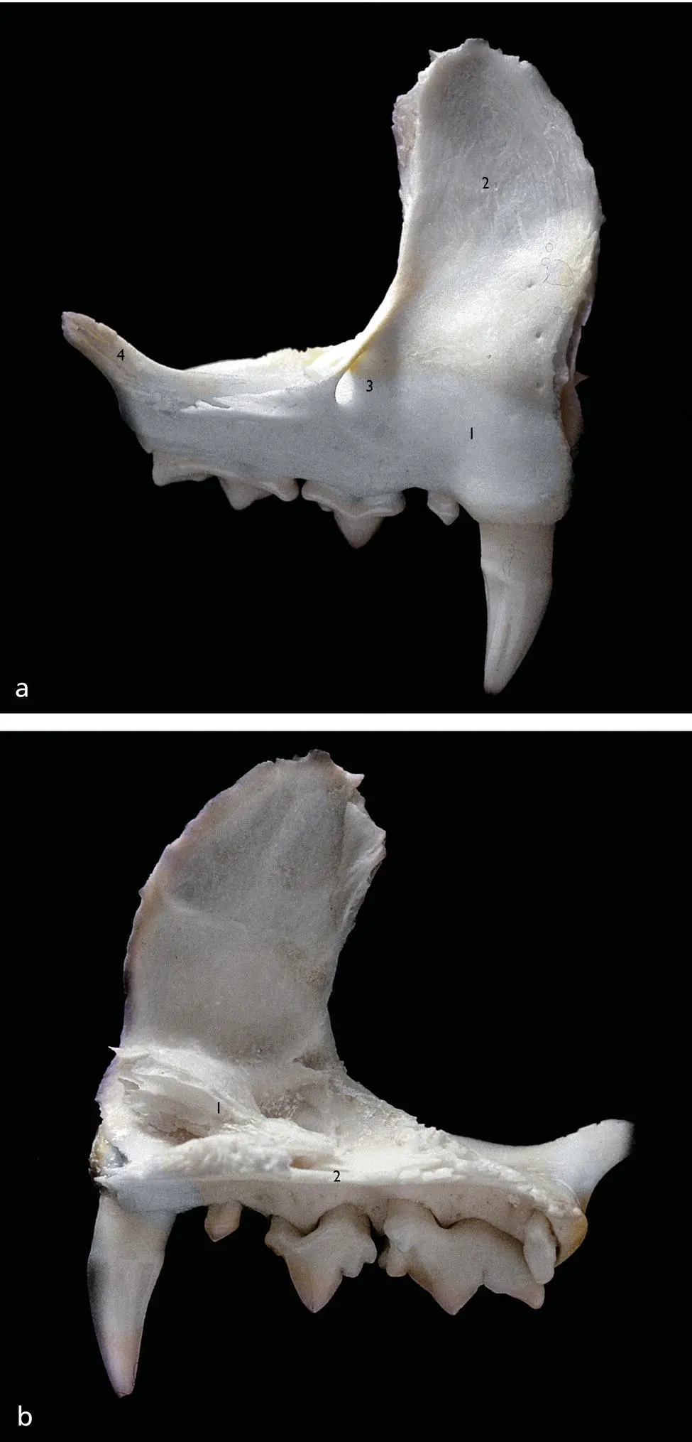
Figure 1.13 (a) Lateral aspect of right maxilla: 1. Alveolar process; 2. Frontal process; 3. Infraorbital canal; 4. Zygomatic process. (b) Medial aspect of the right maxilla: 1. Maxillotubinates; 2. Palatine process.
Читать дальшеИнтервал:
Закладка:
Похожие книги на «Feline Dentistry»
Представляем Вашему вниманию похожие книги на «Feline Dentistry» списком для выбора. Мы отобрали схожую по названию и смыслу литературу в надежде предоставить читателям больше вариантов отыскать новые, интересные, ещё непрочитанные произведения.


Обсуждение, отзывы о книге «Feline Dentistry» и просто собственные мнения читателей. Оставьте ваши комментарии, напишите, что Вы думаете о произведении, его смысле или главных героях. Укажите что конкретно понравилось, а что нет, и почему Вы так считаете.