Jan Bellows - Feline Dentistry
Здесь есть возможность читать онлайн «Jan Bellows - Feline Dentistry» — ознакомительный отрывок электронной книги совершенно бесплатно, а после прочтения отрывка купить полную версию. В некоторых случаях можно слушать аудио, скачать через торрент в формате fb2 и присутствует краткое содержание. Жанр: unrecognised, на английском языке. Описание произведения, (предисловие) а так же отзывы посетителей доступны на портале библиотеки ЛибКат.
- Название:Feline Dentistry
- Автор:
- Жанр:
- Год:неизвестен
- ISBN:нет данных
- Рейтинг книги:3 / 5. Голосов: 1
-
Избранное:Добавить в избранное
- Отзывы:
-
Ваша оценка:
Feline Dentistry: краткое содержание, описание и аннотация
Предлагаем к чтению аннотацию, описание, краткое содержание или предисловие (зависит от того, что написал сам автор книги «Feline Dentistry»). Если вы не нашли необходимую информацию о книге — напишите в комментариях, мы постараемся отыскать её.
delivers a comprehensive exploration of the specific considerations required to provide dental care to cats that emphasizes their unique needs.
The updated Second Edition includes brand-new material and approximately 300 new images illustrating diseases, conditions, and procedures discussed within the book. The new edition combines the pathology and treatment information to provide additional context which helps make it more clinically relevant. The book also offers:
A thorough introduction to feline oral assessment, including anatomy, oral examinations, radiology, and charting Comprehensive explorations of dental pathology and treatment in cats, including necessary equipment and materials and anesthesia and pain control Practical discussions of dental pathology prevention in felines, including plaque and tartar control Perfect for veterinary general practitioners and veterinary students,
will also be useful to veterinary technicians seeking a one-stop, visual resource on feline-specific dentistry.
Feline Dentistry — читать онлайн ознакомительный отрывок
Ниже представлен текст книги, разбитый по страницам. Система сохранения места последней прочитанной страницы, позволяет с удобством читать онлайн бесплатно книгу «Feline Dentistry», без необходимости каждый раз заново искать на чём Вы остановились. Поставьте закладку, и сможете в любой момент перейти на страницу, на которой закончили чтение.
Интервал:
Закладка:
The infraorbital canal is located apical to the maxillary third and fourth premolars below the orbit. Compared to the dog, the cat's infraorbital canal is shorter and usually less than five millimeters in diameter.
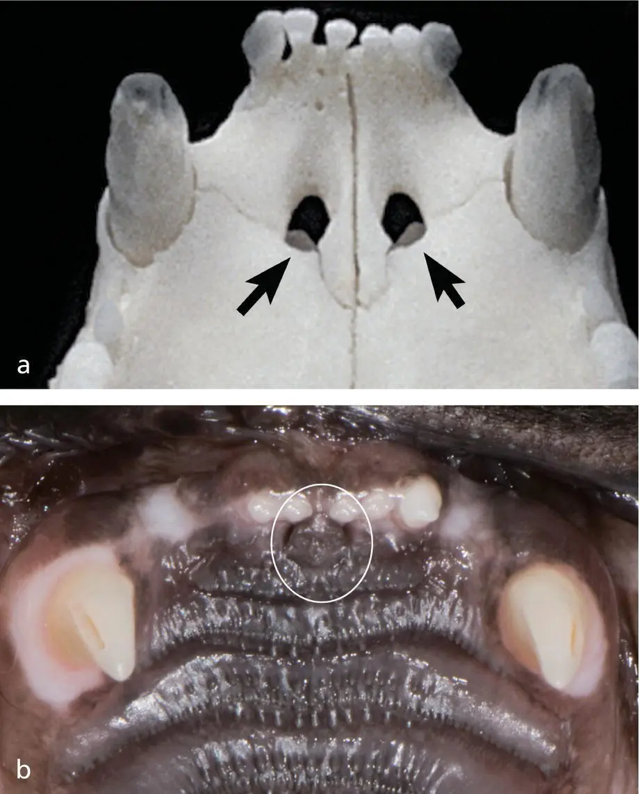
Figure 1.14 (a) Palatine fissures. (b) Incisive papilla.
1.15 Mandibles
The large bones articulating with the skull that support the lower teeth are the mandibles. Each mandible is composed of a horizontal body and a vertical ramus. The body supports the lower teeth. The ramus has three processes: coronoid, condylar, and angular. The condylar process articulates with the cranium in the temporomandibular joint (TMJ) ( Figure 1.15a–c).
The mandibles are connected to each other by a strong fibrocartilaginous joint at the mandibular symphysis. The nerves and vascular supply to the mandibular teeth enter the mandibular canal ventrally on the lingual aspect of the angle of the mandible and course rostrally exiting at the caudal, middle, and rostral mental foramina to supply the rostral mandible, chin, lip, buccal gingiva, and mucosa ( Figure 1.16a–c). The tongue and some of the muscles of the hyoid apparatus occupy the intermandibular space.
1.16 Temporomandibular Joint
The head of the condylar process of the mandibular ramus articulates with the base of the zygomatic process of the squamous part of the temporal bone (mandibular fossa) at the TMJ: a transversely elongated (cigar‐shaped) synovial joint ( Figure 1.17). The retroarticular process is a caudoventral extension of the mandibular fossa which helps prevent caudal luxation of the mandible ( Figure 1.18).
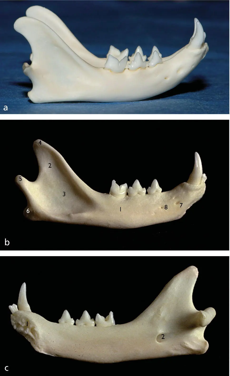
Figure 1.15 (a) Lower jaw. (b) Right mandible buccal aspect: 1. Mandibular body; 2. Mandibular ramus; 3. Masseteric fossa; 4. Coronoid process; 5. Condylar process; 6. Angular process; 7. Middle mental foramen; 8. Caudal mental foramen. (c) Right mandible lingual aspect: 1. Mandibular symphysis articular surface; 2. Mandibular foramen.
The articular cartilage of the TMJ is fibrocartilaginous tissue, with a fibrocartilaginous disc separating the joint into two non‐communicating compartments.
The insertion of the masseter muscle reaches the ventral and rostral aspect of the joint capsule. There is a thin, cartilaginous intra‐articular disc dividing the joint into dorsal and ventral compartments. This disc reduces friction by providing a double synovial film.
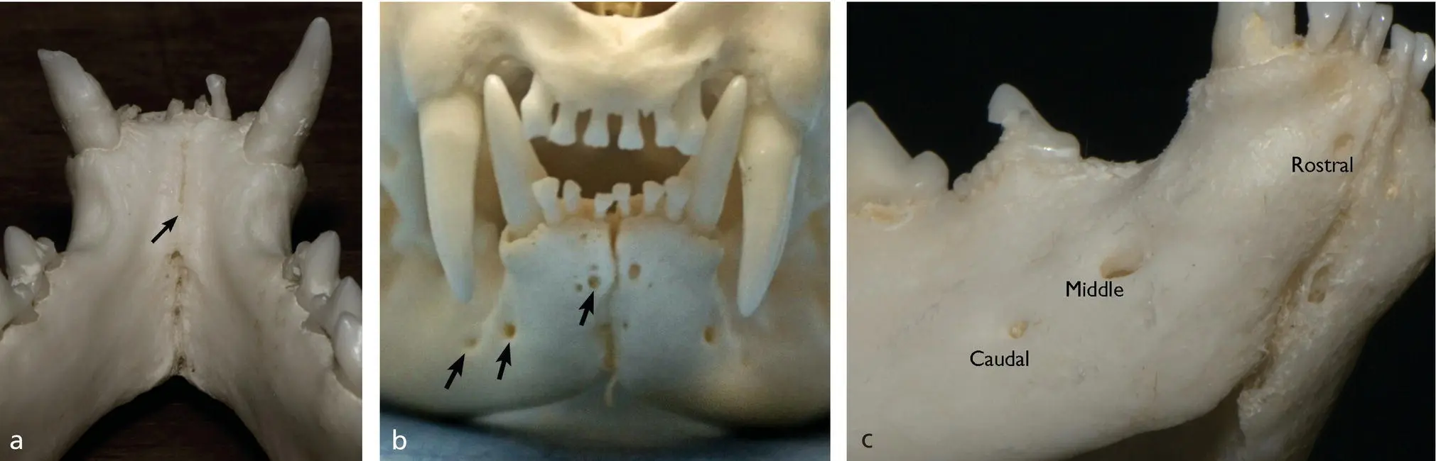
Figure 1.16 (a) Mandibular symphysis dorsal view. (b) Mandibular symphysis rostral view showing the mental foramina (arrows). (c) Mental foramina.
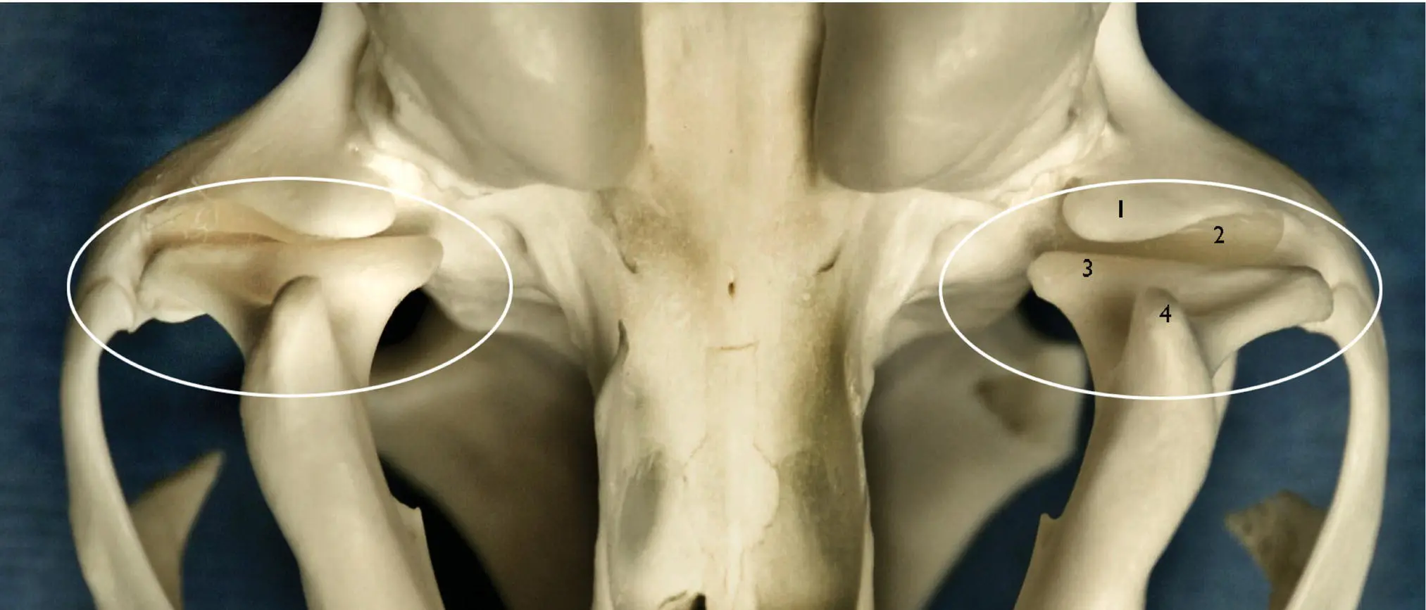
Figure 1.17 Temporomandibular joints ventral view: 1. Retroarticular process; 2. Mandibular fossa; 3. Condylar process; 4. Angular process.
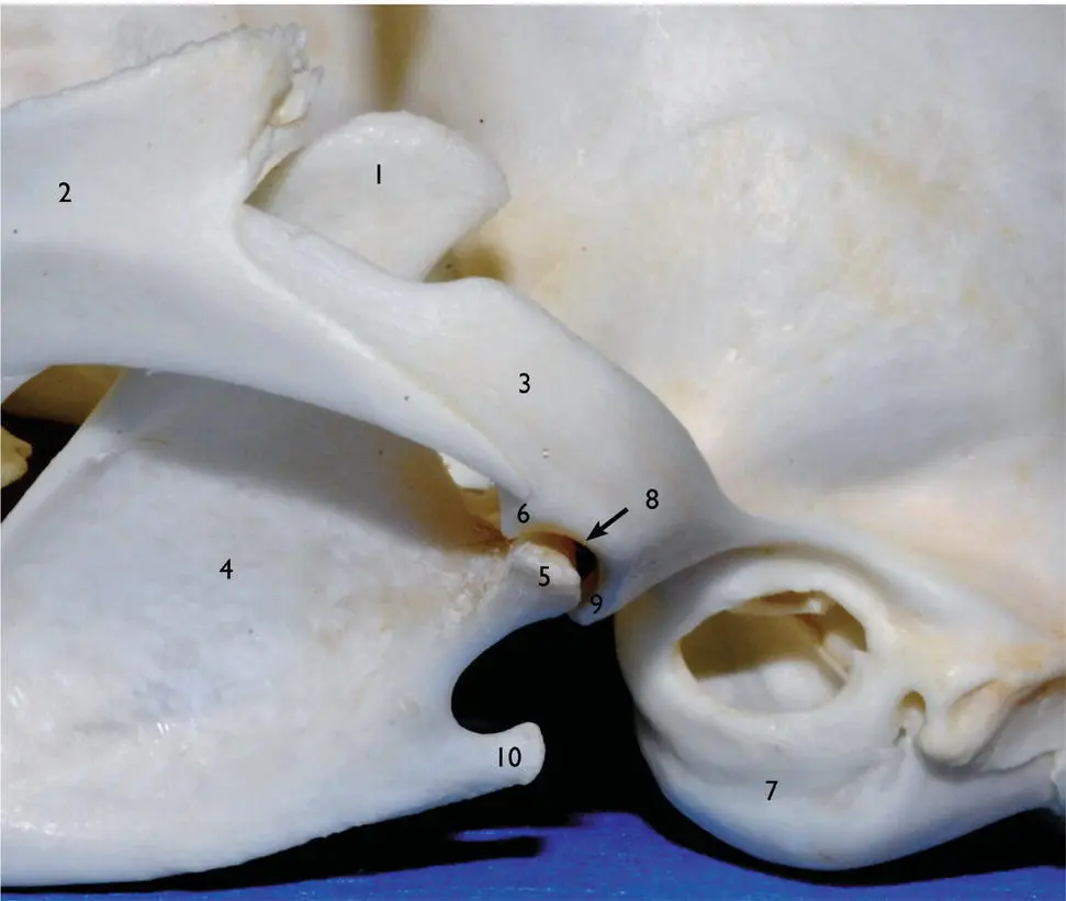
Figure 1.18 Lateral aspect of the left temporomandibular joint: 1. Coronoid process; 2. Zygomatic arch; 3. Zygomatic process of the temporal bone; 4. Mandibular ramus; 5. Condylar process; 6. Articular eminence; 7. Tympanic bulla; 8. Mandibular fossa; 9. Retroarticular process; 10. Angular process.
Temporomandibular Joint Nomenclature
Articular disc:A flat structure composed of fibrocartilaginous tissue and positioned between the articular surfaces of the condylar process of the mandible and mandibular fossa of the temporal bone, separating the joint capsule into dorsal and ventral compartments.
Condylectomy (TMJC):Resection of the condylar process of the mandible.
Mandibular condyle:A convex prominence at the end of the condylar process of the mandible that articulates with the mandibular fossa.
Mandibular fossa:A concave depression in the temporal bone that articulates with the mandibular condyle.
Open‐mouth jaw locking (OMJL):Inability to close the mouth due to locking of the coronoid process of the mandible ventrolateral to the ipsilateral zygomatic arch.
Partial coronoidectomy (CORP):Partial removal of the coronoid process of the mandible.
Partial zygomectomy (ZYGP):Partial removal of the zygomatic arch.
Retroarticular process:A projection of the temporal bone that protrudes ventrally from the caudal end of the zygomatic arch and carries part of the mandibular fossa.
Temporomandibular joint:The area where the condylar process of the mandible articulates with the mandibular fossa of the temporal bone.
Temporomandibular joint ankylosis (TMJA):Fusion between the bones forming the temporomandibular joint or those in close proximity, resulting in progressive inability to open the mouth; removal of bone in ankylotic areas is abbreviated as TMJAR.
Temporomandibular joint dysplasia (TMJD):Dysplasia of soft or hard tissues forming the temporomandibular joint.
Temporomandibular joint fracture (TMJFX):Fracture of one or more bony structures forming the temporomandibular joint.
Temporomandibular joint luxation (TMJLUX):Displacement of the condylar process of the mandible; manual or surgical reduction of temporomandibular joint luxation is abbreviated as TMJLUXR.
1.17 Teeth
Normally, there are 26 deciduous and 30 permanent teeth in the cat's oral cavity.
1.17.1 Dental Formula
Dental formulas (upper number indicates the maxillary teeth and the lower number the mandibular teeth) are as follows:
The deciduous dental formula for kittens is 2 × (I3/I3, C1/C1, P3/P2) = 26 teeth.
The permanent dental formula for adult cats is 2 × (I3/I3, C1/C1, P3/P2, M1/M1) = 30 teeth.
All of the incisors and canine teeth have one root. The maxillary second premolar, if present, normally has one root; however, studies have shown nearly 60% of the maxillary second premolars have two (sometimes partially fused) roots. The maxillary third premolar has two roots and the maxillary fourth premolar normally has three roots. The maxillary first molars, are single‐rooted in 35% of the cases, partly fused roots, in 35% of the cases and two roots in approximately 28% of the cases.
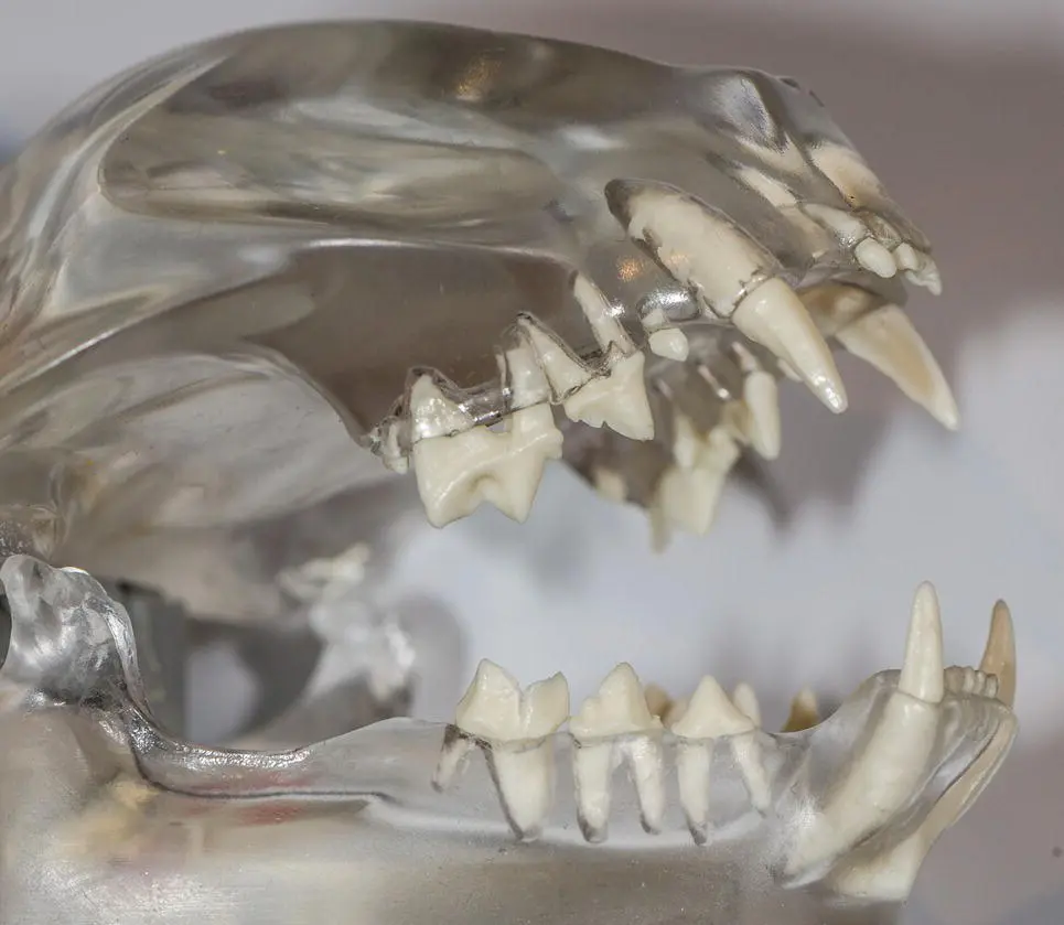
Figure 1.19 Cat model demonstrating normal tooth anatomy.
The mandibular cheek teeth in a cat (third and fourth premolars and first molars) normally have two roots Figure 1.19.
Интервал:
Закладка:
Похожие книги на «Feline Dentistry»
Представляем Вашему вниманию похожие книги на «Feline Dentistry» списком для выбора. Мы отобрали схожую по названию и смыслу литературу в надежде предоставить читателям больше вариантов отыскать новые, интересные, ещё непрочитанные произведения.


Обсуждение, отзывы о книге «Feline Dentistry» и просто собственные мнения читателей. Оставьте ваши комментарии, напишите, что Вы думаете о произведении, его смысле или главных героях. Укажите что конкретно понравилось, а что нет, и почему Вы так считаете.