Jan Bellows - Feline Dentistry
Здесь есть возможность читать онлайн «Jan Bellows - Feline Dentistry» — ознакомительный отрывок электронной книги совершенно бесплатно, а после прочтения отрывка купить полную версию. В некоторых случаях можно слушать аудио, скачать через торрент в формате fb2 и присутствует краткое содержание. Жанр: unrecognised, на английском языке. Описание произведения, (предисловие) а так же отзывы посетителей доступны на портале библиотеки ЛибКат.
- Название:Feline Dentistry
- Автор:
- Жанр:
- Год:неизвестен
- ISBN:нет данных
- Рейтинг книги:3 / 5. Голосов: 1
-
Избранное:Добавить в избранное
- Отзывы:
-
Ваша оценка:
Feline Dentistry: краткое содержание, описание и аннотация
Предлагаем к чтению аннотацию, описание, краткое содержание или предисловие (зависит от того, что написал сам автор книги «Feline Dentistry»). Если вы не нашли необходимую информацию о книге — напишите в комментариях, мы постараемся отыскать её.
delivers a comprehensive exploration of the specific considerations required to provide dental care to cats that emphasizes their unique needs.
The updated Second Edition includes brand-new material and approximately 300 new images illustrating diseases, conditions, and procedures discussed within the book. The new edition combines the pathology and treatment information to provide additional context which helps make it more clinically relevant. The book also offers:
A thorough introduction to feline oral assessment, including anatomy, oral examinations, radiology, and charting Comprehensive explorations of dental pathology and treatment in cats, including necessary equipment and materials and anesthesia and pain control Practical discussions of dental pathology prevention in felines, including plaque and tartar control Perfect for veterinary general practitioners and veterinary students,
will also be useful to veterinary technicians seeking a one-stop, visual resource on feline-specific dentistry.
Feline Dentistry — читать онлайн ознакомительный отрывок
Ниже представлен текст книги, разбитый по страницам. Система сохранения места последней прочитанной страницы, позволяет с удобством читать онлайн бесплатно книгу «Feline Dentistry», без необходимости каждый раз заново искать на чём Вы остановились. Поставьте закладку, и сможете в любой момент перейти на страницу, на которой закончили чтение.
Интервал:
Закладка:
Figure 1.20 Modified Triadan system incisor tooth numbering.
1.17.2 Tooth Types
Teeth are categorized by location and form. There are four types of teeth in the cat:
Incisorsare small teeth located between the canines. They are used for prehension. Incisors are referred to as right/left, maxillary/mandibular, first, second, and third incisors.
When using the modified Triadan system, right maxillary incisors are numbered 101, 102, and 103 starting from the first incisor, and left maxillary incisors are numbered 201, 202, and 203. The left mandibular incisors are numbered 301, 302, 303, and the right mandibular incisors are 401, 402, 403 ( Figure 1.20).
Caninesare single‐rooted teeth located rostrally in the mouth caudolateral to the incisors. They are used for piercing and biting. Canines are referred to as right/left, maxillary/mandibular canines. The crowns of the maxillary and mandibular canine teeth have vertical grooves ( Figures 1.21and 1.22).
When using the modified Triadan system, the right and left maxillary canines are numbered 104 and 204, respectively. The root and crown of the maxillary canines help to hold the upper lip outward, so that when the mouth is closed, the coronal tip of the mandibular canine slides into the vestibule without traumatizing the upper lip. The left and right mandibular canines are numbered 304 and 404, respectively (in the modified Triadan system, all the canines end in 4 and first molars in 9) ( Figures 1.23– 1.25).
Premolarsare located caudal to the canines. There are normally three maxillary and two mandibular premolars in the cat. Proper nomenclature of feline premolars is based on the archetypal carnivore model, which has a full dentition of 44 teeth (12 incisors, four canines, 16 premolars, and 12 molars).
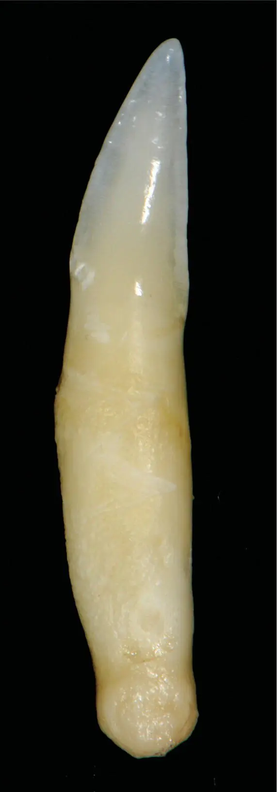
Figure 1.21 Extracted canine tooth.
The premolar behind the maxillary canine is termed the right or left maxillary second premolar ( Figure 1.26a,b). Using the modified Triadan system, the second premolars are referred to as tooth 106 (right) or 206 (left). The second premolar has one root or two fused roots. The third premolars (107, 207) have two roots. The fourth premolars (108, 208) have three roots (mesiobuccal, mesiopalatal, and distal) ( Figure 1.28).
The premolar behind the mandibular canine is termed the left or right mandibular third premolar (307, 407), followed by the fourth premolar (308, 408), which has two roots ( Figures 1.27a,b).
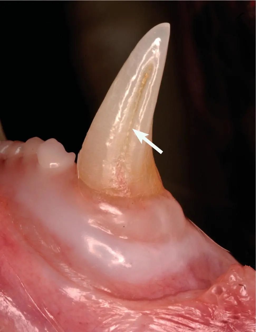
Figure 1.22 Mandibular canine vertical groove.
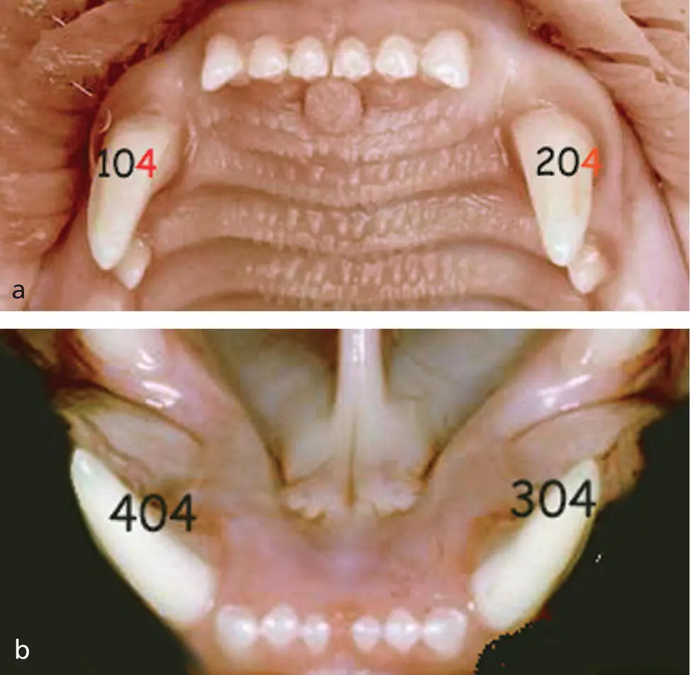
Figure 1.23 Modified Triadan canine tooth numbering: (a) Maxillary canines. (b) Mandibular canines.
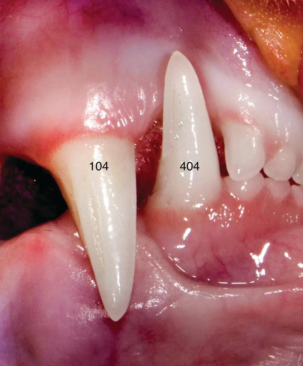
Figure 1.24Normal right maxillary/mandibular canine teeth orientation in the closed mouth.
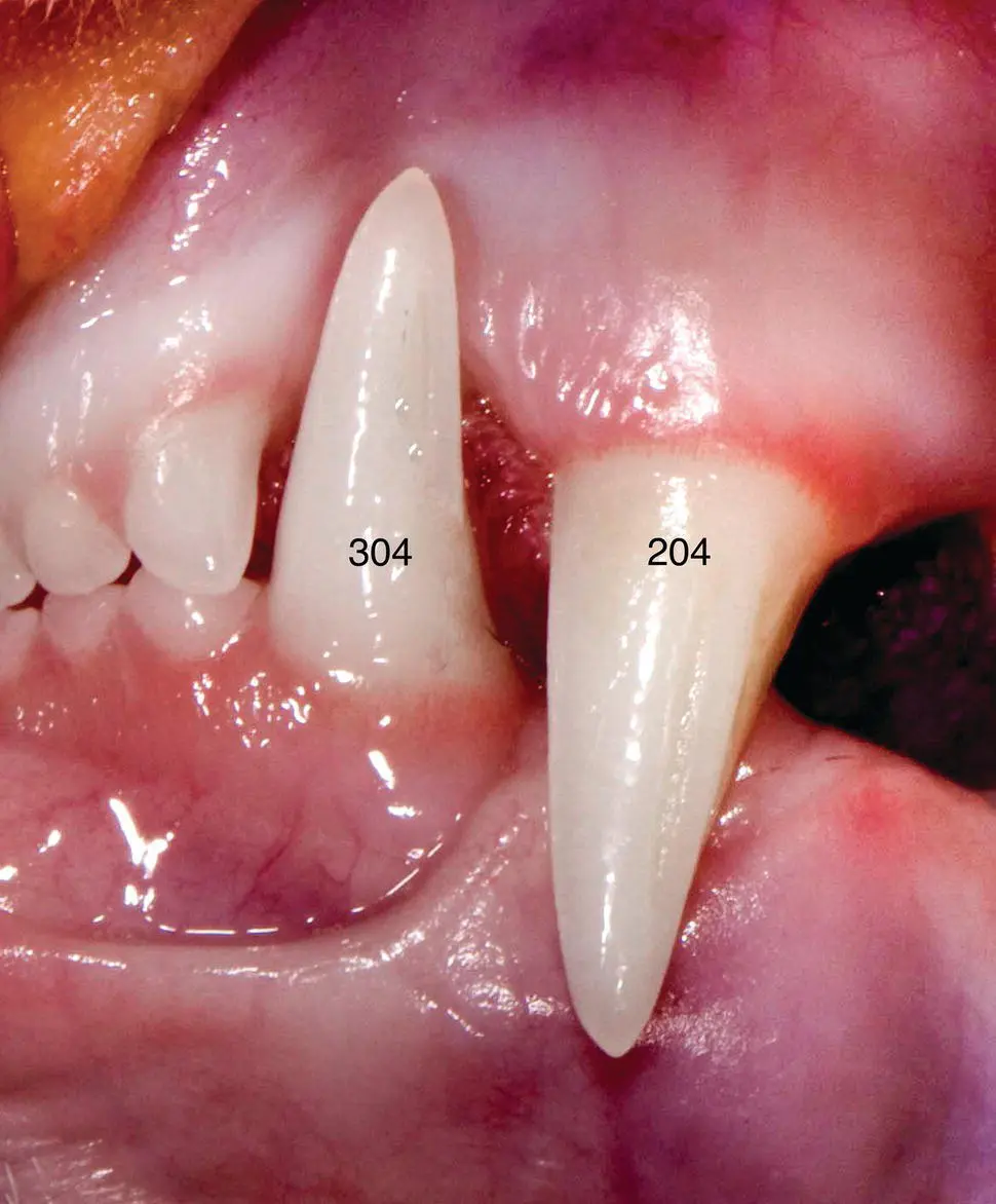
Figure 1.25 Normal left maxillary/mandibular canine teeth orientation in the closed mouth.
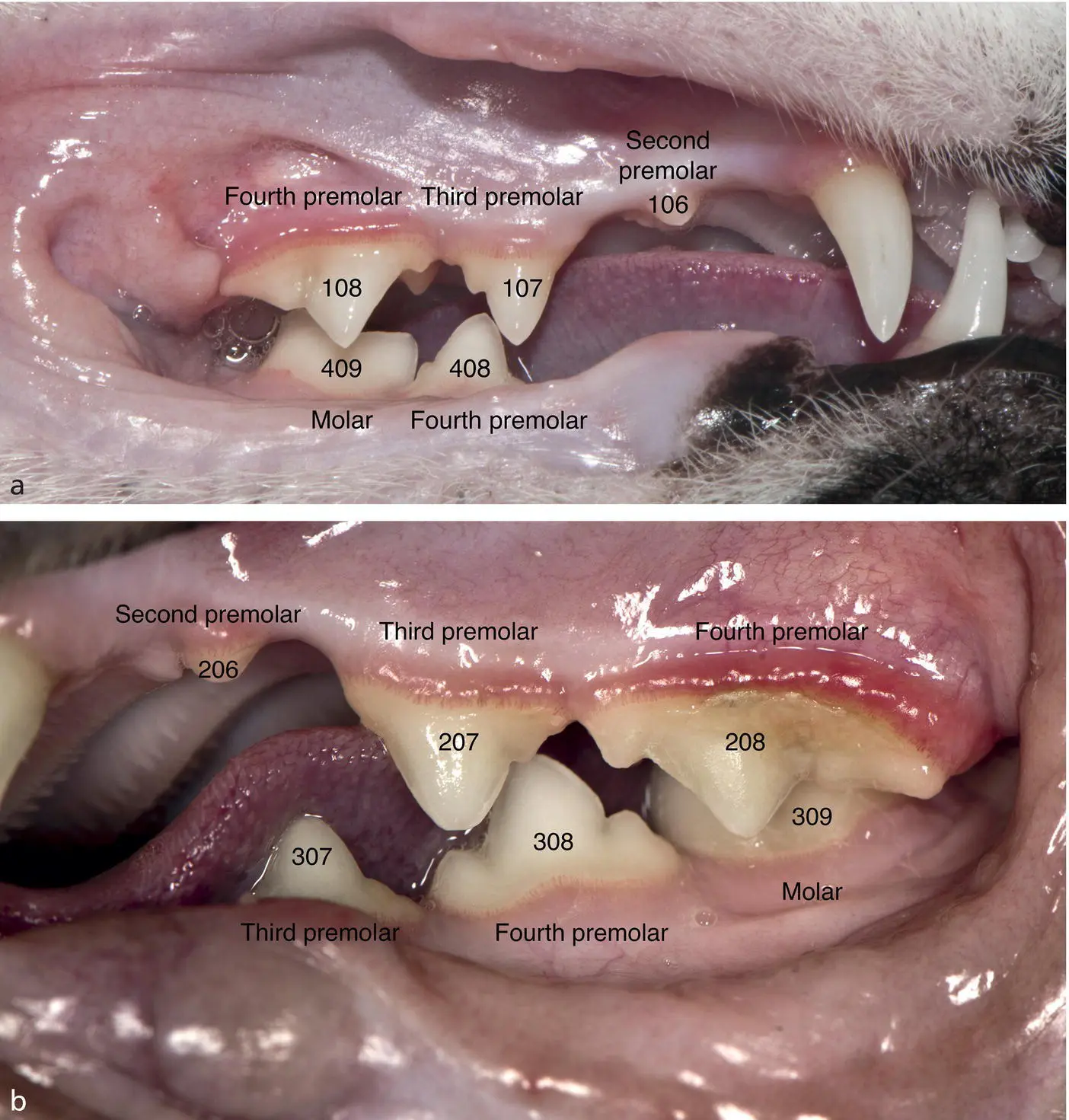
Figure 1.26 (a) Right maxillary and mandibular canine, premolars and molar (b) Left maxillary and mandibular canines, premolars and molar.
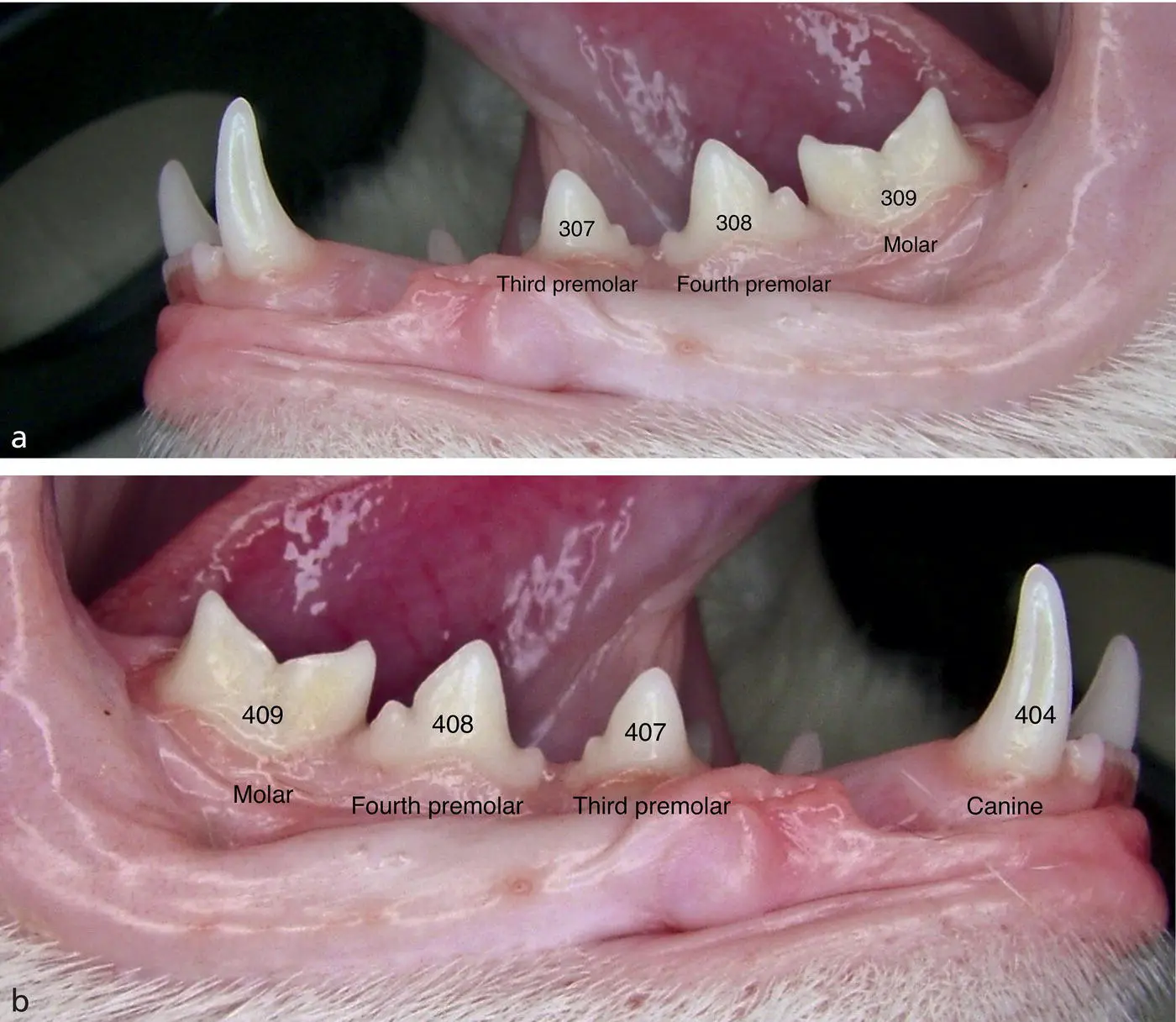
Figure 1.27 (a) Left mandibular, canine, premolars, and molar (b) Right mandibular canine, premolars, and molar.
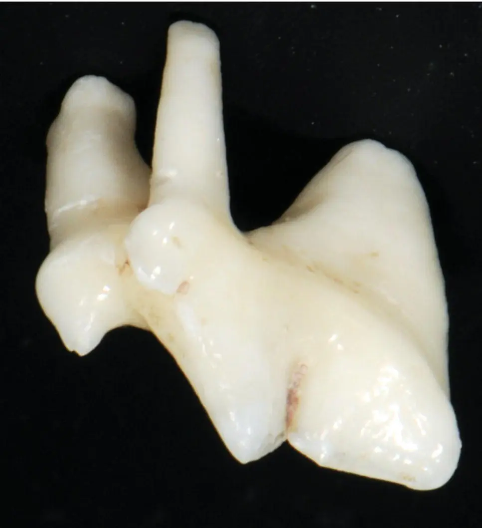
Figure 1.28 Maxillary fourth premolar.
Molarsare located caudal to the premolars. There is one set in the maxilla termed right or left maxillary first molar (109, 209) and one set in the mandible termed left or right mandibular first molar (309, 409). The mandibular first molar has one large mesial root, a smaller distal root which angles caudally and rarely, a third root ( Figure 1.29a,b).
The tooth's anatomical crown is visible to the naked eye. The root is located in the alveolus encased in the alveolar process beneath the gingiva. The cribriform plate (lamina dura) lines the alveolus.
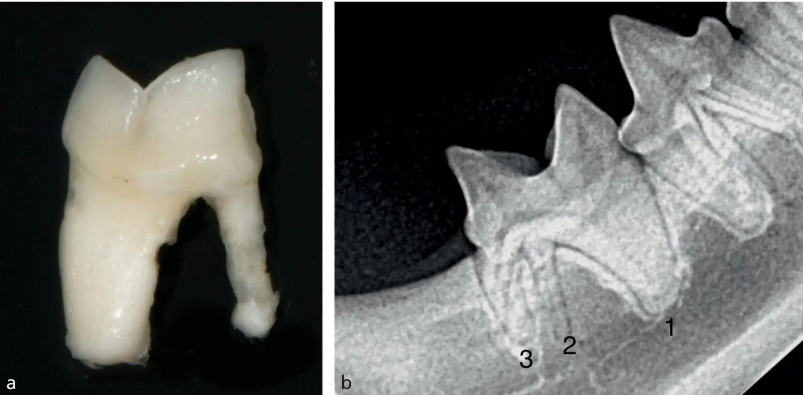
Figure 1.29 (a) Dissected left mandibular first molar. (b) Radiograph of an atypical three‐rooted mandibular molar.
1.18 Variation in Tooth Number and Morphology
Abnormalities in the number of teeth in cats can be inherited or result from disturbances during the initial stages of tooth formation. Anodontia, complete absence of all teeth, and oligodontia, decreased number of teeth, are uncommon conditions in cats. Supernumerary teeth are more common and may result in crowding and misalignment of teeth, which may predispose to the development of periodontal disease. The mandibular fourth premolars are the most common supernumerary teeth observed in the cat. Supernumerary teeth that result in crowding should be extracted early ( Figure 1.30a–d).
Persistence of primary (deciduous) teeth occurs in cats much less frequently than in dogs. The terms “retained” and “persistent” are often used synonymously however there is a difference. Persistent primary teeth are teeth that have failed to exfoliate whereas retained teeth are those that have failed to erupt.
Persistent primary teeth should be extracted as soon as they are diagnosed to make sufficient room for the permanent teeth to erupt into their normal positions. When persistent deciduous teeth are not removed, permanent teeth can deflect lingually (mandibular canine teeth) or rostrally (maxillary canine teeth) ( Figure 1.31).
Gemination has been observed in deciduous as well as in permanent dentition. It is an attempt to form two teeth from one enamel organ. This results in a structure with two completely or incompletely separated crowns each with a single pulp chamber and a shared root canal. Occasionally, complete cleavage or twinning occurs resulting in two teeth from one enamel organ. The etiology is unknown, but trauma has been suggested as a possible cause, though a familial tendency has been observed ( Figure 1.32a,b).
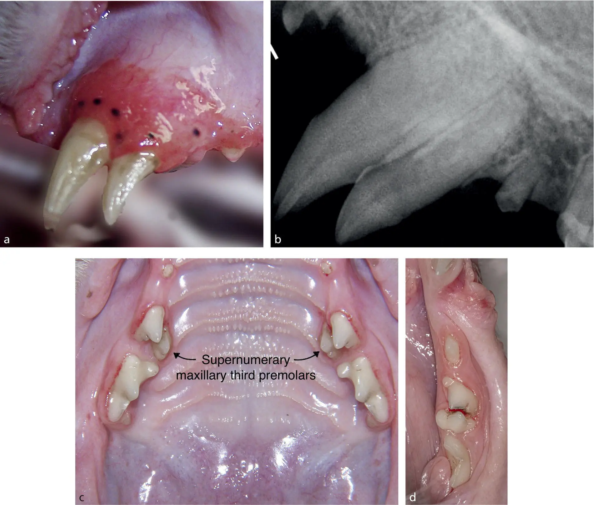
Figure 1.30 (a) Supernumerary maxillary left canine tooth. (b) Radiograph confirming supernumerary tooth versus deciduous. (c) Supernumerary maxillary third premolars causing crowding. (d) Supernumerary mandibular fourth premolar resulting in advanced periodontal disease.
Читать дальшеИнтервал:
Закладка:
Похожие книги на «Feline Dentistry»
Представляем Вашему вниманию похожие книги на «Feline Dentistry» списком для выбора. Мы отобрали схожую по названию и смыслу литературу в надежде предоставить читателям больше вариантов отыскать новые, интересные, ещё непрочитанные произведения.


Обсуждение, отзывы о книге «Feline Dentistry» и просто собственные мнения читателей. Оставьте ваши комментарии, напишите, что Вы думаете о произведении, его смысле или главных героях. Укажите что конкретно понравилось, а что нет, и почему Вы так считаете.