Jan Bellows - Feline Dentistry
Здесь есть возможность читать онлайн «Jan Bellows - Feline Dentistry» — ознакомительный отрывок электронной книги совершенно бесплатно, а после прочтения отрывка купить полную версию. В некоторых случаях можно слушать аудио, скачать через торрент в формате fb2 и присутствует краткое содержание. Жанр: unrecognised, на английском языке. Описание произведения, (предисловие) а так же отзывы посетителей доступны на портале библиотеки ЛибКат.
- Название:Feline Dentistry
- Автор:
- Жанр:
- Год:неизвестен
- ISBN:нет данных
- Рейтинг книги:3 / 5. Голосов: 1
-
Избранное:Добавить в избранное
- Отзывы:
-
Ваша оценка:
Feline Dentistry: краткое содержание, описание и аннотация
Предлагаем к чтению аннотацию, описание, краткое содержание или предисловие (зависит от того, что написал сам автор книги «Feline Dentistry»). Если вы не нашли необходимую информацию о книге — напишите в комментариях, мы постараемся отыскать её.
delivers a comprehensive exploration of the specific considerations required to provide dental care to cats that emphasizes their unique needs.
The updated Second Edition includes brand-new material and approximately 300 new images illustrating diseases, conditions, and procedures discussed within the book. The new edition combines the pathology and treatment information to provide additional context which helps make it more clinically relevant. The book also offers:
A thorough introduction to feline oral assessment, including anatomy, oral examinations, radiology, and charting Comprehensive explorations of dental pathology and treatment in cats, including necessary equipment and materials and anesthesia and pain control Practical discussions of dental pathology prevention in felines, including plaque and tartar control Perfect for veterinary general practitioners and veterinary students,
will also be useful to veterinary technicians seeking a one-stop, visual resource on feline-specific dentistry.
Feline Dentistry — читать онлайн ознакомительный отрывок
Ниже представлен текст книги, разбитый по страницам. Система сохранения места последней прочитанной страницы, позволяет с удобством читать онлайн бесплатно книгу «Feline Dentistry», без необходимости каждый раз заново искать на чём Вы остановились. Поставьте закладку, и сможете в любой момент перейти на страницу, на которой закончили чтение.
Интервал:
Закладка:
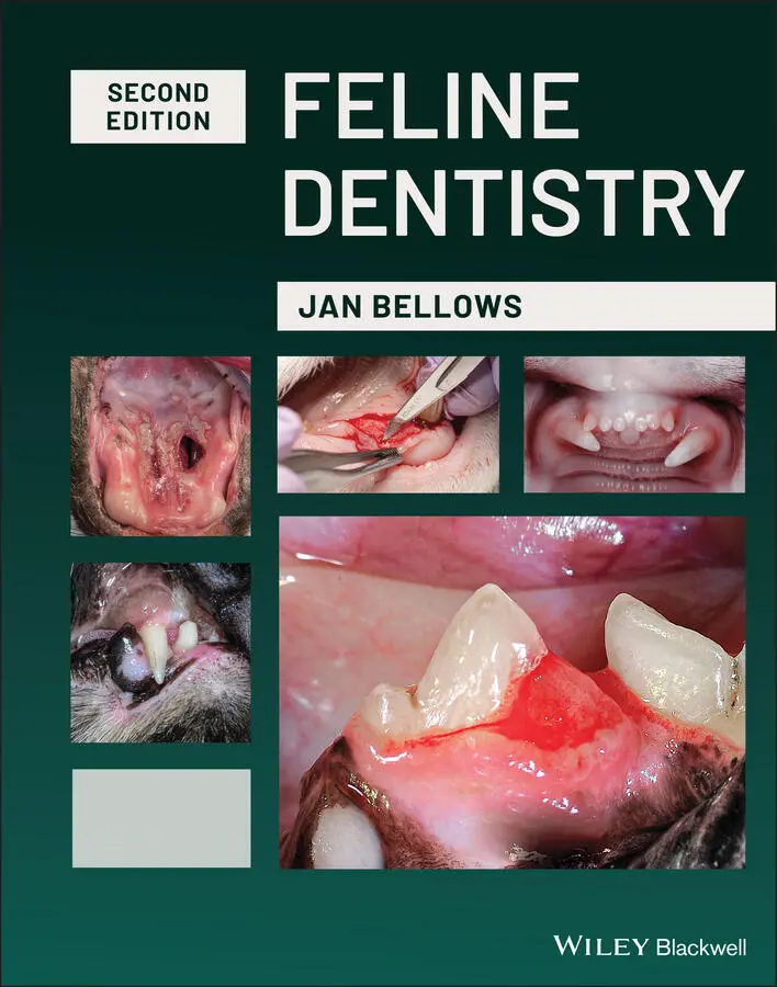
Oral mucosa can be categorized based on function and histology:
1 Masticatory mucosa – keratinized stratified squamous epithelium, found on the dorsum of the tongue, hard palate, and attached gingiva.
2 Lining mucosa – nonkeratinized stratified squamous epithelium, found almost everywhere else in the oral cavity, including:Buccal mucosa – inside lining of the cheeksLabial mucosa – inside lining of the lipAlveolar mucosa – covering the alveoli extending to the buccal/labial mucosa.
3 Specialized mucosa – in the regions of the taste buds on lingual papillae on the dorsal surface of the tongue. It contains nerve endings for general sensory and taste perception as well as filiform keratin projections on the hard palate mucosa.
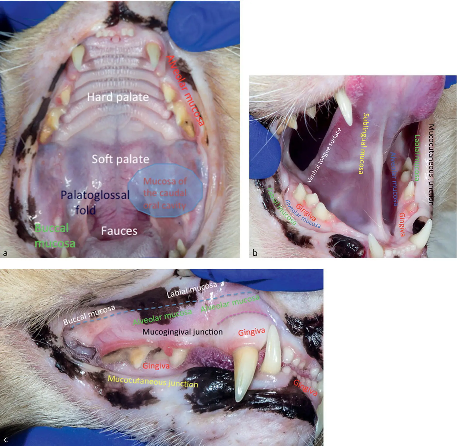
Figure 1.1 (a) Maxilla. (b) Maxillae, mandibles and sublingual areas. (c) Right maxilla and mandible.
1.3 Muscles
The muscles of mastication that close the jaws are the temporal, masseter, and medial and lateral pterygoid muscles, all of which are innervated by the mandibular nerve (the only motor branch of the trigeminal nerve). The digastricus muscle opens the mouth. Its rostral belly is innervated by the mandibular branch of the trigeminal nerve, while its caudal belly is innervated by the facial nerve. The body (the rostral two‐thirds) of the tongue is attached ventrally to the midline of the floor of the mouth by the lingual frenulum.
1.4 Tongue
The tongue has important functions in grooming, eating, drinking, and vocalization. The tongue is composed of both striated intrinsic and extrinsic muscles. The body of the tongue comprises the rostral two‐thirds, the root comprises the caudal one‐third and is attached to the hyoid apparatus. The genioglossus muscle depresses and is the only muscle to protrude the tongue, the hyoglossus and styloglossus both depress and retract the tongue.
The dorsal surface of the tongue is covered by keratinized stratified squamous epithelium that forms papillae which are responsible for taste, temperature control (through transfer of saliva from the mouth to fur) and grooming. The dorsal aspect of the lingual mucosa is specialized, having five types of papillae. They are filiform, fungiform, foliate, vallate, and conical. Filiform and fungiform papillae occupy the dorsal surface of the tongue body. The vallate papillae separate the tongue body and root dorsally. Vallate, foliate, and conical papillae occupy the tongue root ( Figure 1.2).
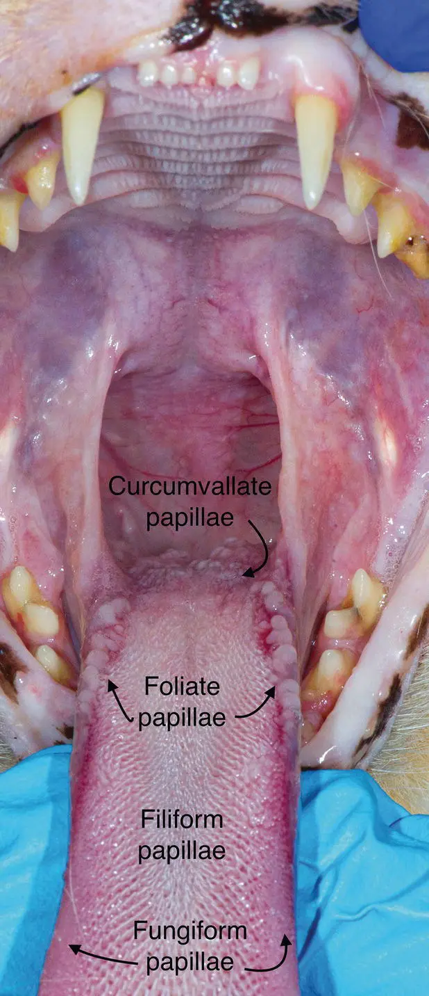
Figure 1.2 Tongue papillae.
Pillars of mucosa and the palatoglossal folds extend to the soft palate at the base of the tongue ( Figure 1.3).
The ventral tongue surface contains less cornified mucosa. The lingual frenulum connects the rostral two‐thirds of the tongue to the floor of the mouth within the intermandibular space.
1.5 Innervation
Sensory input is received from maxillary and mandibular divisions of the trigeminal nerve. The maxillary branch leaves the trigeminal ganglion, then exits the cranial cavity through the foramen rotundum, courses through the alar canal and the pterygopalatine fossa to enter the infraorbital canal. Just before entering the caudal limit of the infraorbital canal, the nerve branches into the major and minor palatine nerves. These nerves innervate the hard and soft palates and the nasopharynx. The palatine nerves are desensitized by the maxillary nerve block.
Figure 1.3 Caudal oral cavity.
The maxillary branch of the trigeminal nerve also branches out into the caudal maxillary alveolar nerve, which innervates the maxillary first molar, the buccal gingiva, and mucosa. This area is blocked by the infraorbital nerve block.
After giving off the caudal maxillary alveolar nerve, the maxillary nerve enters the infraorbital canal, where it is called the infraorbital nerve. While the infraorbital nerve is traversing the infraorbital canal, it gives off two more branches that exit ventrally from the canal. The middle maxillary alveolar nerve innervates the premolars and associated buccal gingiva. The rostral maxillary alveolar nerve supplies the canines, incisors, and associated buccal gingiva. The remaining fibers of the infraorbital nerve then exit the rostral extent of the infraorbital canal to innervate the lateral and dorsal cutaneous structures of the rostral maxilla and upper lip. The middle maxillary alveolar, rostral maxillary alveolar, and the infraorbital nerves are anesthetized by the rostral infraorbital nerve block.
The mandibular division of the trigeminal nerve arises from the trigeminal ganglion, exits the cranium via the foramen ovale, and divides into multiple branches. The divisions include the sensory buccal nerves, lingual nerve, and mandibular (inferior alveolar) nerve. The buccal nerves receive stimuli from the facial musculature, skin and mucosa of the cheek, and buccal gingiva along the caudal mandible.
The hypoglossal nerve innervates the tongue, the floor of the mouth, the lingual gingiva, and the mandibular salivary gland. The mandibular nerve enters the mandible on the lingual side, via the mandibular foramen. The nerve then courses rostrally within the mandibular canal to innervate the mandibular teeth to the midline. This nerve can be blocked by the mandibular (inferior alveolar) nerve block. Rostral to the third premolar tooth, the mandibular nerve gives off mental nerve branches. These branches exit through the mental foramina (rostral, middle, and caudal) and innervate the cutaneous areas of the chin and lip, and the rostral buccal gingiva and mucosa. These nerves are blocked by use of mental nerve blocks (usually the middle mental foramen is used).
1.6 Blood Supply and Lymphatic Drainage
The external carotid arteries branch out into the maxillary arteries. They further supply the mandibular (inferior alveolar) arteries, which enter the mandibular foramina on the medial sides of the mandibles and then course rostrally in the mandibular canals, where they exit through the mental foramina. The maxillary arteries also give rise to the major palatine arteries, which anastomose with the infraorbital arteries. The infraorbital arteries exit at the infraorbital foramina to supply the rostral muzzle. The lingual artery, a branch of the external carotid artery, supplies the tongue and mucosa of the floor of the oral cavity. The mandibular canal contains the inferior alveolar artery, vein, and nerve (the neurovascular bundle).
Lymph from the oral cavity drains into the parotid, mandibular, lateral, and medial retropharyngeal, superficial, and deep cervical lymph nodes. The mandibular lymph nodes are located rostral to the mandibular salivary gland. They are not lobulated like the salivary gland and therefore can be distinguished from them. They, along with the parotid lymph nodes, drain the entire head. They are more superficially located than the parotid lymph nodes and therefore are more easily palpated.
1.7 Salivary Glands
The major salivary glands in the cat include the parotid, zygomatic, mandibular, and sublingual. The mandibular salivary gland is located caudal to the ramus of the mandible and ventral to the parotid gland. The sublingual salivary gland lies in close approximation to the rostral aspect of the mandibular gland.
Читать дальшеИнтервал:
Закладка:
Похожие книги на «Feline Dentistry»
Представляем Вашему вниманию похожие книги на «Feline Dentistry» списком для выбора. Мы отобрали схожую по названию и смыслу литературу в надежде предоставить читателям больше вариантов отыскать новые, интересные, ещё непрочитанные произведения.


Обсуждение, отзывы о книге «Feline Dentistry» и просто собственные мнения читателей. Оставьте ваши комментарии, напишите, что Вы думаете о произведении, его смысле или главных героях. Укажите что конкретно понравилось, а что нет, и почему Вы так считаете.