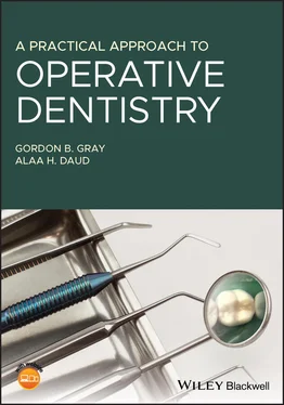
1 Cover
2 Title Page A Practical Approach to Operative Dentistry Gordon B. Gray BDS, MSc, DDS, PGCertEd Senior Clinical Lecturer in Restorative Dentistry (Retired) Dental Clinical Dean Bristol Dental School Bristol, United Kingdom Alaa H. Daud BDS, LDS RCS (Eng.), MFDS RCS (Edin.), MFDS RCS (Eng.), FDTFEd, MSc, PGCertEd, FHEA Senior Clinical Lecturer in Restorative Dentistry Bristol Dental School Bristol, United Kingdom
3 Copyright Page
4 Preface
5 About the Companion Website
6 Section I: Restorative Dentistry 1 Instruments Diagnosis Operative Management Instrument Tray Handpieces and Burs Matrix Systems Further Reading 2 Isolation Methods of Isolation Further Reading 3 Dental Charting Charting Notations Further Reading 4 Minimally Invasive Dentistry Protocol for Using Minimally Invasive Dentistry Diagnosis Alternative Cavity Classification System Assessment of Caries Risk Reduction in Cariogenic Bacteria Arresting Active Lesions Remineralisation of Carious Lesions and Monitoring Restoration of Cavities Using Minimal Cavity Designs Repairing Defective Restorations Monitoring Cavity Preparation MID for Pits and Fissures MID for Interproximal Lesions Other Examples of a More Minimal Approach to Treatment Further Reading
7 Section II: Intra‐Coronal Restorations 5 Pit and Fissure Caries Dental Probe Visual Method Visual Method with Magnification Transillumination Bitewing Radiograph Electronic Methods of Fissure Caries Diagnosis Enamel Biopsy Caries Risk Assessment Categorising Fissure Lesions and Selecting a Management Option Sealant Restorations Evidence Based Dentistry International Caries Classification and Management System Clinical Guide to Restoring a Tooth Using a Sealant Restoration Technique Clinical Guide to Restoring a Posterior Tooth with a Composite Resin A Clinical Case Further Reading 6 Posterior Approximal Restorations Tunnel Restorations Removal of the Marginal Ridge Finishing the Restoration The Extensive Restoration Clinical Guide to Restoring an Interproximal Lesion with a Self‐Retentive Box Clinical Guide to Restoring a Class II Lesion with Composite Resin A Clinical Case Clinical Guide to Restoring a Class II Lesion with Amalgam Clinical Guide to Restoring an Extensive Class II Lesion Further Reading 7 Restorations in Anterior Teeth Cavity Design Restoration of Anterior Interproximal Caries Restoration of a Fractured Incisor A Clinical Case Further Reading 8 Restoration of Lesions in Cervical Third Aetiology Factors Increasing Incidence of Lesions Direct Replacement Restorations Cervical Glass Ionomer Cement (GIC) and Compomer Restorations Compomer Restorations Further Reading
8 Section III: Indirect Restorations 9 Indirect Restorations Principles of Crown Preparation Assessment of a Patient for an Onlay or Crown MOD Gold Onlay Full Gold Crown A Clinical Case of Porcelain Onlay Further Reading 10 Porcelain Fused to Metal and All‐Ceramic Crowns PFM Crown All‐Ceramic Crown Clinical Sequence for All‐Ceramic Crown Further Reading
9 Index
10 End User License Agreement
1 Chapter 1 Figure 1.1 The air turbine handpiece is well balanced and operates in excess... Figure 1.2 An air driven or electric motor can be used to drive the slower‐s... Figure 1.3 A range of steel single use burs for intra‐coronal use are shown.... Figure 1.4 A range of friction grip burs for intra‐coronal and extra‐coronal... Figure 1.5 Handpieces are held using the pen grip to exert maximum control o... Figure 1.6 When a handpiece is substituted for the pen, maximum control of t... Figure 1.7 It is important that a finger rest is employed when a handpiece i... Figure 1.8 A modern version of a sectional matrix band that is held in place... Figure 1.9 A clear polyether matrix strip can be used when anterior teeth ar... Figure 1.10 A metal AutoMatrix can be useful when restoring very heavily bro... Figure 1.11 A Siqveland matrix band and holder in place on tooth 37. The sta... Figure 1.12 A Tofflemire matrix band and holder in position on tooth 1.4, wh...
2 Chapter 2Figure 2.1 Rubber dam can be used to isolate a single tooth for such procedu...Figure 2.2 Alternatively, rubber dam can be used to isolate a number of teet...Figure 2.3 An ink stamp is useful to mark the position of the teeth in the d...Figure 2.4 The recess slots on the tip of the forceps are placed into the ho...
3 Chapter 4Figure 4.1 Note the steep cuspal inclines and the deep fissures. These can b...Figure 4.2 The last standing molar tooth in the above pictures had two separ...Figure 4.3 A tunnel preparation on the distal surface of the lower left firs...Figure 4.4 A proximal slot has been prepared in the distal of upper left sec...Figure 4.5 The interproximal space is opened using an orthodontic separator ...Figure 4.6 This is an example of a cantilever adhesive bridge. The fit surfa...Figure 4.7 This is a fixed design of adhesive bridge that utilises two unres...Figure 4.8 The buccal cusp of this maxillary left premolar tooth fractured a...Figure 4.9 The amalgam restoration was removed and a chamfer finishing line ...
4 Chapter 5Figure 5.1 A section through a fissure with a toothbrush bristle superimpose...Figure 5.2 A dental probe superimposed on a section of tooth that shows deca...Figure 5.3 The fissures in this mandibular molar tooth are stained and show ...Figure 5.4 The use of magnification in visual diagnosis is of considerable h...Figure 5.5 The use of FOTI can be a useful adjunct in diagnosis.Figure 5.6 It is not always easy to see a radiolucency just below the enamel...Figure 5.7 Dürr Dental have developed a method of fluorescing the tooth with...Figure 5.8 A small enamel biopsy has been performed on this mandibular molar...Figure 5.9 The DIAGNOdent from KaVo uses natural fluorescence of the tooth t...
5 Chapter 6Figure 6.1 This bitewing radiograph shows a large carious lesion on the mesi...Figure 6.2 The classification by Foster is based on research showing that 92...Figure 6.3 Diagnosing an interproximal cavity can be demanding and is best d...
6 Chapter 7Figure 7.1 This extracted tooth clearly shows the central position of a whit...Figure 7.2 There are a variety of methods to diagnose interproximal caries i...Figure 7.3 An alternative and contemporary classification for caries is show...
7 Chapter 9Figure 9.1 The picture on the left shows some two‐surface intra‐coronal rest...Figure 9.2 This figure demonstrates the various marginal finish lines for cr...
8 Chapter 10Figure 10.1 (a) The above patient has undergone extensive rehabilitation wit...Figure 10.2 Multiple copings can be milled at the same time from a block of ...Figure 10.3 This patient has all‐ceramic crowns placed on maxillary anterior...
1 Cover Page
2 A Practical Approach to Operative Dentistry
3 Copyright
4 Preface
5 About the Companion Website
6 Table of Contents
7 Begin Reading
8 Index
9 WILEY END USER LICENSE AGREEMENT
1 iii
2 iv
3 ix
4 xi
5 1
6 3
7 4
8 5
9 6
10 7
11 8
12 9
13 10
14 11
15 12
16 13
17 14
18 15
19 16
20 17
21 18
22 19
23 20
24 21
25 23
26 24
27 25
28 26
29 27
30 28
31 29
32 30
33 31
34 32
35 33
36 34
37 35
38 36
39 37
40 38
41 39
42 40
43 41
44 43
45 44
46 45
47 46
48 47
49 48
50 49
51 50
52 51
53 52
54 53
55 54
56 55
57 56
58 57
59 58
60 59
61 60
62 61
63 62
Читать дальше
