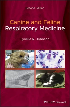
1 Cover
2 Preface Preface Students and practicing veterinarians are often challenged by patients with respiratory disease because the clinical signs are so similar among different disease processes within and beyond the respiratory system. Animals with cardiac and systemic diseases often present with respiratory signs, which means that all cases require thought and introspection on appropriate localization of disease as well as the underlying pathophysiology. The most critical piece of the puzzle is a comprehensive respiratory and systemic examination, and this book has been designed to provide easily understood pathways to the diagnosis. It is my hope that this contribution to the veterinary literature conveys highly specific details on respiratory examination, diagnostics, and diseases in a clinically relevant, logical, and easy‐to‐read fashion. This extensively updated edition includes new knowledge that has been generated on respiratory diagnostic testing and diseases such as Norwich terrier upper airway syndrome and epiglottic retroversion. My goal with this text was to integrate relevant anatomy, physiology, and pathophysiology in a rational and readable manner that is immediately clinically applicable for the busy practitioner and inquiring student. I approached this task recognizing that the major comprehensive textbooks in veterinary medicine contain excellent chapters on respiratory disorders. My book aims to provide an authoritative, cohesive, and complete discussion in a user‐friendly, single‐author volume. The first section deals with the common presenting signs demonstrated by patients with respiratory disease (nasal discharge, loud breathing, cough, tachypnea, and exercise intolerance) as well as a new section on differentiating cardiac from respiratory disease. The next section contains detailed how‐to descriptions of the most important diagnostic methods. I then devote an extensive chapter to therapeutic options, with special reference to new guidelines for management of infectious respiratory diseases. The remainder of the book has thorough explanations of individual diseases, divided into chapters dealing with disorders of the nose, airways, lung parenchyma, pleura, and pulmonary vasculature. Each chapter follows the same easy‐to‐read order, with diseases subdivided by etiology into structural, infectious, inflammatory, and neoplastic disorders. I hope that this second edition of my textbook will instill confidence in students and practitioners as they identify and manage respiratory conditions of dogs and cats.
3 Acknowledgments Acknowledgments I remain grateful to the clients and patients who have both tested and expanded my knowledge. This work was completed with the support of my colleagues at UC Davis, who have afforded me the opportunity and freedom to pursue respiratory medicine as my passion. Clinicians and house officers in all services have supported my clinical efforts as well as my research, and my departmental colleagues and the School’s leadership have been both encouraging and accommodating This book is the result of years of discovery and clinical effort, and it would not have been possible without the inspiration from colleagues and collaborators in the USA and worldwide who share a fascination with and great knowledge of respiratory medicine. The veterinarians I have met through the American and European Colleges of Veterinary Internal Medicine and the Veterinary Comparative Respiratory Society have motivated me to continue my search for knowledge.
4 1 Localization of Disease 1 Localization of Disease Clinical signs that provide clues to the existence of respiratory disease include nasal discharge, cough, respiratory noise, tachypnea, difficulty breathing, or exercise intolerance. The first step in making a diagnosis is the accurate localization of the anatomic origin of disease within the respiratory tract: the nasal cavity, upper or lower airway, lung parenchyma, or pleural space. Achieving appropriate anatomic localization of the site of dysfunction will allow construction of an accurate list of differential diagnoses, will facilitate efficient diagnostic testing, and will allow rational empiric therapy while waiting for test results.
Nasal Discharge Loud Breathing Cough Tachypnea Exercise Intolerance Differentiating Cardiac from Respiratory Disease Additional Considerations References
5 2 Respiratory DiagnosticsGeneral Diagnostic Imaging Airway Sampling Respiratory Endoscopy Sample Analysis References
6 3 Respiratory TherapeuticsDrug Therapy Routes of Administration Adjunct Therapy References
7 4 Nasal DisordersStructural Diseases Infectious Diseases Inflammatory Diseases Other Nasal Diseases References
8 5 Diseases of AirwaysStructural Disorders Infectious Diseases Inflammatory Disorders Neoplastic Disorders References
9 6 Parenchymal DiseaseStructural Diseases Infectious Diseases Inflammatory Disorders References
10 7 Pleural and Mediastinal DiseaseStructural Disorders Infectious Disorders Neoplastic Disorders Other Pleural Disorders References
11 8 Vascular DisordersStructural Disorders Infectious Disorders Miscellaneous Disorders References
12 Glossary
13 Index
14 End User License Agreement
1 Chapter 1 Table 1.1 Causes of nasal discharge in dogs and cats. Table 1.2 Respiratory causes of cough in dogs and cats.
2 Chapter 2 Table 2.1 Normal blood gas values for dogs and cats. Table 2.2 Respiratory causes of hypoxemia. Table 2.3 Airway sampling techniques for various lung patterns. Table 2.4 General guidelines for performingbronchoalveolar lavage (BAL). Table 2.5 Lower respiratory tract flora found in healthy dogs and cats. Table 2.6 Characteristics of pleural fluid.
3 Chapter 3Table 3.1 Gram‐stain characteristics and antibiotic susceptibility for common...Table 3.2 Side effects of commonly used antibiotics.Table 3.3 Antifungal drug therapy.
4 Chapter 5Table 5.1 Congenital forms of laryngeal paralysis.Table 5.2 List of pathogens currently identified as involved incanine infecti...Table 5.3 Clinical characteristics, diagnostic methods, and treatment for par...
5 Chapter 6Table 6.1 Bacterial organisms commonly found in dogs with lower respiratory t...Table 6.2 Bacterial organisms commonly found in cats with lower respiratory t...Table 6.3 Characteristics of common fungal organisms.
6 Chapter 7Table 7.1 Organisms commonly identified in animals with pyothorax.
1 Chapter 1 Figure 1.1 Nasal airflow can be assessed by occluding one nostril and assess... Figure 1.2 Palpation during ocular retropulsion can suggest the presence of ... Figure 1.3 In the normal animal, palpation of the soft palate will readily d... Figure 1.4 Prior to thoracic auscultation, the laryngeal and cervical trache... Figure 1.5 Each region of the thorax should be percussed to detect regional ...
2 Chapter 2 Figure 2.1 Coagulation cascade. FDPs, fibrin degradation products. Figure 2.2 Pulse oximetry measures hemoglobin saturation with oxygen, which ... Figure 2.3 (a and b) The urinary catheter used to collect an airway sample d... Figure 2.4 A three‐way stopcock is turned on to the urinary catheter used to... Figure 2.5 A suction trap can be secured below the patient during the trache... Figure 2.6 An over‐the‐needle catheter can be used as a cannula to perform a... Figure 2.7 (a) To initiate a transtracheal wash, the needle‐through‐catheter... Figure 2.8 The short catheter has been seated between the tracheal rings and... Figure 2.9 Visualization of the choanae is obtained by inserting a flexible ... Figure 2.10 Image of the normal nasopharynx in (a) a dog and (b) a cat. The ... Figure 2.11 The sheath (∼5 mm outer diameter) for the telescope portion of t... Figure 2.12 Normal nasal turbinates in (a) the dog and (b) the cat. Figure 2.13 Diagnostic yield of a nasal biopsy sample is improved when the s... Figure 2.14 Nasal flush can be performed by inserting a syringe tip into the... Figure 2.15 (a) Right‐angle forceps can be used to place a Foley catheter ab... Figure 2.16 A periodontal probe is inserted along the gum lines surrounding ... Figure 2.17 Necropsy image of the larynx showing the corniculate processes o... Figure 2.18 A bronchoscopy adapter allows passage of a 5.0 mm endoscope thro... Figure 2.19 Endoscopic view of the normal canine trachea. Figure 2.20 Endoscopic view of the carina illustrating the openings into the... Figure 2.21 Distal airway openings in the dog exhibit relatively sharp bifur... Figure 2.22 Bronchoalveolar lavage (BAL) is performed by gently wedging the b... Figure 2.23 (a) To initiate thoracocentesis, the skin is tented and the cath... Figure 2.24 Ultrasound guidance is used to obtain a lung biopsy using a Temn... Figure 2.25 Cytocentrifuge of a normal bronchoalveolar lavage sample (40×) r...
Читать дальше


