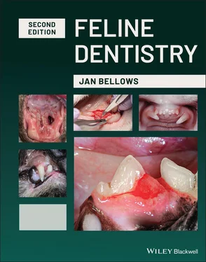
Cats are not dogs. Small dogs are plagued primarily with various degrees of periodontal disease (gingivitis and periodontitis). Large dogs more commonly present with gingivitis, fractured teeth, and oral masses.
Cats also are affected by periodontal disease and fractured teeth, but their main oral pathologies include tooth resorption, oropharyngeal inflammation, and maxillofacial cancer. Plaque prevention products and techniques covered in this text also differ from those used in dogs. Cats deserve their fair due, a book on dentistry dedicated solely to their species.
A global goal in writing this text is to introduce to some and reinforce to others a paradigm shift eliminating the terminology “doing a dentistry,” “performing a prophy,” or “Max is in for a dental.” Replacing the old terminology with “Max is in for a professional oral prevention, assessment and treatment visit,” which better represents what we do as veterinary dentists.
Prevention of periodontal disease is aimed at controlling plaque. Prevention is as important as the assessment and treatment steps. Without prevention, there is little doubt that periodontal disease will either continue or worsen. Plaque control methods must be specifically tailored to the patient and client in order to be effective.
Assessment involves evaluation of the patient before the anesthetic procedure and includes medical and dental history, feeding management, home oral hygiene, and physical and laboratory testing. Once the patient is anesthetized, a tooth-by-tooth examination is conducted to create a treatment plan.
Treatment with the goal of eliminating non-functional abnormalities uncovered during assessment is next. The treatment plan often can be accomplished within one anesthetic visit. In some instances, multiple visits or lifelong therapy are indicated.
Through daily use of the oral prevention, assessment and treatment process, patients can get the best in veterinary dentistry, which is our ultimate goal.
A genuine effort has been made to assure that the dosages and information included in this text are correct, but errors may occur, and it is recommended that the reader refer to the original reference or the approved labeling information of the product for additional information. Dosages should be confirmed prior to use or dispensing of medications.
Jan Bellows, D.V.M.
Diplomate, American Veterinary Dental College
Fellow, Academy Veterinary Dentistry
Diplomate, American Board of Veterinary Practitioners
(canine and feline specialties)

Our daughter Lauren and our grandson Elliot’s cat Jasper.
1 Anatomy
Tooth Nomenclature
Alveolar jugum(plural, juga): The palpable convexity of the buccal alveolar bone overlying a large tooth root.
Alveolar margin:Often incorrectly referred to as alveolar crest.
Alveolus (A):Socket in the jaw for a tooth root or reserve crown (plural: alveoli).
Ameloblasts:Epithelial cells involved in the formation of enamel (amelogenesis).
Anatomical crown (CR/AC):That part of a tooth that is coronal to the cementoenamel junction (or anatomical root).
Anatomical root (RO/AR):That part of a tooth that is apical to the cementoenamel junction (or anatomical crown).
Angular process:Caudoventral process (in carnivora).
Apex (AP):End of the root or reserve crown (plural: apices).
Apical delta:Multiple apical foramina forming a branching pattern at the apex of a mature tooth reminiscent of a river delta when sectioned and viewed through a microscope and occurring in some brachyodont teeth.
Apical foramen:Opening at the apex of a tooth, through which neurovascular structures pass to and from the dental pulp.
Articular disk:A flat structure composed of fibrocartilaginous tissue and positioned between the articular surfaces of the condylar process of the mandible and mandibular fossa of the temporal bone, separating the joint capsule into dorsal and ventral compartments; often incorrectly referred to as menisci.
Body of the mandible:The part that houses the teeth; often incorrectly referred to as horizontal ramus.
Canine (C):Canine tooth.
Cementoenamel junction:Area of a tooth where cementum and enamel meet.
Clinical crown (CR/CC):That part of a tooth that is coronal to the gingival margin; also called erupted crown in equines.
Clinical root (RO/CR):That part of a brachyodont tooth that is apical to the gingival margin.
Condylar process:Consists of mandibular head and mandibular neck; often incorrectly referred to as condyloid process.
Coronoid process:Process for the attachment of the temporal muscle.
Crown (C):Coronal portion of a tooth.
Deciduous:Deciduous and permanent are the anatomically correct terms to denote the two generations of teeth in diphyodont species.
Deciduous dentition period:That period during which only deciduous teeth are present.
Deciduous tooth (DT):Primary tooth which is replaced by a permanent (secondary) tooth.
Dental arch:Referring to the curving structure formed by the teeth in their normal position; upper dental arch formed by the maxillary teeth, lower dental arch formed by the mandibular teeth.
Enamel (E):Mineralized tissue covering the crowns of brachyodont teeth.
Facial vascular notch:Shallow indentation on the ventral aspect of the mandible, rostral to the angular process (absent in carnivores).
Incisive bones:The paired bones that house the maxillary incisors, located rostral to the maxillary bones, are the incisive bones, not the premaxilla.
Incisive part:The part that houses the incisors.
Incisors:Incisors will be referred to as (right or left) (maxillary or mandibular) first, second, or third incisors numbered from the midline.
Interarch:Referring to between the upper and lower dental arches.
Intermandibular joint (mandibular symphysis):Median connection of the bodies of the right and left mandibles (in adult Sus and Equus replaced by a synostosis), consisting of intermandibular synchondrosis and intermandibular suture.
Intermandibular suture:The larger part of the intermandibular joint formed by connective tissue.
Intermandibular synchondrosis:The smaller part of the intermandibular joint formed by cartilage.
Interquadrant:Referring to between the left and right upper or lower jaw quadrants.
Jaw quadrant:Referring to the left or right upper or lower jaw.
Lingual/Palatal:Lingual: The surface of a mandibular or maxillary tooth facing the tongue is the lingual surface. Palatal can also be used when referring to the lingual surface of maxillary teeth.
Mandible/mandibular (M/N):Referring to the lower jaw; all animals have two mandibles, not one; removing one entire mandible is a total mandibulectomy not a hemimandibulectomy.
Mandibular angle:Angle between the body and ramus of the mandible.
Mandibular canal:Contains a neurovascular bundle; often incorrectly referred to as the medullary cavity of the mandible.
Читать дальше
