Jan Bellows - Feline Dentistry
Здесь есть возможность читать онлайн «Jan Bellows - Feline Dentistry» — ознакомительный отрывок электронной книги совершенно бесплатно, а после прочтения отрывка купить полную версию. В некоторых случаях можно слушать аудио, скачать через торрент в формате fb2 и присутствует краткое содержание. Жанр: unrecognised, на английском языке. Описание произведения, (предисловие) а так же отзывы посетителей доступны на портале библиотеки ЛибКат.
- Название:Feline Dentistry
- Автор:
- Жанр:
- Год:неизвестен
- ISBN:нет данных
- Рейтинг книги:3 / 5. Голосов: 1
-
Избранное:Добавить в избранное
- Отзывы:
-
Ваша оценка:
Feline Dentistry: краткое содержание, описание и аннотация
Предлагаем к чтению аннотацию, описание, краткое содержание или предисловие (зависит от того, что написал сам автор книги «Feline Dentistry»). Если вы не нашли необходимую информацию о книге — напишите в комментариях, мы постараемся отыскать её.
delivers a comprehensive exploration of the specific considerations required to provide dental care to cats that emphasizes their unique needs.
The updated Second Edition includes brand-new material and approximately 300 new images illustrating diseases, conditions, and procedures discussed within the book. The new edition combines the pathology and treatment information to provide additional context which helps make it more clinically relevant. The book also offers:
A thorough introduction to feline oral assessment, including anatomy, oral examinations, radiology, and charting Comprehensive explorations of dental pathology and treatment in cats, including necessary equipment and materials and anesthesia and pain control Practical discussions of dental pathology prevention in felines, including plaque and tartar control Perfect for veterinary general practitioners and veterinary students,
will also be useful to veterinary technicians seeking a one-stop, visual resource on feline-specific dentistry.
Feline Dentistry — читать онлайн ознакомительный отрывок
Ниже представлен текст книги, разбитый по страницам. Система сохранения места последней прочитанной страницы, позволяет с удобством читать онлайн бесплатно книгу «Feline Dentistry», без необходимости каждый раз заново искать на чём Вы остановились. Поставьте закладку, и сможете в любой момент перейти на страницу, на которой закончили чтение.
Интервал:
Закладка:
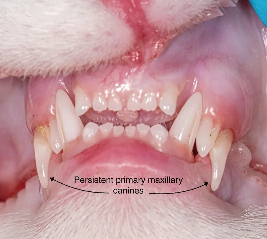
Figure 1.31 Persistent maxillary primary canine teeth, note that the secondary canines are malpositioned rostrally.
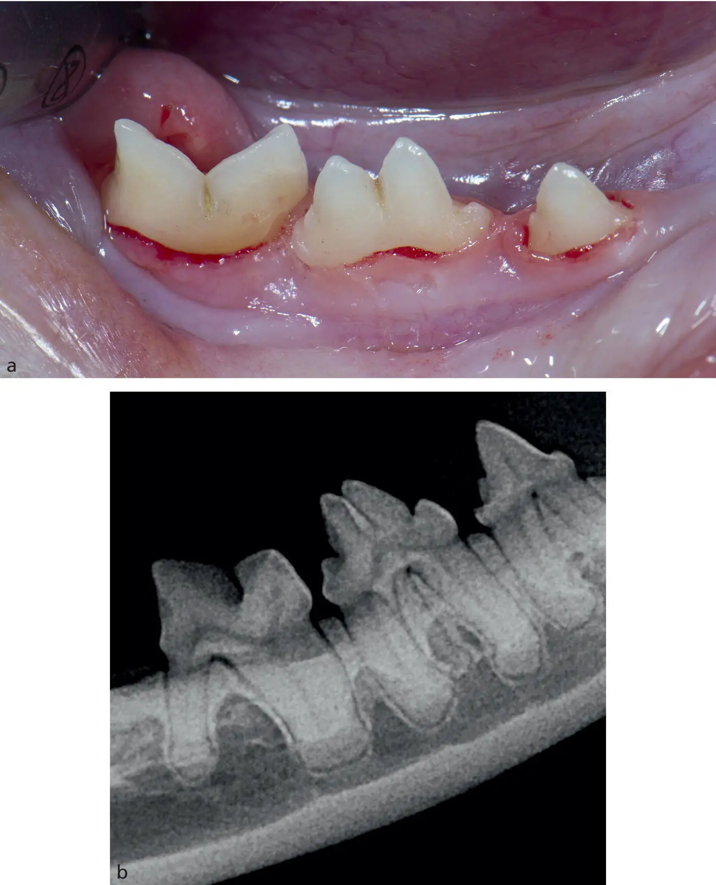
Figure 1.32 (a) Abnormally shaped right mandibular fourth premolar consistent with gemination. (b) Radiograph confirmation of one tooth with two incompletely separated crowns, and 3 roots.
Fusion is the joining of two tooth germs, resulting in a single large tooth. Fusion may involve the entire length of the tooth or only the roots, depending on the stage of development of the teeth at the time of the union. The root canal can be shared or separate. The etiology is unknown, but trauma and a familial tendency have both been suggested as possible causes. Fusion is also observed in deciduous as well as in permanent dentition. Occasionally, it is difficult to differentiate fusion of supernumerary teeth from gemination ( Figure 1.33a,b).
Concrescence is a condition of teeth where the cementum overlying the roots of at least two teeth join together. It involves only two teeth.
1.19 Composition
1.19.1 Enamel
The exterior surface of the healthy crown is covered by a thin layer of enamel, a hard inorganic substance (96% inorganic) formed by ameloblasts within the tooth bud before eruption. A study found that the enamel thickness in most cat teeth ranges from <0.1 to 0.3 mm. In dogs, the range for most teeth was <0.1 to 0.6 mm whereas in humans, the enamel on occlusal tables is usually 1–2 mm thick. Enamel when damaged is incapable of repair once the tooth has erupted.
1.19.2 Dentin
Dentin is located beneath the enamel and cementum and composes the majority of the mature tooth mass. Dentin is a specialized connective tissue of mesenchymal origin and is the second hardest tissue in the body after enamel. It is 70% inorganic and 30% organic (water, collagen, and mucopolysaccharide).
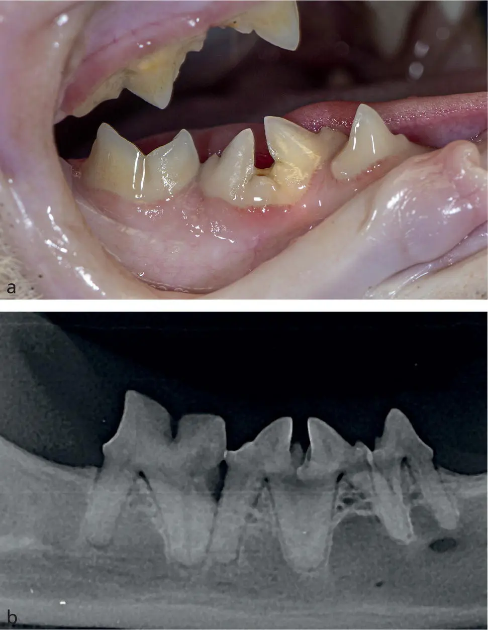
Figure 1.33 (a) Suspected fusion of supernumerary mandibular fourth premolars. (b) Radiograph confirmation of fusion.
Dentin is porous; each square millimeter contains over 40 000 dentinal tubules that communicate between the pulp and the dentin‐enamel or dentin‐cementum junctions. If there is near‐pulp exposure from trauma or resorption, bacteria can travel through the exposed dentinal tubules to the pulp. If untreated, inflammation may spread up and/or down the pulp, eventually causing irreversible necrosis. Odontoblast processes extend into the dentinal tubules. Near exposure can also transmit painful stimuli (heat, cold, pressure) due to afferent nerve fibers within the tubules adjacent to the odontoblastic processes. Toxic products from damaged tissue and microorganisms in the tissue perpetuate inflammation.
During pre‐eruptive development and during eruption, the odontoblasts produce primary dentin. Once the tooth has developed to its final length, the odontoblasts produce secondary dentin, causing the dentinal walls to thicken toward the pulp cavity. Primary dentin is deposited until root formation is complete, then secondary dentin accounts for all subsequent dentinogenesis throughout life. This effectively decreases the width of the pulp cavity as the cat ages. Reparative or tertiary dentin is produced in response to thermal, mechanical, occlusal, or chemical trauma to the odontoblasts. The pulp chamber in cats lies closer to the enamel than in dogs. For this reason, any tooth fracture in the cat should be treated aggressively, since most require endodontic therapy or extraction.
Two microscopic features of the dentin known as vasodentin and osteodentin may occasionally exist. Vasodentin is characterized by microscopic vascular inclusions within the outer third of the dentin. It is found to have vascular channels and dentinal tubules coursing through randomly. Osteodentin, unlike vasodentin, is most often found in the dentin adjacent to the root canal. Osteodentin resembles tertiary dentin which occurs secondary to trauma. Previous studies have demonstrated the presence of these two peculiar microscopic structures in cats which have tooth resorption. However, vasodentin and osteodentin have also been found in teeth free of resorption, making a cause‐and‐effect relationship difficult to confirm.
1.19.3 Pulp
The pulp, located in the center of the tooth, is composed of connective tissue, nerves, lymph and blood vessels, collagen, and odontoblasts which form dentin throughout the tooth's life. The pulp cavity consists of a pulp chamber located in the crown and a root canal in the root. In kittens, the root apex formation is not complete, and the apex is open. As cats age, closure of the apex occurs through the activity of the root sheath, and pulpal volume decreases due to formation of dentin by the odontoblasts. The latter process continues throughout life. In a fully mature tooth, an apical delta containing minute openings allowing the passage of vessels and nerves is present at the root apex ( Figures 1.34and 1.35).
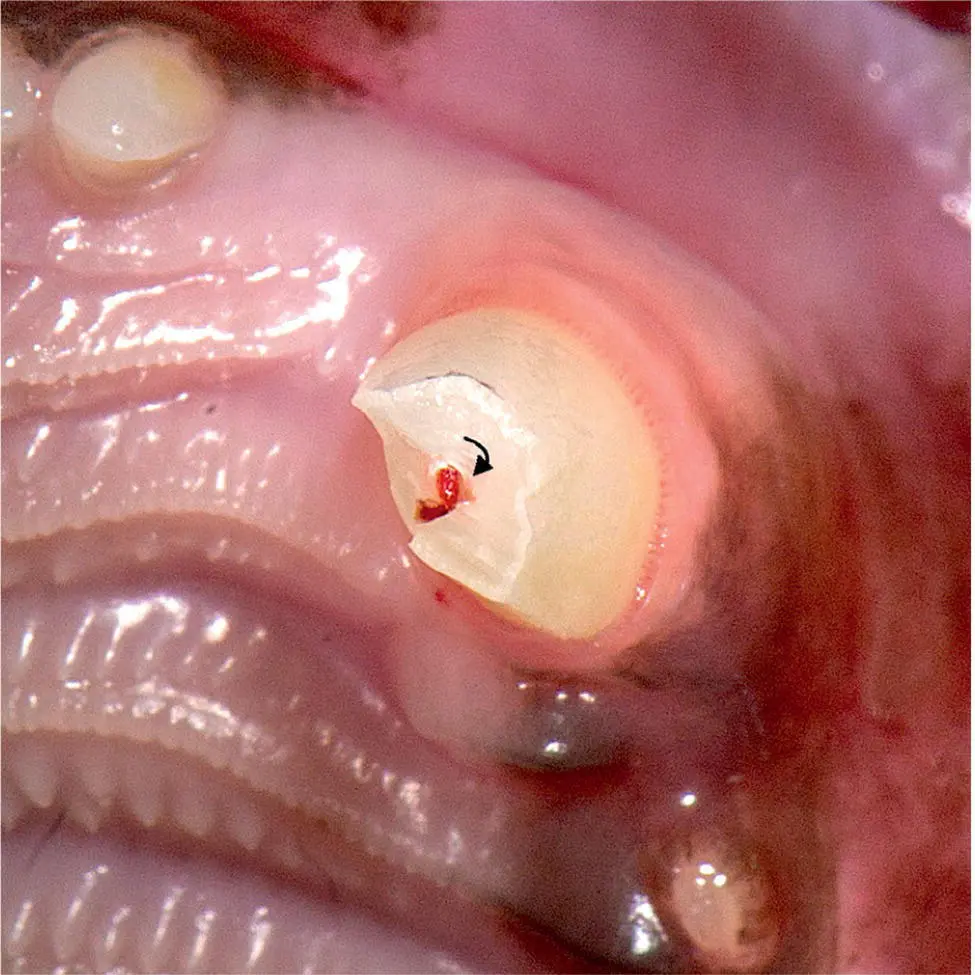
Figure 1.34 Enamel, dentin, and pulp exposed in an acutely fractured canine tooth.
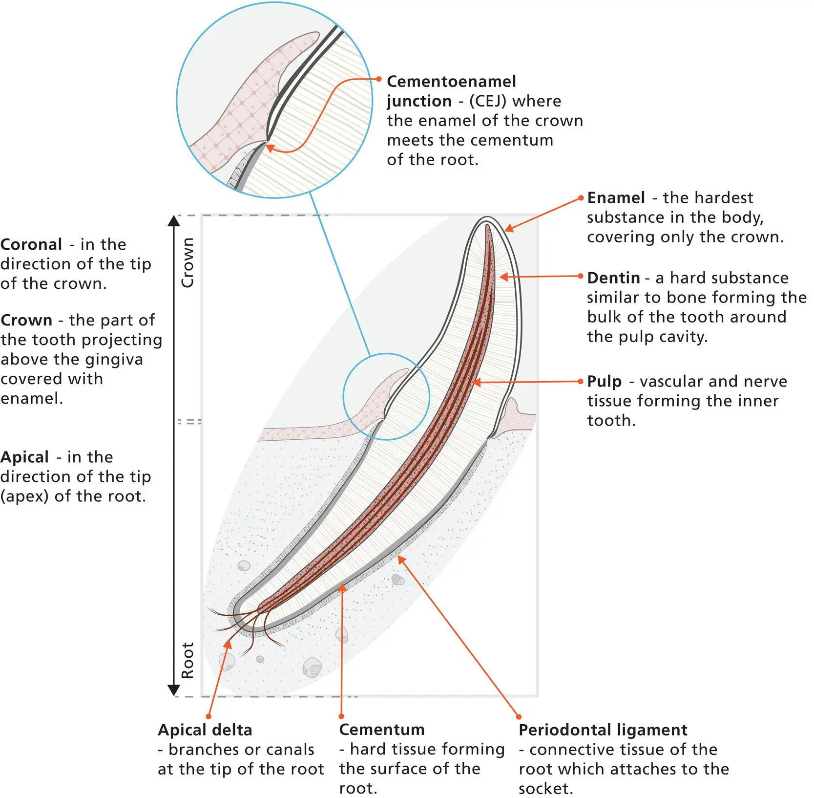
Figure 1.35 Canine tooth and surrounding structures.
1.20 Tooth Eruption
The maxillary teeth generally erupt before their mandibular counterparts. Eruption of the incisors precedes that of the canines. The premolars and molars erupt last.
There are normally 26 deciduous teeth. Between 11 and 15 days, the incisors erupt; at 17–19 days, the canines erupt; between 24 and 30 days, all premolars erupt. The secondary (adult) upper first molars erupt later, between 37 and 60 days. At 60 days, the deciduous dentition is complete. By seven months, the permanent teeth should be fully erupted ( Table 1.1).
Table 1.1 Approximate age by which permanent teeth erupt (in days).
| Upper | Lower | |
|---|---|---|
| Incisors | ||
| Central | 103 | 113 |
| Middle | 114 | 119 |
| Lateral | 135 | 132 |
| Canine | 153 | 149 |
| Premolar | ||
| Second | 150 | |
| Third | 168 | 173 |
| Fourth | 151 | 174 |
| Molar | 162 | 130 |
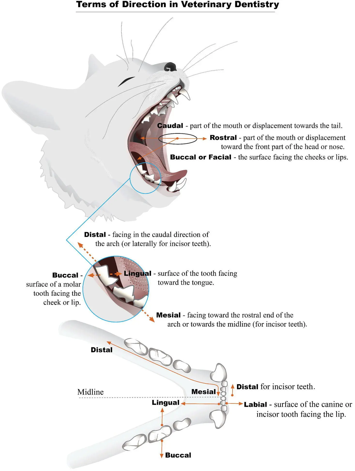
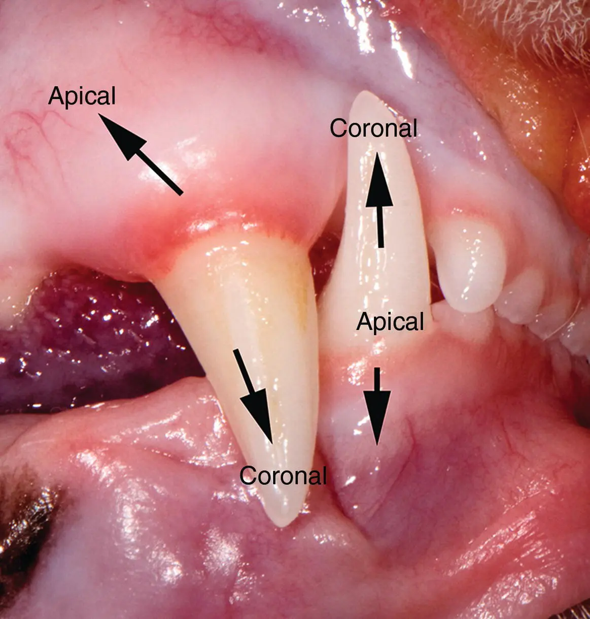
Figure 1.36 Directions in the oral cavity.
Интервал:
Закладка:
Похожие книги на «Feline Dentistry»
Представляем Вашему вниманию похожие книги на «Feline Dentistry» списком для выбора. Мы отобрали схожую по названию и смыслу литературу в надежде предоставить читателям больше вариантов отыскать новые, интересные, ещё непрочитанные произведения.


Обсуждение, отзывы о книге «Feline Dentistry» и просто собственные мнения читателей. Оставьте ваши комментарии, напишите, что Вы думаете о произведении, его смысле или главных героях. Укажите что конкретно понравилось, а что нет, и почему Вы так считаете.