Joe Mayhew - Large Animal Neurology
Здесь есть возможность читать онлайн «Joe Mayhew - Large Animal Neurology» — ознакомительный отрывок электронной книги совершенно бесплатно, а после прочтения отрывка купить полную версию. В некоторых случаях можно слушать аудио, скачать через торрент в формате fb2 и присутствует краткое содержание. Жанр: unrecognised, на английском языке. Описание произведения, (предисловие) а так же отзывы посетителей доступны на портале библиотеки ЛибКат.
- Название:Large Animal Neurology
- Автор:
- Жанр:
- Год:неизвестен
- ISBN:нет данных
- Рейтинг книги:5 / 5. Голосов: 1
-
Избранное:Добавить в избранное
- Отзывы:
-
Ваша оценка:
Large Animal Neurology: краткое содержание, описание и аннотация
Предлагаем к чтению аннотацию, описание, краткое содержание или предисловие (зависит от того, что написал сам автор книги «Large Animal Neurology»). Если вы не нашли необходимую информацию о книге — напишите в комментариях, мы постараемся отыскать её.
delivers a practical and complete reference for veterinarians, veterinary trainees and scientists dealing with large animal neurology. The book is vividly illustrated in full colour and contains many clinical photographs and detailed line drawings to highlight the concepts discussed within.
Organised into three parts,
offers practitioners and students straightforward guides on how to perform neurologic examinations for domestic large animal species, including neonates. It also discusses the presenting clinical syndromes caused by common nervous system diseases, as well as giving details of the specific neurologic diseases of large domestic animals.
The book includes:
A thorough introduction to the evaluation of large animal neurologic patients, including discussions of neuroanatomy, neurologic evaluation, ancillary diagnostic aids, and the important pathologic responses of the nervous system Comprehensive exploration of 26 presenting clinical problems, including behaviour disorders, seizures, epilepsy, sleep disorders, blindness, strabismus, monoplegia, wobblers, tetraplegia, pruritus and cauda equina syndrome Detailed coverage of the specific diseases, including those of genetic, infectious, nutritional, toxic and metabolic cause, and the many diseases with multifactorial and with unknown cause Perfect for all equine and farm animal veterinarians, veterinary neurologists, as well as trainees in the field, is also an ideal resource for undergraduate veterinary students, animal pathologists, and neuroscience researchers.
Large Animal Neurology — читать онлайн ознакомительный отрывок
Ниже представлен текст книги, разбитый по страницам. Система сохранения места последней прочитанной страницы, позволяет с удобством читать онлайн бесплатно книгу «Large Animal Neurology», без необходимости каждый раз заново искать на чём Вы остановились. Поставьте закладку, и сможете в любой момент перейти на страницу, на которой закончили чтение.
Интервал:
Закладка:
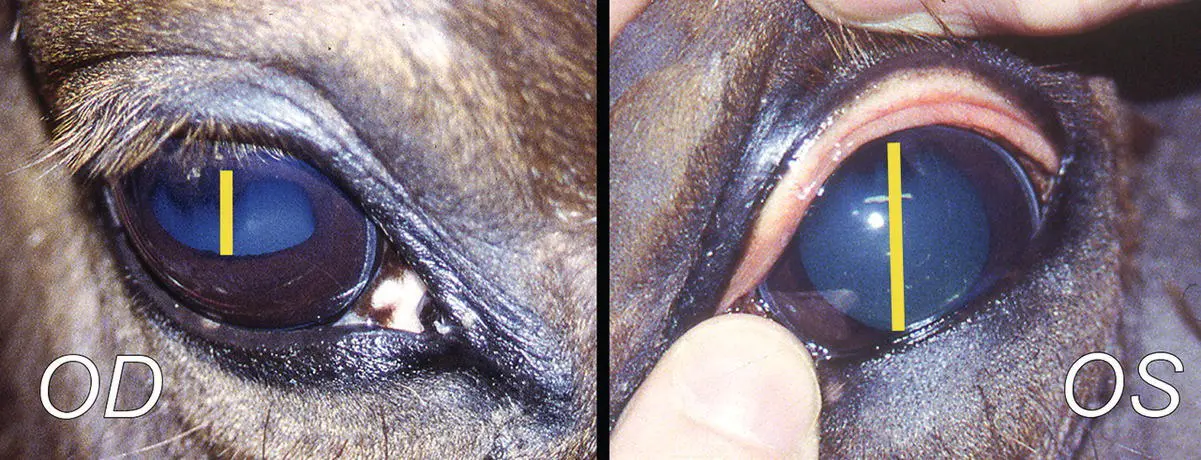
Figure 13.2 This case of polyneuritis involved the trigeminal nerves, and quite distinctly the ophthalmic branch on the left side was clinically affected. The latter resulted in conjunctival and corneal hypalgesia demonstrated here in the left eye (OS). Likely because of perineuritis, and the fact that branches of the ophthalmic nerve are confluent with the oculomotor fibers and the ciliary ganglion, a degree of pupillary dilation was also present in the left eye (OS) compared with the right (OD) (yellow bars).
Medullary (and cranial cervical spinal cord) diseases such as equine protozoal myeloencephalitis and listeriosis in ruminants are the more common central causes of decreased facial sensation due to involvement of the sensory tract and/or nucleus of the trigeminal nerve in the caudal medulla oblongata and cranial cervical spinal cord ( Figure 13.3).
Central pathways for sensory perception from the face pass from the trigeminal sensory nucleus to the contralateral thalamus, contralateral internal capsule, and somesthetic, sensory cerebral cortex, mostly in the frontoparietal lobe. Lesions in these structures rostral to CNs V and VII can result in degrees of contralesional facial hypalgesia or analgesia but no interruption to the facial (CN V sensory → CN VII motor) reflex ( Figure 13.4). This facial hypalgesia or analgesia is most evident when the noxious stimulus is applied to the sensitive nasal septal membranes on the side opposite to such rostral lesions. Several severe, particularly focal or selective, diseases of the thalamus, internal capsule, and forebrain including cerebral abscess, cholesterinic granuloma, Fusarium verticillioides ( ex. moniliforme ) leukoencephalomalacia, intracarotid injection, and Sarcocystis neurona encephalitis have resulted in this contralesional facial hypalgesic and analgesic syndrome.
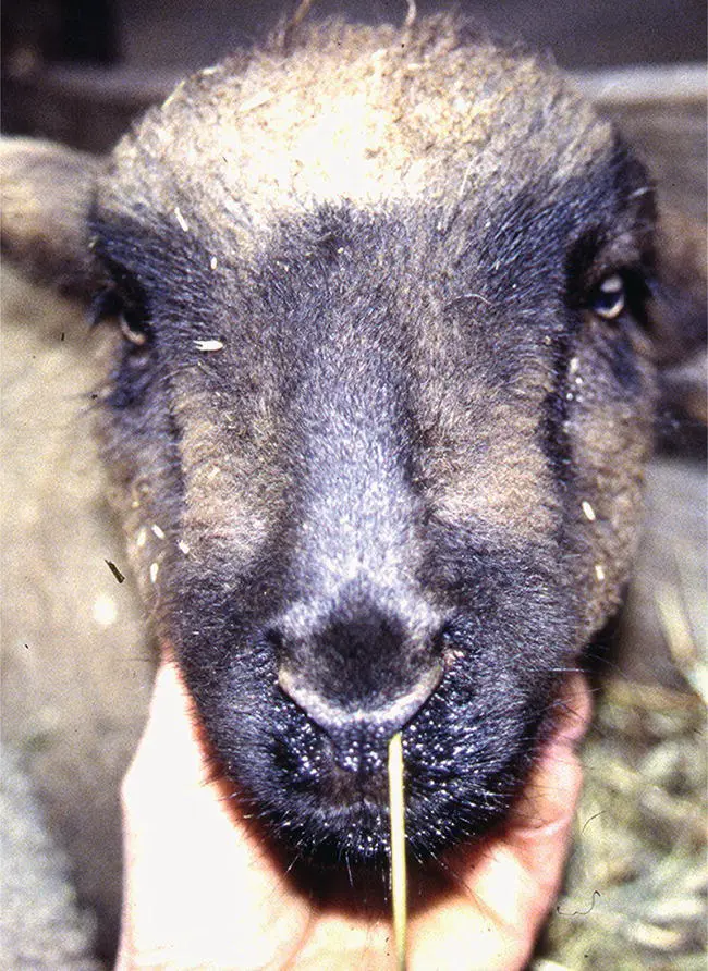
Figure 13.3 Oblivious to the presence of a straw stuck in its left nasal cavity, this lamb suffering from listeriosis is demonstrating facial analgesia. There were accompanying facial and vestibulocochlear nerve deficits with ataxia and weakness. Indeed, involvement of the sensory branch of the trigeminal nerve is a very common sign with listeriosis and possibly reflects one of the routes of bacterial infection reaching the medulla oblongata in that disease.
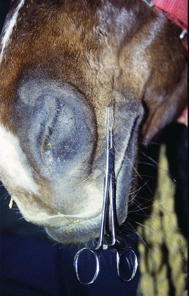
Figure 13.4 Definitive facial hypalgesia and analgesia are best detected over the distal face as shown and particularly on the nasal membranes. When this is found, the main question to arise is whether it is due to a central forebrain (thalamus or cerebral hemisphere) lesion or it relates to an afferent sensory lesion of the trigeminal distribution. In this horse with a lesion in the forebrain causing facial analgesia, the facial reflexes remained intact and voluntary movement of the face was good. With peripheral sensory CN Vs lesions, the loss of facial sensation can be very well demarcated particularly with more distal lesions. Also, facial reflexes will be reduced or absent to the same degree as the hypalgesia or analgesia. The central distribution of the afferent fibers from the trigeminal nerve is quite spread out in the medulla oblongata and cranial cervical spinal cord within the sensory trigeminal nucleus and its tract. Thus, a central medullary lesion causing dense analgesia over the face would entail a very extensive lesion such that there would be many other signs of brain disease such as recumbency and (semi)coma. Thus, dense facial sensory loss alone, due to an afferent lesion, is most likely in the peripheral trigeminal nerve or ganglion.
Equine protozoal myeloencephalitis caused by S. neurona and filariid nematode encephalitis have caused ipsilesional facial hypalgesia and no interference to the facial (CN V sensory → CN VII motor) reflexes with brainstem lesions involving pontine and midbrain sites, but with no lesion(s) in thalamus or cerebrum. It is likely that the ascending impulses for conscious sensory perception from the face cross in the region of the rostral brainstem to join the contralateral medial lemniscus on its path to the ventral caudal lateral thalamic nucleus. Also, it is probable that facial sensory input is perceived not only at the somatosensory cerebral cortex, but also at the level of the thalamus as a few newborn ruminants with no cerebral cortices (agenesis) appeared to react with avoidance movements when the nasal septal region was stimulated strongly.
Facial analgesia can be seen in cattle with extensive basilar empyema adjacent to and invading the sensory branches of the trigeminal nerves along with accompanying signs of dropped mandible, head and neck extension, bradycardia, peripheral blindness, dilated pupils, and facial, tongue and pharyngeal paralysis. The full syndrome results from extension to involve CNs II, III, V, IX, X, XI and XII, the ventral brainstem, and the hypothalamus.
Facial and body regional hypersensitivity to firm stimuli (hyperesthesia) but not to light touch stimuli (allodynia) is seen with trigeminal and diffuse neuritis in early stages of diffuse bacterial meningitis. Trigeminal neuritis is the presumed diagnosis in the syndrome of pernicious head rubbing in horses, wherein there is an acquired, profound irritation such that the patient severely self‐traumatizes the side of the face. Such a syndrome has been replicated during (presumed) regrowth of infraorbital nerve axons following compression or cauterization of that nerve to attempt to ameliorate signs of headshaking in horses. 4Facial hyperesthesia with allodynia to light touch ( Figure 13.5) has been seen in association with fluctuating signs of facial hyporeflexia and hypalgesia to firm stimuli. This has occurred in cases of paranasal sinusitis and following sinus surgery when the trigeminal nerve within or adjacent to the sphenopalatine sinus and within the infraorbital canal have been involved. 5,6The syndrome has resolved in a few cases as the sinusitis was controlled. The absence of true, stable allodynia alone does seem to differentiate these cases from cases of regional pain syndrome with prominant allodynia as well as from the enigmatic syndrome of headshaking with no facial hypalgesia. (see Chapter 28).
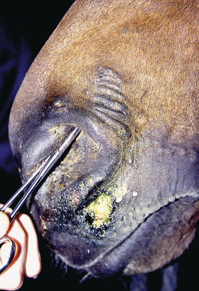
Figure 13.5 Accompanying the obvious nasal and distal facial hypalgesia to firm pressure in this case, there was evidence of nasal and facial allodynia to light touch in the form of self‐inflicted injury to the affected area of the face as indicated here. This horse was suffering from a squamous cell carcinoma affecting the left side of the sphenopalatine sinus region and the trigeminal nerve. It rubbed its distal face on objects spontaneously and with stimulation to the face, despite the demonstrable hypalgesia, but did not demonstrate headshaking.
References
1 1 Samuel JL, Kelly WR and Vanselow BA. Intracranial invasion by bovine ocular squamous cell carcinoma via cranial nerves. Vet Rec 1987; 121(18): 424–425.
2 2 Rebhun WC. Diseases of the bovine orbit and globe. J Am Vet Med Assoc 1979; 175(2): 171–175.
Читать дальшеИнтервал:
Закладка:
Похожие книги на «Large Animal Neurology»
Представляем Вашему вниманию похожие книги на «Large Animal Neurology» списком для выбора. Мы отобрали схожую по названию и смыслу литературу в надежде предоставить читателям больше вариантов отыскать новые, интересные, ещё непрочитанные произведения.


Обсуждение, отзывы о книге «Large Animal Neurology» и просто собственные мнения читателей. Оставьте ваши комментарии, напишите, что Вы думаете о произведении, его смысле или главных героях. Укажите что конкретно понравилось, а что нет, и почему Вы так считаете.