Joe Mayhew - Large Animal Neurology
Здесь есть возможность читать онлайн «Joe Mayhew - Large Animal Neurology» — ознакомительный отрывок электронной книги совершенно бесплатно, а после прочтения отрывка купить полную версию. В некоторых случаях можно слушать аудио, скачать через торрент в формате fb2 и присутствует краткое содержание. Жанр: unrecognised, на английском языке. Описание произведения, (предисловие) а так же отзывы посетителей доступны на портале библиотеки ЛибКат.
- Название:Large Animal Neurology
- Автор:
- Жанр:
- Год:неизвестен
- ISBN:нет данных
- Рейтинг книги:5 / 5. Голосов: 1
-
Избранное:Добавить в избранное
- Отзывы:
-
Ваша оценка:
Large Animal Neurology: краткое содержание, описание и аннотация
Предлагаем к чтению аннотацию, описание, краткое содержание или предисловие (зависит от того, что написал сам автор книги «Large Animal Neurology»). Если вы не нашли необходимую информацию о книге — напишите в комментариях, мы постараемся отыскать её.
delivers a practical and complete reference for veterinarians, veterinary trainees and scientists dealing with large animal neurology. The book is vividly illustrated in full colour and contains many clinical photographs and detailed line drawings to highlight the concepts discussed within.
Organised into three parts,
offers practitioners and students straightforward guides on how to perform neurologic examinations for domestic large animal species, including neonates. It also discusses the presenting clinical syndromes caused by common nervous system diseases, as well as giving details of the specific neurologic diseases of large domestic animals.
The book includes:
A thorough introduction to the evaluation of large animal neurologic patients, including discussions of neuroanatomy, neurologic evaluation, ancillary diagnostic aids, and the important pathologic responses of the nervous system Comprehensive exploration of 26 presenting clinical problems, including behaviour disorders, seizures, epilepsy, sleep disorders, blindness, strabismus, monoplegia, wobblers, tetraplegia, pruritus and cauda equina syndrome Detailed coverage of the specific diseases, including those of genetic, infectious, nutritional, toxic and metabolic cause, and the many diseases with multifactorial and with unknown cause Perfect for all equine and farm animal veterinarians, veterinary neurologists, as well as trainees in the field, is also an ideal resource for undergraduate veterinary students, animal pathologists, and neuroscience researchers.
Large Animal Neurology — читать онлайн ознакомительный отрывок
Ниже представлен текст книги, разбитый по страницам. Система сохранения места последней прочитанной страницы, позволяет с удобством читать онлайн бесплатно книгу «Large Animal Neurology», без необходимости каждый раз заново искать на чём Вы остановились. Поставьте закладку, и сможете в любой момент перейти на страницу, на которой закончили чтение.
Интервал:
Закладка:
3 3 Guard CL, Rebhun WC and Perdrizet JA. Cranial tumors in aged cattle causing Horner's syndrome and exophthalmos. Cornell Vet 1984; 74(4): 361–365.
4 4 Roberts V. Trigeminal‐mediated headshaking in horses: prevalence, impact, and management strategies. Vet Med Auckl 2019; 10: 1–8.
5 5 Tucker R, Windley ZE, Abernethy AD, et al. Radiographic, computed tomographic and surgical anatomy of the equine sphenopalatine sinus in normal and diseased horses. Equine Vet J 2016; 48(5): 578–584.
6 6 McCann JL, Dixon PM and Mayhew IG. Clinical anatomy of the equine sphenopalatine sinus. Equine Vet J 2004; 36(6): 466–472.
14 Facial paralysis and facial spasm
Paralysis of the muscles of facial expression, or facial paralysis, is a very common problem in large animals with neurologic disorders and is a common part of the syndromes caused by temporohyoid osteoarthropathy. 1–3Dysfunction of the somatic efferent fibers of the facial nerve produces decreased spontaneous and reflex movements of the ear, eyelids, lips, and external nares. The ear, upper eyelid, and lips on the side of the lesion droop and the muzzle tends to be pulled to the opposite side with a unilateral lesion, making it usually easy to recognize in horses ( Figure 14.1). This is less so in other species that tend to have little facial expression, naturally stiff or droopy ears and firm muzzles. However, when quite mild and when the patient is excited, facial muscle tone can be remarkably normal so that the syndrome may be very subtle to observe. In this situation, returning to observe the patient during a resting state, or use of mild sedation, can help uncover the signs. Ptosis of the upper eyelid is a feature of facial paralysis in all large domestic animals and is prominent in horses. It is due to the paralysis of the strong levator anguli oculi medialis muscle in these species that is innervated by the facial nerve CN VII. It is possible that the bulk of atonic supraorbital muscles and the paralysis of the frontalis muscle also contribute to the ptosis. Unfortunately, when upper eyelid ptosis is seen in large animals, the immediate thought is one of it being Horner syndrome ( Figure 10.4). However, facial paralysis is vastly more common than sympathetic denervation of the eyelids in large animals and always must be considered the most likely cause.
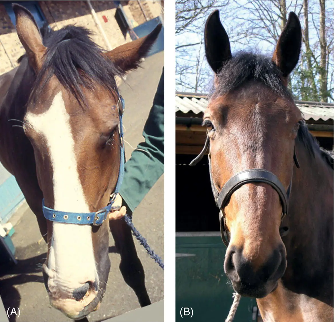
Figure 14.1 Total left facial paralysis is seen in this horse (A) suffering from direct penetrating trauma to the base of the cranium including the left middle and inner ears. The horse in (B) suffered direct injury to the buccal branches of the left facial nerve resulting in mild deviation of the muzzle to the right.
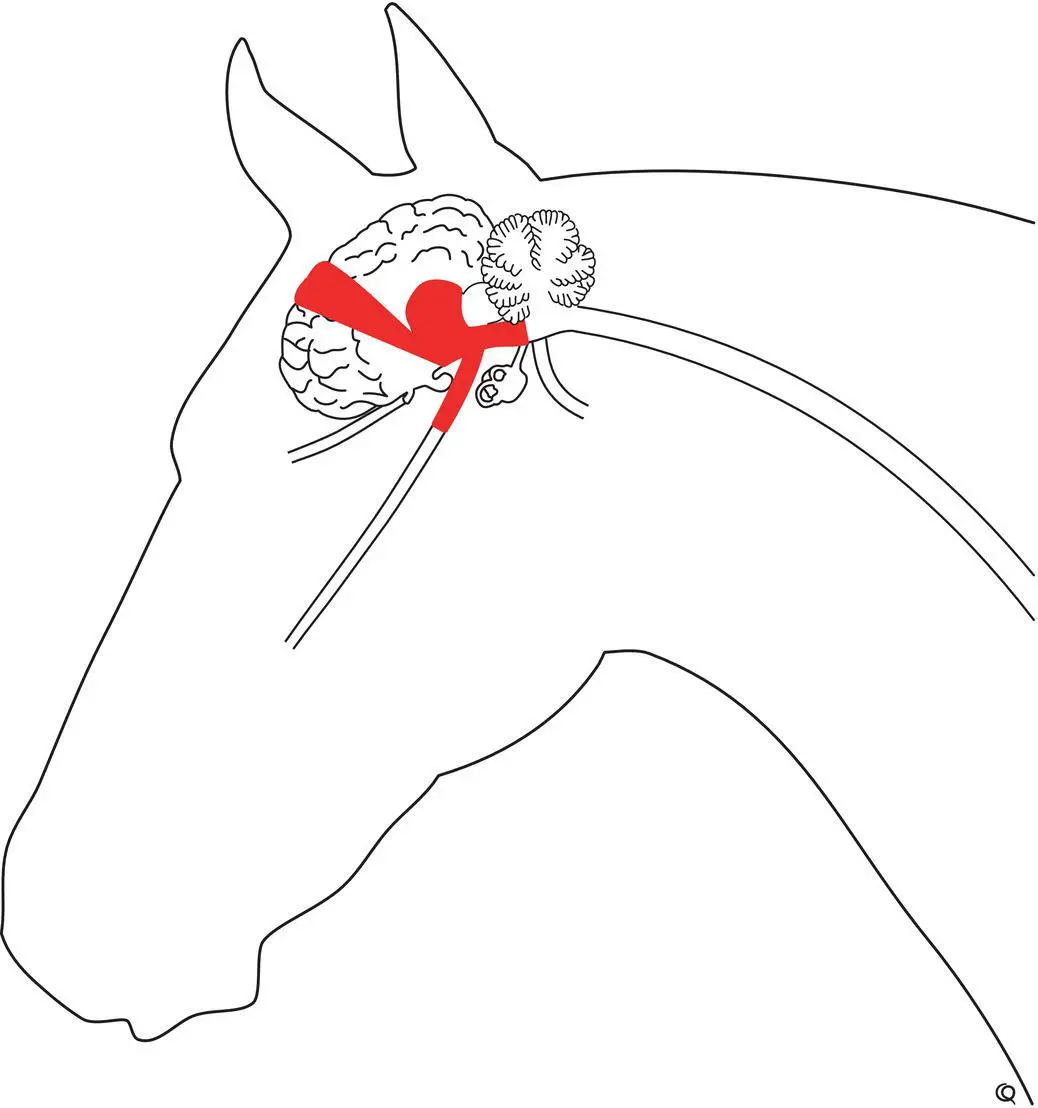
Parasympathetic visceral efferent fibers pass with the somatic fibers to exit the brainstem, pass through the internal acoustic meatus, and then leave the main facial nerve within the facial canal to innervate the lachrymal gland (and palatine and nasal glands) via the major petrosal nerve and pterygoid ganglion. 4When these parasympathetic fibers in the facial nerve are damaged, then the loss of lachrymal tear production ensues resulting in keratoconjunctivitis sicca.
Damage to the central motor pathways in the frontal cortex, internal capsule, crus cerebri, and brainstem that control the facial nucleus and nerve can result in abnormal facial expression ( Figure 14.2). This occurs without flaccid facial paralysis. There is still tone in the muscles of facial expression, and facial reflexes (CN V sensory → CN VII motor) are intact and may even be hyperactive. However, the expression may be bland or grimacing on one or both sides. Needle electromyographic examination of the facial muscles does not reveal denervation because the facial motor nucleus and nerve are still intact. Large, focal cerebral lesions such as hematoma, S. neurona encephalitis, and abscess have produced such signs of supranuclear facial motor dysfunction that, unlike in humans, appear to be ipsilesional, at least to the muzzle and lips of the distal face.
Trauma and bacterial infection of the middle and inner ears are the commonest cause of peripheral facial nerve paralysis, 5–9most particularly in young patients. With distal, peripheral facial nerve involvement, usually one or two branches of the nerve, not all three nerve branches (auricular, palpebral, and buccal), are involved. Pressure on the side of the face as a result of a tight halter or from recumbency damages buccal branches, paralyzing just the nares and lips; however, the ear and upper eyelid may droop because of separate but direct auricular and palpebral nerve trauma. Brainstem lesions, particu larly those caused by equine protozoal myeloencephalitis and listeriosis, can selectively involve facial nuclei in the brainstem and can mimic a peripheral lesion by producing selective, partial facial paresis. Neuritis of polyneuritis equi and equine Lyme neuroborreliosis are causes of unilateral facial paresis, 10–12and proliferative inflammatory lesions in and around the facial nerves of calves may be the result of partly treated and chronic bacterial otitis. 13–16
Figure 14.2 In comparison to facial weakness, these two horses (A and B) have, at times, very good tone and movement in their face but at rest have a bland facial expression (A). Sometimes with voluntary effort and with facial reflex testing, their face shows hypertonic expression and hyperreflexia (B). Both horses had lesions in the forebrain without facial nuclear or facial nerve involvement. The first (A) had a vascular lesion involving much of the left forebrain, and the second had thalamic lesions of equine protozoal myeloencephalitis on the left side. It does appear that the distal facial, functional abnormalities as seen in these horses are ipsilesional to the damage to the central motor pathways to the facial nuclei.
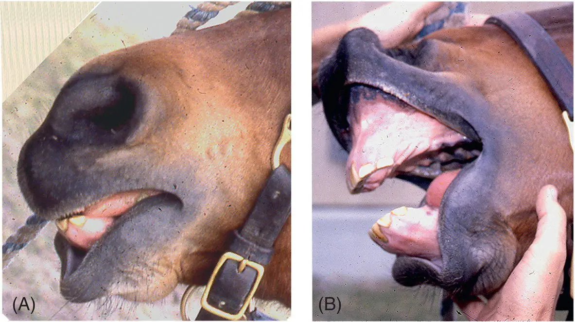
Figure 14.3 Facial paralysis is seen as marked loss of facial expression and movement with drooping of the ear, and upper eyelid, and of the lips (A and B). Bilateral facial weakness seen here can be less evident due to the relative facial symmetry present (B). Collapse of alar folds with inspiratory restriction (A) occurs far more with bilateral than unilateral paralysis in horses. Also, mild degrees of facial paresis can be difficult to see especially in patients such as calves and piglets that do not normally have prominent facial expression and facial muscle tone. Re‐examination when a patient is fully relaxed or sedated can help determine degrees of partial facial weakness. Palpating for decreased muscle tone and observing and palpating for reduced facial reflexes are also very useful to confirm degrees of facial paresis.
Large animals accommodate to unilateral facial paralysis very well, but bilateral facial paralysis ( Figure 14.3) results in difficulty in prehending food and in keeping it in the mouth. Horses with this syndrome do sequester food in the flaccid cheek pouches and drop a lot of food while eating. Temporary tarsorrhaphy may help alleviate damage to the cornea from the lack of tears, but permanent facial paralysis may necessitate enucleation of the eyeball because of keratitis sicca and exposure ophthalmitis. 3Exercising horses may require false nostril surgery as a result of an obstruction to inspiratory airflow. 17Chronic paralysis with muscle atrophy and fibrous contracture of the face can cause twisting of the muzzle and nares back across the midline toward the paralyzed side.
Читать дальшеИнтервал:
Закладка:
Похожие книги на «Large Animal Neurology»
Представляем Вашему вниманию похожие книги на «Large Animal Neurology» списком для выбора. Мы отобрали схожую по названию и смыслу литературу в надежде предоставить читателям больше вариантов отыскать новые, интересные, ещё непрочитанные произведения.


Обсуждение, отзывы о книге «Large Animal Neurology» и просто собственные мнения читателей. Оставьте ваши комментарии, напишите, что Вы думаете о произведении, его смысле или главных героях. Укажите что конкретно понравилось, а что нет, и почему Вы так считаете.