David Linden - Touch
Здесь есть возможность читать онлайн «David Linden - Touch» весь текст электронной книги совершенно бесплатно (целиком полную версию без сокращений). В некоторых случаях можно слушать аудио, скачать через торрент в формате fb2 и присутствует краткое содержание. Год выпуска: 2014, ISBN: 2014, Издательство: Penguin Books Ltd, Жанр: Психология, Биология, sci_popular, на английском языке. Описание произведения, (предисловие) а так же отзывы посетителей доступны на портале библиотеки ЛибКат.

- Название:Touch
- Автор:
- Издательство:Penguin Books Ltd
- Жанр:
- Год:2014
- ISBN:9780241184059
- Рейтинг книги:4 / 5. Голосов: 1
-
Избранное:Добавить в избранное
- Отзывы:
-
Ваша оценка:
Touch: краткое содержание, описание и аннотация
Предлагаем к чтению аннотацию, описание, краткое содержание или предисловие (зависит от того, что написал сам автор книги «Touch»). Если вы не нашли необходимую информацию о книге — напишите в комментариях, мы постараемся отыскать её.
Dual-function receptors in our skin make mint feel cool and chili peppers hot. Without the brain’s dedicated centers for emotional touch, an orgasm would feel more like a sneeze—convulsive, but not especially nice. From skin to nerves to brain, the organization of our body’s touch circuits is a complex and often counterintuitive system that affects everything from our social interactions to our general health and development.
In Touch, neuroscientist and bestselling author David J. Linden explores this critical interface between our bodies and the outside world, between ourselves and others. Along the way, he answers such questions as: Why do women have more refined detection with their fingertips than men? Is there a biological basis for the use of acupuncture to relieve pain? How do drugs like Ecstasy heighten and motivate sensual touch? Why can’t we tickle ourselves? Linking biology and behavioral science, Touch offers an entertaining and enlightening answer to how we feel in every sense of the word.
Touch — читать онлайн бесплатно полную книгу (весь текст) целиком
Ниже представлен текст книги, разбитый по страницам. Система сохранения места последней прочитанной страницы, позволяет с удобством читать онлайн бесплатно книгу «Touch», без необходимости каждый раз заново искать на чём Вы остановились. Поставьте закладку, и сможете в любой момент перейти на страницу, на которой закончили чтение.
Интервал:
Закладка:
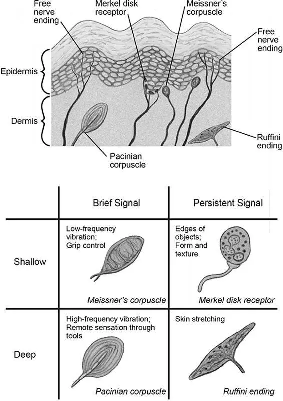
Figure 2.3Four types of sensor for mechanical stimuli found in glabrous skin. Merkel disks are located in the deepest part of the epidermis, where it borders the dermis at the peaks of the primary epidermal ridges. Meissner’s corpuscles are found just across this border in the shallowest parts of the dermis but in the troughs between epidermal ridges, while Pacinian corpuscles and Ruffini endings are located deeper in the dermis. The nerve fibers receiving signals from Meissner’s and Pacinian corpuscles send electrical signals to the brain briefly, only at the beginning and end of a sustained touch, while those that receive signals from Ruffini endings and Merkel cells signal persistently, throughout the touch stimulus. Also shown are free nerve endings, which are sensors for certain chemicals, temperature, pain, and itch. Those will be discussed in later chapters.
Merkel disks are present at a very high density in the skin of the lips and the fingertips, at a low density on other glabrous regions, and at a very low density on hairy skin. They are sensitive to very small forces that produce skin indentations of about 0.05 millimeter, and continue to respond more strongly (firing spikes at a higher rate) in a linear fashion, until they fire maximally in response to indentations of about 1.5 millimeters. Electrical recordings made from single nerve fibers conveying Merkel disk signals reveal that these fibers continue to fire spikes as long as the skin remains indented. 16Artificial electrical stimulation of a single Merkel nerve fiber running through the upper arm causes subjects to report a sensation like a “soft painting brush held tangentially against the skin.” 17
Merkel disks allow us to distinguish individual surface features with our fingertips, like the rough-textured ridges on the edge of the quarter. Crucially, the ability of Merkel disks to distinguish tactile features flows from their particular structure, location, and connections. Because Merkels are located in a relatively shallow layer of the skin, they can respond to small indentations produced by textured surfaces. And because they are densely packed in the fingertips, and each is innervated by a single nerve fiber, this array of sensors can resolve the difference between two features on the surface of an object that are only about 0.7 millimeter apart. 18
Okay, now that you’ve identified the quarter, you grip it between your thumb and forefinger and prepare to maneuver it toward the coin slot. How do you determine the amount of force to exert with this pincer motion? You don’t want to use maximal bone-crushing force for everything you grip—which might not be bad for holding a quarter, but would be disastrous for clutching an egg or a child’s hand—nor do you want to use so little force that the quarter slips out of your grasp. Ideally, you’d like to use the minimal amount of force needed to hold the quarter securely. For this job you rely mostly on another skin sensor, called the Meissner’s corpuscle (figure 2.3). Like the Merkel disks, the Meissner’s corpuscles are located at the border between the dermis and the epidermis. 19They reside just on the dermal side of the border, but in the troughs between the ridges, where the epidermis is thinnest. Each Meissner’s corpuscle consists of a coiled arrangement of nerve fiber endings, intermingled with layers of nonneuronal cells called Schwann cells. Together these form a bulbous encapsulated structure, the corpuscle, which is tethered to nearby skin cells by structural cables made from the protein collagen. 20Meissner’s corpuscles are physically deformed by the tug of these cables when the skin is indented and pop back into shape when the indenting object is removed.
The array of Meissner’s corpuscles in the fingertips is even denser than that of Merkel disks, and they are located even closer to the skin surface. These properties might lead one to believe that Meissner’s corpuscles are also built to convey information about fine features of objects, such as texture, edges, and curvature. However, when electrical recordings are made from the nerve fibers that innervate Meissner’s corpuscles, we find completely different responses. First, Meissner fibers fire spikes only at the very beginning and the very ending of a prolonged skin indentation: when the outer capsule is initially deformed and then again when it pops back into place. This means that, unlike Merkel disks, Meissner’s corpuscles don’t respond well to steady force on the skin, but rather are strongly activated by faint low-frequency vibration that repeatedly indents and reforms the capsule. Second, a single nerve fiber conveys and collects signals from many Meissner’s corpuscles, spread over about 10 square millimeters of skin surface. Even though Meissner’s corpuscles are found in the fingertips at great density, electrical recordings show that they cannot distinguish the finest features of objects. The convergent wiring of the Meissner system is built to be exquisitely sensitive to tiny, rapid skin movements but to localize those movements with only moderate precision.
What does all this have to do with gripping your quarter properly? It turns out that when you grip and move an object, there are microscopic slips of that object along your skin. These microslips are detected by the Meissner system, which sends electrical signals to neurons in the spinal cord that contract the relevant finger muscles to increase gripping force until the microslips stop. This allows you to manipulate objects with delicacy, using the minimal force for the job at hand. Because Meissner-mediated grip control is a spinal cord circuit, it works as a reflex and does not directly enter your conscious awareness—you don’t have to think about gripping the quarter slightly harder as you move it from your pocket to the coin slot, as it just happens naturally.
To appreciate how the anatomy and physiology of the Meissner’s array in the fingertips make precision grip control possible, let’s indulge in a little science fiction. Imagine an alternate human biology in which Meissner’s corpuscles signaled throughout a period of skin indentation rather than just at its beginning and end. If this were the case, they would become sensitive to ongoing force, rather than insensitive, as they are in our fingers. In that alternate-skin universe, the Meissner’s corpuscle responses to the large steady forces required to grip an object would overwhelm the small signals produced by local microslip vibrations. The useful signal about grip efficiency would drown in a sea of noise, and fine grip control would fail. Without fine grip control, we would have been unable to develop tool use and, very likely, human culture as we know it. Sometimes even the tiniest, obscure-seeming details of biology turn out to be critically important.
By now you’re ready to place the quarter in the meter’s slot. As you insert the coin, you begin feeling sensations transmitted through it as it collides with the internal walls of the slot and you will use this tactile feedback subconsciously to alter the trajectory of your arm, hand, and fingers in order to insert the quarter smoothly. For this portion of the task, the most important sensor is called the Pacinian corpuscle. 21Pacinian corpuscles look weirdly lovely (figure 2.3), each consisting of a single nerve fiber ending wrapped in many concentric layers of supporting cells with fluid-filled spaces. In cross-section, they look like an onion or an entry in one of those engineering contests for high school students that involves designing a lightweight casing to protect an egg dropped off the roof of a building. 22There are about 350 Pacinian corpuscles per finger, located in the deeper regions of the dermis. Electrical recordings from Pacinian nerve fibers reveal that, like Meissner’s corpuscles, they don’t respond well to continuous force but fire spikes only at the onset and offset of skin indentation. Although they are also poor at resolving the surface features of an object, Pacinians are extremely sensitive to tiny vibrations and have almost no spatial localization: A single Pacinian corpuscle in the fingertip, by virtue of its layered wrapping and deep location, can be activated by vibration occurring anywhere on the finger. In a way, the properties of the Meissner’s corpuscle (sensitive to small vibrations but insensitive to steady force or fine spatial detail) are even more extreme in the Pacinian corpuscle. Pacinian corpuscles are most sensitive to high-frequency vibration in the range of 200 to 300 hertz, at which they can detect skin motion as small as 0.00001 millimeter (two hundred times smaller than the diameter of a tiny vellus hair).
Читать дальшеИнтервал:
Закладка:
Похожие книги на «Touch»
Представляем Вашему вниманию похожие книги на «Touch» списком для выбора. Мы отобрали схожую по названию и смыслу литературу в надежде предоставить читателям больше вариантов отыскать новые, интересные, ещё непрочитанные произведения.


Обсуждение, отзывы о книге «Touch» и просто собственные мнения читателей. Оставьте ваши комментарии, напишите, что Вы думаете о произведении, его смысле или главных героях. Укажите что конкретно понравилось, а что нет, и почему Вы так считаете.