Point-of-Care Ultrasound Techniques for the Small Animal Practitioner
Здесь есть возможность читать онлайн «Point-of-Care Ultrasound Techniques for the Small Animal Practitioner» — ознакомительный отрывок электронной книги совершенно бесплатно, а после прочтения отрывка купить полную версию. В некоторых случаях можно слушать аудио, скачать через торрент в формате fb2 и присутствует краткое содержание. Жанр: unrecognised, на английском языке. Описание произведения, (предисловие) а так же отзывы посетителей доступны на портале библиотеки ЛибКат.

- Название:Point-of-Care Ultrasound Techniques for the Small Animal Practitioner
- Автор:
- Жанр:
- Год:неизвестен
- ISBN:нет данных
- Рейтинг книги:5 / 5. Голосов: 1
-
Избранное:Добавить в избранное
- Отзывы:
-
Ваша оценка:
Point-of-Care Ultrasound Techniques for the Small Animal Practitioner: краткое содержание, описание и аннотация
Предлагаем к чтению аннотацию, описание, краткое содержание или предисловие (зависит от того, что написал сам автор книги «Point-of-Care Ultrasound Techniques for the Small Animal Practitioner»). Если вы не нашли необходимую информацию о книге — напишите в комментариях, мы постараемся отыскать её.
New chapters in
cover ultrasound-guided nerve blocks, musculoskeletal, brain imaging, and applications of focused ultrasound techniques in cats, exotics and marine mammals—making it an essential purchase for veterinarians wanting to incorporate point-of-care ultrasound techniques into their veterinary practices.
Presents a standardized approach to point-of-care ultrasound as an extension of the physical exam, including trauma, non-trauma, and monitoring applications Includes coverage of new techniques for focused ultrasound exams, including lung, anesthesia and ultrasound guided nerve blocks, transcranial brain imaging, musculoskeletal, volume status evaluation, and rapid assessment for treatable forms of shock Adds cats, exotic and wildlife mammals, and marine mammals to the existing canine coverage Emphasizes the integration of point-of-care ultrasound techniques for optimizing patient care and accurate patient assessment Offers access to a companion website with supplementary video clips showing many clinically relevant didactic examples The second edition of
is an excellent resource for veterinary practitioners, ranging from the general practitioner to nearly all clinical specialists, including internal medicine, oncology, cardiology, emergency and critical care, anesthesiology, ophthalmology, exotics, and zoo medicine specialists, and veterinary students.
Point-of-Care Ultrasound Techniques for the Small Animal Practitioner — читать онлайн ознакомительный отрывок
Ниже представлен текст книги, разбитый по страницам. Система сохранения места последней прочитанной страницы, позволяет с удобством читать онлайн бесплатно книгу «Point-of-Care Ultrasound Techniques for the Small Animal Practitioner», без необходимости каждый раз заново искать на чём Вы остановились. Поставьте закладку, и сможете в любой момент перейти на страницу, на которой закончили чтение.
Интервал:
Закладка:
In summary, the four primary AFAST views are part of the AFS system. The AFAST 5th bonus view (HR5th and SR5th views depending on which lateral recumbency is being used) is not part of the AFAST AFS system, but important for acquiring information for that respective retroperitoneal space and any obvious soft tissue or parenchymal abnormalities of the kidney and its adjacent liver and spleen dependent on positioning. In cats and smaller dogs when using enough depth, both kidneys are often seen through the SR (or HR) view (Lisciandro et al. 2009; Lisciandro 2011, 2012, 2014a; McMurray et al. 2016) (see Figure 6.21).
Pearl:By imaging using the AFAST target organ approach, sonographic anatomy is better recognized, and the sonographer is building POCUS skills on every AFAST exam, with the only pressure being the recognition of free fluid.
Probe Maneuvering is Standardized
All AFAST acoustic views are imaged in longitudinal (sagittal) orientation with the marker of the probe always directed toward the head of the patient; with muscle memory, most sonographers quickly learn this repetitive probe motion. In longitudinal (sagittal) orientation target organs are more recognizable than in transverse orientation, especially for the nonradiologist sonographer ( Figure 6.7). The single longitudinal (sagittal) orientation is supported by the original veterinary FAST study in which when longitudinal (sagittal) and transverse orientation were compared, they matched in 397 out of 400 views (Boysen et al. 2004). In time, rotating the probe to transverse orientation may be considered as part of each AFAST view as an add‐on skill but is unnecessary.
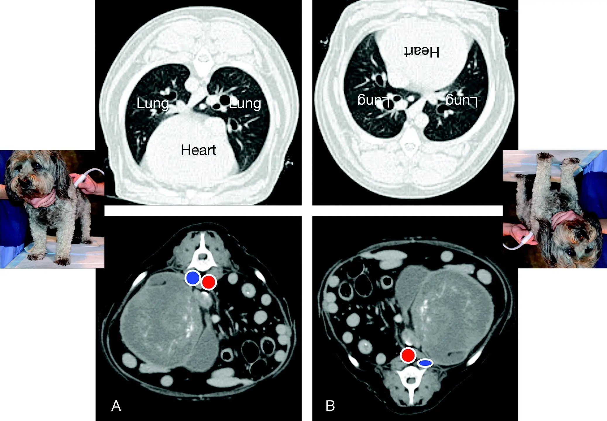
Figure 6.5. Never use dorsal recumbency without doing a Global FAST to assess stability. The use of dorsal recumbency on a hemodynamically fragile or unstable patient is potentially catastrophic. In (A) the computed tomography (CT) of a cross‐section of the thorax and abdomen is is compared to (B) in dorsal recumbency. Ventilation is changed from (A) to (B) by moving the most aerated, least perfused, least gravity‐dependent lung fields to a ventral and gravity‐dependent location. Moreover, the weight of the abdominal organs against the diaphragm further adversely affects ventilation. Venous return to the heart is also markedly changed from (A) to (B) in which the CVC ( blue circle ) next to the aorta ( red circle ) becomes compressed in dorsal recumbency from the weight of the abdominal organs. In hemodynamically fragile patients, there is real risk for decompensation.
Source: Reproduced with permission of Dr Gregory Lisciandro, Hill Country Veterinary Specialists and FASTVet.com, Spicewood, TX.
Every AFAST view is a “fan, rock cranially, and return to your starting position” of the probe by fanning through the target organ, rocking cranially, and then returning to your starting point (see Figures 6.2, 6.3, and 6.4).
DH view: Fan through the gallbladder in both directions until the gallbladder disappears in both directions, and then rock cranially to image the “cardiac bump” before returning to your starting point for one final look within the abdominal cavity.
SR view: Fan through the left kidney in both directions until the left kidney disappears in both directions, and then rock cranially to image the head of the spleen (fan on it too) before returning to your starting point of the left kidney for one final look. The kidney is retroperitoneal and the spleen peritoneal. The head of the spleen is reliably imaged in both dogs and cats. Figure 6.6. Correlation of AFAST acoustic windows with a dog in right lateral recumbency to an abdominal radiograph and CT. Sites are named by their target organs. In (A) the AFAST acoustic windows are labeled and correlate in (B) to a radiograph and (C) to computed tomography. The AFAST order is always the same: DH to SR to CC to HRU view. Note that the head is always to the left and tail to the right in both ultrasound and radiography. Computed tomography and radiograph courtesy of Dr Daniel Rodriguez, VETTEM, and Dr Jesús Paredes, CVM, Mexico City, Mexico.Source: Reproduced with permission of Dr Gregory Lisciandro, Hill Country Veterinary Specialists and FASTVet.com, Spicewood, TX. Figure 6.7. Longitudinal orientation for AFAST. (A) Kidney in longitudinal orientation, standard for all the AFAST views, because organs are more readily apparent in longitudinal over transverse scanning planes for most nonradiologist sonographers. (B) The same kidney as in (A) but in transverse orientation. The author keeps it simple – “fan (longitudinal planes), rock (direct the scanning plane cranial) and return to your starting point” is the AFAST mantra. Transverse orientation should be considered as an add‐on AFAST skill but is unnecessary for the detection of free intraabdominal fluid (Boysen et al. 2004). Computed tomography courtesy of Dr Daniel Rodriguez, VETTEM, Mexico City, Mexico.Source: Reproduced with permission of Dr Gregory Lisciandro, Hill Country Veterinary Specialists and FASTVet.com, Spicewood, TX.
CC view: Fan through the urinary bladder in both directions until the bladder disappears in both directions, and then rock cranially to image the “CC pouch”, its most gravity‐dependent region, and back to your starting point for one final look.
HRU view (right lateral recumbency): Fan through the small bowel and spleen in both directions and then rock cranially to image the cranial abdominal region before returning to the “HRU pouch”, its most gravity‐dependent region, for one final look. The left kidney may be viewed with increased depth through a single HR view, especially in cats and smaller dogs (Lisciandro 2014a; McMurray et al. 2016).
SRU view (left lateral recumbency): Fan through the small bowel and spleen in both directions and then rock cranially to image the cranial abdominal region before returning to the “SRU pouch”, its most gravity‐dependent region, for one final look. The right kidney may be viewed with increased depth through a single SR view, especially in cats and smaller dogs (Lisciandro 2014a).
HR5th bonus view (right lateral recumbency): Fan through the right kidney in both directions until the right kidney disappears in both directions and then rock cranially to image the right liver lobes before returning to your starting point of the right kidney for one final look. This view is generally unnecessary if the right kidney is imaged through the SR view.
SR5th bonus view (left lateral recumbency): Fan through the left kidney in both directions until the left kidney disappears in both directions and then rock cranially to image the spleen before returning to your starting point of the left kidney for one final look. This view is generally unnecessary if the left kidney is imaged through the HR view.
Pearl:For both those learning AFAST and experienced AFAST sonographers, only longitudinal (sagittal) orientation is necessary for the detection of free fluid at each view, with this standardization accelerating the learning process by building consistent sonographic expectations of the AFAST target organs. In time, transverse orientation may become an add‐on skill but it is unnecessary.
AFAST Diaphragmatico‐Hepatic View
| Questions Asked at the DH View | |
| Is there any free fluid in the abdominal (peritoneal) cavity? | Yes or no |
| How much free fluid is at the DH view using the AFAST AFS system? | 0, 1/2, 1 |
| Is there any pericardial effusion?Subjective amount? | Yes or noSmall (<1 cm), moderate (1–2 cm), large (>2 cm) (Candotti and Arntfield 2015) aMust be placed into clinical context |
| Is there any pleural effusion?Subjective amount? | Yes or noTrivial, mild, moderate, severe |
| What does the pulmonary‐diaphragmatic surface look like?Are there any lung lesions along the diaphragm? | Unremarkable (dry lung) or abnormalB‐lines and Vet BLUE B‐line scoring, shred sign, tissue sign, nodule sign, wedge sign |
| What does the gallbladder look like? | Unremarkable, halo sign (sonographic striation), abnormalities in its lumen or wall |
| What does the liver look like? | Unremarkable or abnormal |
| What do the caudal vena cava and its associated hepatic veins look like? | Unremarkable (bounce) or abnormal (FAT, flat): Tables 7.6and 36.3 and Figures 36.8 and 36.9 |
| Could I be mistaking an artifact or pitfall for pathology? | Know pitfalls and artifacts |
aThe AFAST target organ approach for parenchymal abnormalities is binary using “unremarkable” or “abnormal” to capture the case for additional imaging and confirmatory testing. Over time, more interpretative skills may be gained through experience and accompanied by additional POCUS study and training.
Читать дальшеИнтервал:
Закладка:
Похожие книги на «Point-of-Care Ultrasound Techniques for the Small Animal Practitioner»
Представляем Вашему вниманию похожие книги на «Point-of-Care Ultrasound Techniques for the Small Animal Practitioner» списком для выбора. Мы отобрали схожую по названию и смыслу литературу в надежде предоставить читателям больше вариантов отыскать новые, интересные, ещё непрочитанные произведения.


Обсуждение, отзывы о книге «Point-of-Care Ultrasound Techniques for the Small Animal Practitioner» и просто собственные мнения читателей. Оставьте ваши комментарии, напишите, что Вы думаете о произведении, его смысле или главных героях. Укажите что конкретно понравилось, а что нет, и почему Вы так считаете.