Point-of-Care Ultrasound Techniques for the Small Animal Practitioner
Здесь есть возможность читать онлайн «Point-of-Care Ultrasound Techniques for the Small Animal Practitioner» — ознакомительный отрывок электронной книги совершенно бесплатно, а после прочтения отрывка купить полную версию. В некоторых случаях можно слушать аудио, скачать через торрент в формате fb2 и присутствует краткое содержание. Жанр: unrecognised, на английском языке. Описание произведения, (предисловие) а так же отзывы посетителей доступны на портале библиотеки ЛибКат.

- Название:Point-of-Care Ultrasound Techniques for the Small Animal Practitioner
- Автор:
- Жанр:
- Год:неизвестен
- ISBN:нет данных
- Рейтинг книги:5 / 5. Голосов: 1
-
Избранное:Добавить в избранное
- Отзывы:
-
Ваша оценка:
Point-of-Care Ultrasound Techniques for the Small Animal Practitioner: краткое содержание, описание и аннотация
Предлагаем к чтению аннотацию, описание, краткое содержание или предисловие (зависит от того, что написал сам автор книги «Point-of-Care Ultrasound Techniques for the Small Animal Practitioner»). Если вы не нашли необходимую информацию о книге — напишите в комментариях, мы постараемся отыскать её.
New chapters in
cover ultrasound-guided nerve blocks, musculoskeletal, brain imaging, and applications of focused ultrasound techniques in cats, exotics and marine mammals—making it an essential purchase for veterinarians wanting to incorporate point-of-care ultrasound techniques into their veterinary practices.
Presents a standardized approach to point-of-care ultrasound as an extension of the physical exam, including trauma, non-trauma, and monitoring applications Includes coverage of new techniques for focused ultrasound exams, including lung, anesthesia and ultrasound guided nerve blocks, transcranial brain imaging, musculoskeletal, volume status evaluation, and rapid assessment for treatable forms of shock Adds cats, exotic and wildlife mammals, and marine mammals to the existing canine coverage Emphasizes the integration of point-of-care ultrasound techniques for optimizing patient care and accurate patient assessment Offers access to a companion website with supplementary video clips showing many clinically relevant didactic examples The second edition of
is an excellent resource for veterinary practitioners, ranging from the general practitioner to nearly all clinical specialists, including internal medicine, oncology, cardiology, emergency and critical care, anesthesiology, ophthalmology, exotics, and zoo medicine specialists, and veterinary students.
Point-of-Care Ultrasound Techniques for the Small Animal Practitioner — читать онлайн ознакомительный отрывок
Ниже представлен текст книги, разбитый по страницам. Система сохранения места последней прочитанной страницы, позволяет с удобством читать онлайн бесплатно книгу «Point-of-Care Ultrasound Techniques for the Small Animal Practitioner», без необходимости каждый раз заново искать на чём Вы остановились. Поставьте закладку, и сможете в любой момент перейти на страницу, на которой закончили чтение.
Интервал:
Закладка:
AFAST misses peritonitis in some dehydrated and hypotensive patients, so should always be repeated post resuscitation and rehydration, with continued serial (repeat) exams as long as necessary until assured that the patient is not a surgical candidate.
The AFAST AFS system is effective in cats, but cats as a species lack a large splenic blood reservoir and thus felines generally do not survive large‐volume bleeds, and larger volume intraabdominal effusions are more likely to be due to uroabdomen.
Understand that serial (repeat) exams using the Global FAST approach is an even better strategy because the thorax, heart, and lung are also surveyed.
Cannot replace a complete detailed abdominal ultrasound.
Cannot replace proper training.
Indications
All blunt and penetrating trauma cases as standard of care for screening for indirect evidence of intraabdominal and retroperitoneal injury.
All collapsed both recovered and unrecovered cases with unexplained hypotension, tachycardia, or mentation changes.
All anemic cases.
All “ain’t doing right” (ADR) cases.
All postinterventional cases including intravenous fluid therapy, postsurgical cases, postpercutaneous procedures, etc. that are at risk for bleeding, infections, vascular complications.
All peritonitis suspects, including acute abdomen, for expedient diagnosis through the detection of free fluid (and subsequent sampling, fluid analysis testing as deemed appropriate) and free air.Preanesthetic screening test.Extension of the physical exam for patients presenting once or twice a year for routine care.
Objectives
Be able to perform the AFAST views and apply its fluid scoring system.
Be able to recognize basic abnormalities of the AFAST target organs in each of its views.
Know how to assign an AFS during AFAST.
Be able to recognize sonographic striation of the gallbladder wall, referred to as the “halo effect” or “double rim effect” or “halo sign,” that can be present in both dogs and cats from different causes.
Be able to recognize retroperitoneal free fluid and distinguish it from intraabdominal fluid.
Know common artifacts at each AFAST view.
Know the major pitfalls at each AFAST view.
Know how to do a focused (POCUS) spleen examination and its importance following the completion of AFAST and after assigning an AFS.
Understand why best practice would be to add on the Global FAST approach for all AFAST and POCUS exams (abdomen, thorax, eye, brain, musculoskeletal, etc.) to make sure that forms of peritonitis and pleuritis, presence of bleeding, and cardiac and pulmonary conditions are not being missed.
How to Perform an AFAST Exam
Ultrasound Settings and Probe Preferences
Standard abdominal settings, presets, with adequate depth to be able to visualize the target organs of each AFAST view.
Curvilinear (microconvex) probe with a range of 5–10 MHz and a maximum depth of 12–15 cm is acceptable for most dogs and cats.
Optimizing Image Quality and Probe–Skin Contact
Hair is generally not shaved but rather parted for the best probe–skin contact with the use of isopropyl alcohol, and/or alcohol‐based hand sanitizer, and/or acoustic coupling gel. The author prefers the use of minimal amounts of isopropyl alcohol to effectively part the hair followed by alcohol‐based hand sanitizer. The strategy is much less noxious to your patient by minimizing the cold wetness of isopropyl alcohol and its fumes, especially when moved to an oxygen cage. Alcohol‐based hand sanitizer has the added benefit of being easily wiped off and it also evaporates quickly. Isopropyl alcohol should not be used if electrical defibrillation is anticipated because it poses a burn/fire hazard. The clinician should be aware that isopropyl alcohol may cause probe head damage (see Figure 4.13).
Pearl:By not shaving (or limiting shaving to small acoustic windows), the cosmetic appearance of the patient is preserved (happier clients), the exam time is lessened, and imaging quality is sufficient with most newer ultrasound machines (median AFAST time <3–3.5 minutes) (Lisciandro et al. 2009; Lisciandro 2011, 2012; Boysen and Lisciandro 2013).
Pearl:Maximize image quality by parting the hair and getting the probe head and its acoustic coupling medium in direct contact with the patient's skin to minimize air trapping, which is your imaging enemy because ultrasound does not transmit through air. Placing the probe head on a wetted mat of hair full of trapped air will produce a poor image (see Figures 5.1and 5.2).
Pearl:Hold the probe in a way that is most comfortable while being able to fan toward and away from the table top while maintaining a longitudinal (sagittal) plane in patients placed in lateral recumbency. Holding the probe like a pencil for many who have learned to scan patients in dorsal recumbency usually becomes problematic at the DH view for the caudal vena cava. Holding the probe on top and keeping the thumb on the probe marker and a finger out to prevent drifting works best.
Patient Positioning
Lateral Recumbency
Right lateral (RL) recumbency is generally preferred over left lateral (LL) recumbency for AFAST because RL recumbency is standard positioning for electrocardiographic and echocardiographic evaluation ( Figures 6.2and 6.3; see also Figure 6.1). Moreover, the left kidney (a window into the retroperitoneal space) at the AFAST SR view and the gallbladder at the DH view (by directing the probe in the gravity‐dependent region or toward the tabletop) are readily imaged. RL recumbency over LL is preferred positioning for abdominocentesis because anatomically the spleen lies more on the left side of dogs and cats. LL recumbency may be used in cases in which injury prohibits RL positioning, or the right retroperitoneal space warrants imaging. In a more recent study evaluating RL versus LL recumbency, abdominal FAST time was shorter in LL recumbency and both kidneys were more reliably imaged (McMurray et al. 2016). However, this is probably due to training bias, as we could argue the same in RL recumbency (Lisciandro 2014a,b), and moreover, TFAST echo views are more easily acquired in RL recumbency. When combining AFAST and TFAST, RL recumbency is clearly advantageous. AFAST on a cat is shown in Chapter 39. See Chapters 17, 19, and 20for echo views.
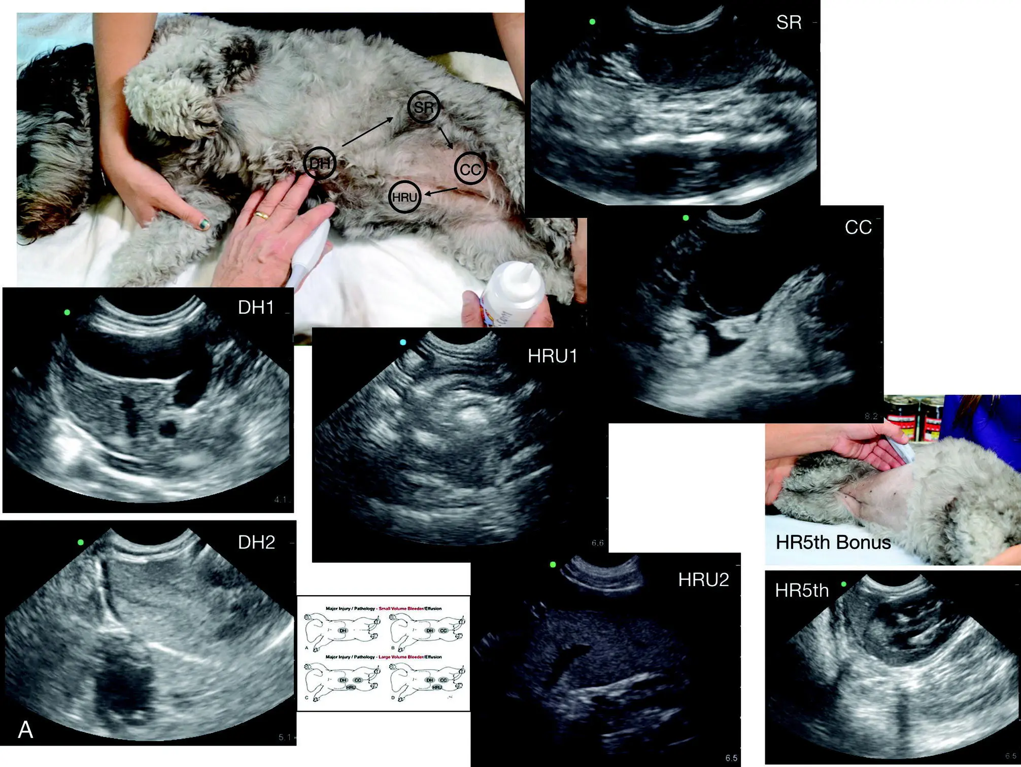
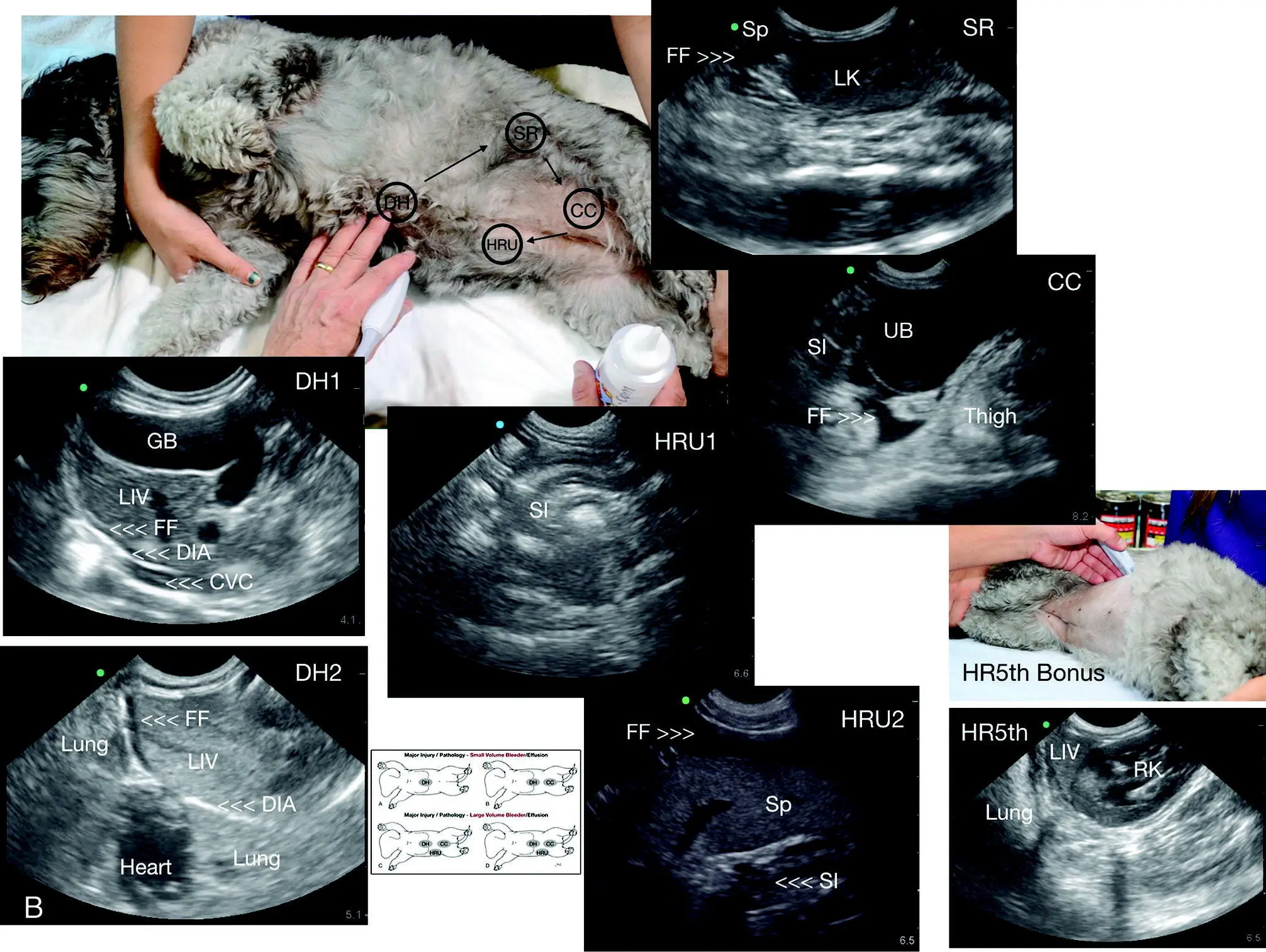
Figure 6.2. AFAST and its five views performed in right lateral recumbency in a dog. (A) AFAST unlabeled and (B) AFAST labeled. The order is always the same as follows: DH to SR to CC to HRU to HR5th bonus view. The final HR5th bonus view is not part of the abdominal fluid score. The AFAST images and their proportionality should look nearly the same regardless of positioning. DH, diaphragmatico‐hepatic view; DH1, DH 1 of 2 views; DH2, DH 2 of 2 views; SR, spleno‐renal view; CC, cysto‐colic view; HRU, hepato‐renal umbilical view; HR5th, hepato‐renal 5th bonus view; HRU1, 1 of 2 views; HRU2, 2 of 2 views. DIA, diaphragm; FF, free fluid; GB, gallbladder; LIV, liver; LK, left kidney; RK, right kidney; SI, small intestine. AFAST views are nearly identical no matter the positioning because the respective target organs are imaged with the same methodology.
Читать дальшеИнтервал:
Закладка:
Похожие книги на «Point-of-Care Ultrasound Techniques for the Small Animal Practitioner»
Представляем Вашему вниманию похожие книги на «Point-of-Care Ultrasound Techniques for the Small Animal Practitioner» списком для выбора. Мы отобрали схожую по названию и смыслу литературу в надежде предоставить читателям больше вариантов отыскать новые, интересные, ещё непрочитанные произведения.


Обсуждение, отзывы о книге «Point-of-Care Ultrasound Techniques for the Small Animal Practitioner» и просто собственные мнения читателей. Оставьте ваши комментарии, напишите, что Вы думаете о произведении, его смысле или главных героях. Укажите что конкретно понравилось, а что нет, и почему Вы так считаете.