Point-of-Care Ultrasound Techniques for the Small Animal Practitioner
Здесь есть возможность читать онлайн «Point-of-Care Ultrasound Techniques for the Small Animal Practitioner» — ознакомительный отрывок электронной книги совершенно бесплатно, а после прочтения отрывка купить полную версию. В некоторых случаях можно слушать аудио, скачать через торрент в формате fb2 и присутствует краткое содержание. Жанр: unrecognised, на английском языке. Описание произведения, (предисловие) а так же отзывы посетителей доступны на портале библиотеки ЛибКат.

- Название:Point-of-Care Ultrasound Techniques for the Small Animal Practitioner
- Автор:
- Жанр:
- Год:неизвестен
- ISBN:нет данных
- Рейтинг книги:5 / 5. Голосов: 1
-
Избранное:Добавить в избранное
- Отзывы:
-
Ваша оценка:
Point-of-Care Ultrasound Techniques for the Small Animal Practitioner: краткое содержание, описание и аннотация
Предлагаем к чтению аннотацию, описание, краткое содержание или предисловие (зависит от того, что написал сам автор книги «Point-of-Care Ultrasound Techniques for the Small Animal Practitioner»). Если вы не нашли необходимую информацию о книге — напишите в комментариях, мы постараемся отыскать её.
New chapters in
cover ultrasound-guided nerve blocks, musculoskeletal, brain imaging, and applications of focused ultrasound techniques in cats, exotics and marine mammals—making it an essential purchase for veterinarians wanting to incorporate point-of-care ultrasound techniques into their veterinary practices.
Presents a standardized approach to point-of-care ultrasound as an extension of the physical exam, including trauma, non-trauma, and monitoring applications Includes coverage of new techniques for focused ultrasound exams, including lung, anesthesia and ultrasound guided nerve blocks, transcranial brain imaging, musculoskeletal, volume status evaluation, and rapid assessment for treatable forms of shock Adds cats, exotic and wildlife mammals, and marine mammals to the existing canine coverage Emphasizes the integration of point-of-care ultrasound techniques for optimizing patient care and accurate patient assessment Offers access to a companion website with supplementary video clips showing many clinically relevant didactic examples The second edition of
is an excellent resource for veterinary practitioners, ranging from the general practitioner to nearly all clinical specialists, including internal medicine, oncology, cardiology, emergency and critical care, anesthesiology, ophthalmology, exotics, and zoo medicine specialists, and veterinary students.
Point-of-Care Ultrasound Techniques for the Small Animal Practitioner — читать онлайн ознакомительный отрывок
Ниже представлен текст книги, разбитый по страницам. Система сохранения места последней прочитанной страницы, позволяет с удобством читать онлайн бесплатно книгу «Point-of-Care Ultrasound Techniques for the Small Animal Practitioner», без необходимости каждый раз заново искать на чём Вы остановились. Поставьте закладку, и сможете в любой момент перейти на страницу, на которой закончили чтение.
Интервал:
Закладка:
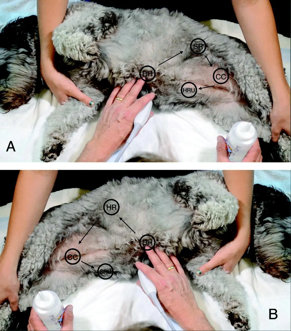
Figure 6.1. AFAST on a dog in right and left lateral recumbency. In (A) AFAST is shown on a dog in right lateral recumbency and in (B) left lateral recumbency. Sites are named by their target organs. The AFAST order is always the same. In right lateral, (1) DH view, (2) SR view, (3) CC view, (4) HRU view. In left lateral recumbency, (1) DH view, (2) HR view, (3) CC view, (4) SRU view. These AFAST views are part of the abdominal fluid scoring (AFS) system and the order ends at the most gravity‐dependent view where abdominocentesis is likely to be performed in higher‐scoring patients. Note that the 5th AFAST bonus view is not shown in these images. The AFAST views are nearly identical sonographically no matter the positioning (lateral recumbency versus standing‐sternal). AFAST target organs are imaged in the same standardized manner regarding probe maneuvering with the “fan, rock (cranially) and return to your starting point” approach.
Source: Reproduced with permission of Dr Gregory Lisciandro, Hill Country Veterinary Specialists and FASTVet.com, Spicewood, TX.
Table 6.1. Changes in methodology from FAST to AFAST.
Source: Reproduced with permission of Dr Gregory Lisciandro, Hill Country Veterinary Specialists, FASTVet.com, Spicewood, TX.
| Parameters | FAST (Boysen 2004) | AFAST (Lisciandro et al. 2009) |
|---|---|---|
| Shaving patient | Shaving | No shaving |
| Primary probe orientation | Longitudinal and transverse | Only longitudinal |
| Primary probe maneuver | Sliding, rotating and sweeping | Fanning and rocking |
| Main probe direction | Toward spine | Gravity‐dependent pouches |
| Lateral a | Left | Right |
| Fluid scoring | No | Yes |
| Naming acoustic views | External locations | Target organs |
| Timing of examination (median time presentation to ultrasound examination) | Post resuscitation (median 1 hour) | Presentation and serially post resuscitation (median <5 minutes) |
aLateral recumbency was a brilliant proposition, being markedly safer than dorsal recumbency (see Figure 6.5).
More recently, a human study showed that in people with prehospital hypotension, the only intervention that prevented the “crump factor,” the phenomenon of a patient decompensating unexpectedly, was the liberal use of FAST examinations (Bilello et al. 2011). The upshot is veterinarians have a better tool, the AFAST and its applied fluid scoring system, to determine within minutes of presentation or during hospitalized care when patients are becoming unstable, to not only “see” if the patient is positive or negative for free fluid, but also the degree of bleeding (or effusion) by easily calculating the patient's AFS (0–4 scale). AFAST and AFS are the missing link to traditional trauma, triage and tracking algorithms, and by adding the target organ approach, a huge amount of clinical information is easily gained within minutes, when minutes count .
The comparison of the original FAST to the subsequent AFAST study is fascinating and some important differences are noted (see Table 6.1) (Boysen et al. 2004; Lisciandro et al. 2009). Remarkably, degree of trauma was very similar, including numbers of pelvic fractures and pneumothoraces ( Table 6.2), suggesting that the overall degree of trauma between studies was comparable and thus inferences may be loosely drawn (Boysen et al. 2004; Lisciandro et al. 2009).
One involves the case management and decision making for blood transfusion(s), and that knowing if the dog at triage was AFS positive affected fluid therapy administration strategies. In other words, intravenous fluid resuscitation was likely titrated more closely to low normal endpoints, such as mean arterial pressure, thus mitigating exacerbation of hemorrhage by lessening the probability of “popping the clot” and diluting clotting factors through overresuscitation in bleeding dogs (Lisciandro et al. 2009). The differences in median time from trauma to FAST/AFAST, median time presentation to FAST/AFAST (240 versus <5 minutes), and numbers of transfusions FAST/AFAST (9 versus 3) support this conclusion. AFAST was performed as part of the physical exam versus FAST, which was a second line test after initial assessment, intravenous fluid resuscitation, and blind abdominocentesis possibly to the dog's detriment by the much higher positive rate (45% versus 27%).
The original FAST study lacked a fluid scoring system, and as simple as the AFAST system is, with a range of 0–4 (AFS 0 negative all AFAST views to a maximum of four being positive for fluid at all four AFAST views), AFS provides an effective tool for decision making (see Table 7.3). This decision making, ranging from intravenous fluid resuscitation strategies to administration of blood transfusion products to the need for exploratory surgery, importantly carries the potential to improve outcome and decrease complications, as shown in people (see Chapter 7) (Blackbourne et al. 2004; Ollerton et al. 2006; Bilello et al. 2011).
Table 6.2. Comparison of FAST and AFAST in dogs.
Source: Reproduced with permission of Dr Gregory Lisciandro, Hill Country Veterinary Specialists, FASTVet.com, Spicewood, TX.
| Parameters | FAST (Boysen et al. 2004) | AFAST (Lisciandro et al. 2009) |
|---|---|---|
| Primary presentations | 65% | 96% |
| FAST positive cases | 45% | 27% |
| Low‐scoring (AFS 1 and 2) small‐volume bleeders | NA | 13 |
| High‐scoring (AFS 3 and 4) large‐volume bleeders | NA | 14 |
| Cases of abdominocentesis prior to AFAST/FAST examination | 16 | 0 |
| Median time trauma to AFAST/FAST examination | 240 minutes | 60 minutes |
| Median time presentation to AFAST/FAST examination | 60 minutesFAST was a secondary evaluation post initial resuscitation | <5 minutesAFAST was a first‐line screening test |
| Median time for AFAST/FAST examination | 6 minutesShaved sites | 3 minutesNo shaving |
| Pelvic fractures | 20 | 22 |
| Pneumothorax | 21 | 22 |
| Appendicular fractures | 15 | 25 |
| Diaphragmatic hernia | NA | 2 |
| Number of blood transfusions | 9 | 3 |
Of note, dogs with pneumothorax (55%), pelvic fractures (40%), and high alanine transaminase (ALT) (>400 U/L) were also more likely to concurrently have or develop hemoabdomen detected by either their initial or serial AFAST examinations than dogs without these findings (Lisciandro et al. 2009; Lisciandro 2014c). The serial use of AFAST is helpful in determining the integrity of the urinary bladder, estimating urinary bladder volume and urine output during resuscitation (Lisciandro et al. 2009; Lisciandro 2011; Lisciandro and Fosgate 2017). Both FAST and AFAST studies documented that when the urinary bladder was imaged with an expected, smooth, rounded contour, it was unlikely to be ruptured, holding advantages over traditional means of palpation, characterization of urine post micturition, and plain radiography (Boysen et al. 2004; Lisciandro et al. 2009; Boysen and Lisciandro 2013).
More recently, a urinary bladder estimation formula for use during AFAST has been published and provides a noninvasive way to estimate urine output when serial calculations are made over time (Lisciandro and Fosgate 2017). AFAST additionally remains useful to survey for intrathoracic trauma, pleural and pericardial effusion, and lung conditions, through the acoustic window of the liver and gallbladder by imaging cranial to the diaphragm in every patient, dependent on patient size, coupled with depth limitations of the ultrasound machine (Boysen et al. 2004; Lisciandro et al. 2009; Lisciandro 2011, 2016a; McMurray et al. 2016).
Читать дальшеИнтервал:
Закладка:
Похожие книги на «Point-of-Care Ultrasound Techniques for the Small Animal Practitioner»
Представляем Вашему вниманию похожие книги на «Point-of-Care Ultrasound Techniques for the Small Animal Practitioner» списком для выбора. Мы отобрали схожую по названию и смыслу литературу в надежде предоставить читателям больше вариантов отыскать новые, интересные, ещё непрочитанные произведения.


Обсуждение, отзывы о книге «Point-of-Care Ultrasound Techniques for the Small Animal Practitioner» и просто собственные мнения читателей. Оставьте ваши комментарии, напишите, что Вы думаете о произведении, его смысле или главных героях. Укажите что конкретно понравилось, а что нет, и почему Вы так считаете.