Katherine Kula - Cephalometry in Orthodontics
Здесь есть возможность читать онлайн «Katherine Kula - Cephalometry in Orthodontics» — ознакомительный отрывок электронной книги совершенно бесплатно, а после прочтения отрывка купить полную версию. В некоторых случаях можно слушать аудио, скачать через торрент в формате fb2 и присутствует краткое содержание. Жанр: unrecognised, на английском языке. Описание произведения, (предисловие) а так же отзывы посетителей доступны на портале библиотеки ЛибКат.
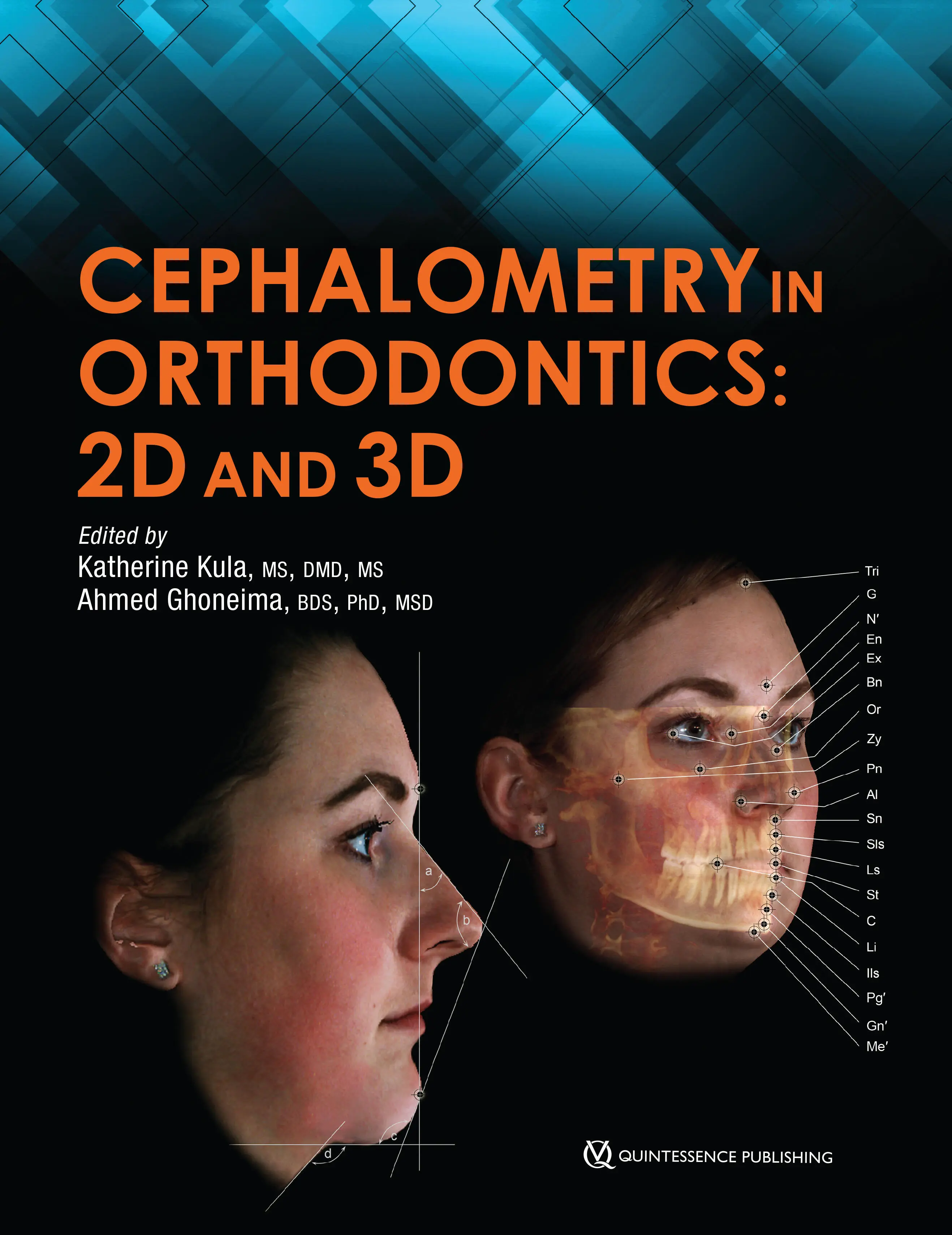
- Название:Cephalometry in Orthodontics
- Автор:
- Жанр:
- Год:неизвестен
- ISBN:нет данных
- Рейтинг книги:4 / 5. Голосов: 1
-
Избранное:Добавить в избранное
- Отзывы:
-
Ваша оценка:
Cephalometry in Orthodontics: краткое содержание, описание и аннотация
Предлагаем к чтению аннотацию, описание, краткое содержание или предисловие (зависит от того, что написал сам автор книги «Cephalometry in Orthodontics»). Если вы не нашли необходимую информацию о книге — напишите в комментариях, мы постараемся отыскать её.
Cephalometry in Orthodontics — читать онлайн ознакомительный отрывок
Ниже представлен текст книги, разбитый по страницам. Система сохранения места последней прочитанной страницы, позволяет с удобством читать онлайн бесплатно книгу «Cephalometry in Orthodontics», без необходимости каждый раз заново искать на чём Вы остановились. Поставьте закладку, и сможете в любой момент перейти на страницу, на которой закончили чтение.
Интервал:
Закладка:
Image reconstruction
Once the appropriate scan is captured, the acquisition computer will generate data as a multiplanar reformation (MPR) providing images in the sagittal, coronal, and axial planes. While this data is captured in a 3D matrix, the MPR images are sequential 2D images. Further reconstruction is needed to generate useful 3D data. Additionally, conventional 2D images—cephalograms and panoramic radiographs—can be derived from the 3D data. As previously stated, there is no magnification in the CBCT-derived cephalograms as opposed to conventionally acquired cephalograms. Numerous studies have quantified the differences between landmark identification in conventional versus CBCT-derived cephalograms. 13–15For the most part, the identification of landmarks has been comparable between the two. Some of the outcomes of these studies suggest that some “landmarks” visualized in two dimensions are not necessarily point landmarks in 3D data. Research is ongoing to better elucidate cephalometric landmarks in 3D data sets.
References
1.Broadbent BH. A new x-ray technique and its application to orthodontia. Angle Orthod 1931;1:45–66.
2.Jacobson A, Jacobson RL (eds). Radiographic Cephalometry: From Basics to 3-D Imaging, ed 2. Chicago: Quintessence, 2006.
3.American Dental Association. Dental Radiographic Examinations: Recommendations For Patient Selection And Limiting Radiation Exposure [PDF]. http://www.ada.org/en/∼/media/ADA/Member%20Center/FIles/Dental_Radiographic_Examinations_2012. Accessed 22 May 2017.
4.Brand JW, Gibbs SJ, Edwards M, et al. Radiation Protection in Dentistry [Report 145]. Bethesda, MD: National Council on Radiation Protection and Measurements, 2003.
5.Ludlow JB, Davies-Ludlow LE, White SC. Patient risk related to common dental radiographic examinations: The impact of 2007 International Commission on Radiological Protection recommendations regarding dose calculation. J Am Dent Assoc 2008;139:1237–1243.
6.Ludlow JB, Davies-Ludlow LE, Brooks SL, Howerton WB. Dosimetry of 3 CBCT devices for oral and maxillofacial radiology: CB Mercuray, NewTom 3G and i-CAT. Dentomaxillofac Radiol 2006;35:219–226.
7.Rottke D, Grossekettler L, Sawada K, Poxleitner P, Schulze D. Influence of lead apron shielding on absorbed doses from panoramic radiography. Dentomaxillofac Radiol 2013;42:20130302.
8.Chadwick J, Prentice RN, Major PW, Lam EW. Image distortion and magnification of 3 digital CCD cephalometric systems. Oral Surg Oral Med Oral Pathol Oral Radiol Endod 2009;107:105–112.
9.McClure SR, Sadowsky PL, Ferreira A, Jacobson A. Reliability of digital versus conventional cephalometric radiology: A comparative evaluation of landmark identification error. Semin Orthod 2005;11:98–110.
10.White SC, Pharoah MJ. Oral Radiology Principles and Interpretation, ed 7. St Louis: Mosby/Elsevier, 2014.
11.Swennen G, Schutyser F, Hausamen J. Three-Dimensional Cephalometry: A Color Atlas and Manual. Berlin: Springer-Verlag, 2006.
12.Clinical recommendations regarding use of cone beam computed tomography in orthodontics. Position statement by the American Academy of Oral and Maxillofacial Radiology. Oral Surg Oral Med Oral Pathol Oral Radiol 2013;116:238–257 [erratum 2013;116:661].
13.Park JW, Kim N, Chang YI. Comparison of landmark position between conventional cephalometric radiography and CT scans projected to midsagittal plane. Korean J Orthod 2008;38:426–436.
14.Chien PC, Parks ET, Eraso F, Hartsfield JK, Roberts WE, Ofner S. Comparison of reliability in anatomical landmark identification using two-dimensional digital cephalometrics and three-dimensional cone beam computed tomography in vivo. Dentomaxillofac Radiol 2009;38:262–273.
15.Zamora N, Llamas JM, Cibrián R, Gandia JL, Paredes V. Cephalometric measurements from 3D reconstructed images compared with conventional 2D images. Angle Orthod 2011;81:856–864.
Конец ознакомительного фрагмента.
Текст предоставлен ООО «ЛитРес».
Прочитайте эту книгу целиком, купив полную легальную версию на ЛитРес.
Безопасно оплатить книгу можно банковской картой Visa, MasterCard, Maestro, со счета мобильного телефона, с платежного терминала, в салоне МТС или Связной, через PayPal, WebMoney, Яндекс.Деньги, QIWI Кошелек, бонусными картами или другим удобным Вам способом.
Интервал:
Закладка:
Похожие книги на «Cephalometry in Orthodontics»
Представляем Вашему вниманию похожие книги на «Cephalometry in Orthodontics» списком для выбора. Мы отобрали схожую по названию и смыслу литературу в надежде предоставить читателям больше вариантов отыскать новые, интересные, ещё непрочитанные произведения.


Обсуждение, отзывы о книге «Cephalometry in Orthodontics» и просто собственные мнения читателей. Оставьте ваши комментарии, напишите, что Вы думаете о произведении, его смысле или главных героях. Укажите что конкретно понравилось, а что нет, и почему Вы так считаете.