Katherine Kula - Cephalometry in Orthodontics
Здесь есть возможность читать онлайн «Katherine Kula - Cephalometry in Orthodontics» — ознакомительный отрывок электронной книги совершенно бесплатно, а после прочтения отрывка купить полную версию. В некоторых случаях можно слушать аудио, скачать через торрент в формате fb2 и присутствует краткое содержание. Жанр: unrecognised, на английском языке. Описание произведения, (предисловие) а так же отзывы посетителей доступны на портале библиотеки ЛибКат.
- Название:Cephalometry in Orthodontics
- Автор:
- Жанр:
- Год:неизвестен
- ISBN:нет данных
- Рейтинг книги:4 / 5. Голосов: 1
-
Избранное:Добавить в избранное
- Отзывы:
-
Ваша оценка:
Cephalometry in Orthodontics: краткое содержание, описание и аннотация
Предлагаем к чтению аннотацию, описание, краткое содержание или предисловие (зависит от того, что написал сам автор книги «Cephalometry in Orthodontics»). Если вы не нашли необходимую информацию о книге — напишите в комментариях, мы постараемся отыскать её.
Cephalometry in Orthodontics — читать онлайн ознакомительный отрывок
Ниже представлен текст книги, разбитый по страницам. Система сохранения места последней прочитанной страницы, позволяет с удобством читать онлайн бесплатно книгу «Cephalometry in Orthodontics», без необходимости каждый раз заново искать на чём Вы остановились. Поставьте закладку, и сможете в любой момент перейти на страницу, на которой закончили чтение.
Интервал:
Закладка:
All radiographic imaging is predicated by differential absorption of the x-ray beam by the region of interest. Multiple exposure factors need to be adjusted depending on the patient’s size and bone density. These exposure factors—kVp, mA, and exposure time—are discussed below.
Kilovoltage peak (kVp)
Kilovoltage refers to the energy or penetrating power of the x-ray beam. Peak simply refers to the highest energy in a polyenergetic beam. The mean beam energy is generally considered to be one-third of the peak. As kilovoltage increases, the beam energy and penetrating power also increase. Conversely, lower kilovoltage produces lower beam energy and generates photons that are more likely to be absorbed by the region of interest. Kilovoltage should be increased for patients with large or dense facial bones and decreased for patients with small or less dense facial bones. Most cephalometric units function in a range of 70 to 90 kVp. CBCT units function between 90 and 120 kVp.
Milliamperage (mA) and exposure time
Milliamperage is the determinant of the tube current and controls the number of photons of x-radiation that are produced in the tube head. It is often adjusted because of the density of the soft tissues of the head and neck, and it is often reported together with exposure time (seconds). Both mA and exposure time have a direct relationship with output. It is important to remember that mAs = mAs. This simply means that as long as the product of mA and exposure time remains constant, the output of the machine will also remain constant. For example, if mA is 5 and the exposure time is 0.5 seconds, the mAs is 2.5. If the milliamperage is 10, the exposure time would need to be decreased to 0.25 seconds to maintain output.
Collimation/Soft Tissue Filtration
The shape and size of the primary beam of x-radiation is controlled by collimation of the beam. The radiation beam should be collimated to the size of the image receptor. Collimating the beam to the size of the receptor decreases the exposure and dose received by the patient. The cephalometric unit should have a mechanism to filter the soft tissues of the nose and lips. Generally, the x-ray beam is generated to penetrate bony structures and will burn out soft tissue structures. The soft tissue filter attenuates the beam prior to it contacting the patient, providing some radiation protection to the patient and decreasing the energy of the beam so that the soft tissues will be enhanced in the cephalogram.
Image Distortion/Magnification
A 2D cephalogram will contain some image distortion in the form of differential magnification because a 3D object is being imaged using diverging radiation rays. Structures away from the image receptor will be magnified much more than objects that are positioned close to the image receptor. Magnification is calculated by dividing the distance from the source of radiation to the image receptor (SID) by the distance from the source to the object of interest (SOD). Based on this calculation, it is easy to see that the right and left sides of the skull will be different sizes in a lateral cephalogram. Because there is a potential for distortion just from projection geometry, it is essential to either record the distance from the center of the cephalostat to the image receptor or to establish a standard distance when evaluating sequential cephalograms.
Image Receptors
Film-based systems
Film-based cephalometry employs indirect-exposure radiographic film positioned between two intensifying screens. Intensifying screens convert x-radiation into light. Indirect-exposure film is more sensitive to light than it is to x-radiation. As a consequence, the use of intensifying screens and indirect-exposure radiographic film allows for very low exposure times. Different types of intensifying screens emit different wavelengths of light. Care must be taken to match the spectral sensitivity of the film to the light emitted from the intensifying screens. Rare-earth intensifying screens emit either green or blue light. Traditional or par screens emit purple light. If intensifying screens emit a green light, you must use green-sensitive film to produce an acceptable image. Figure 2-4shows examples of film-based lateral and PA cephalograms.
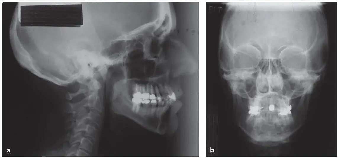
Fig 2-4Examples of film-based lateral (a) and PA (b) cephalograms.
Darkroom procedures
As with any film-based imaging, chemical processing must be performed to convert the latent or chemical image into a visible image. All film processors go through the same steps: development, fixation rinse, and drying. The function of the developer is to convert the silver ions on the film into metallic silver. The process of fixation stops the development process and renders an archival image. The quality of correctly processed film images will not change over time; unfortunately, however, most images are not correctly processed. Quality assurance in the darkroom is essential for quality film-based imaging. Quality assurance pertains to many components of the darkroom—lighting as well as the activity of the processing chemistry. Processing chemistry must be replenished every day. Developer and fixer activity diminish due to workload rather than time, so it is essential to have an ongoing program of assessing the activity of the chemistry. Finally, because direct- and indirect-exposure films require different safelight filters, make sure that the safelight in the darkroom does not fog the film prior to processing.
Digital systems
Digital receptors are divided into two groups: indirect digital and direct digital systems. PSP plates are considered to be indirect digital sensors, whereas CCD and complementary metal oxide semiconductors (CMOS) are considered to be direct digital sensors. There are many advantages to the use of digital receptors ( Box 2-1). In addition to reduced exposure, a huge advantage is the ability to enhance images once they are captured. Electronic image storage and image transmission are also advantages of digital receptors compared with film-based systems. While automated analysis can be performed on a film-based image that is converted into a digital image through a process called analog to digital conversion, data is lost in the process, whereas with digital images automated analysis can be performed without any lost data. Staff efficiency is also increased with the use of digital receptors: There is no downtime spent waiting for the image to be processed.
Box 2-1Advantages and disadvantages of digital cephalometric imaging
Advantages
•Exposure reduction
•Image enhancement
•Digital image storage
•Automated analysis
•Image transmission
•Increased staff efficiency
Disadvantages
•High initial cost
•Differences in projection geometry
There are two potential disadvantages to the use of digital receptors: (1) cost and (2) differences in projection geometry (see Box 2-1). There is no doubt that the initial cost of a digital cephalometric unit is higher than the cost of a film-based system. However, when one factors in the costs of film, processing chemistry, and lost staff efficiency, the difference in initial cost is recouped rather quickly. The issue of differences in projection geometry is covered in the section entitled “Digital Versus Conventional Cephalometry.”
PSP plates
PSP plates are image receptors that convert x-radiation into an electrical charge contained within the imaging plate. PSP plates come in all sizes (from 0 to an 8 × 10-inch plate) for cephalometry. The PSP plate is placed in the 8 × 10-inch cassette with the intensifying screens removed. The imaging plate is coated with europium-activated barium fluorohalide. The electronic information is converted into a visible image by subjecting the phosphor plate to a helium-neon laser. The PSP plate in turn emits a blue-violet light at 400 nm that is captured by the scanner and converted into a digital image. As a final step, the plate must be exposed to white light to remove the latent image; this step is performed in most scanners automatically. PSP plates are considered to be indirect digital images because the x-ray data is captured as analog or continuous data and converted into digital data in the scanner. This is the same reason that film-based images that are scanned as digital images are considered to be indirect digital images.
Читать дальшеИнтервал:
Закладка:
Похожие книги на «Cephalometry in Orthodontics»
Представляем Вашему вниманию похожие книги на «Cephalometry in Orthodontics» списком для выбора. Мы отобрали схожую по названию и смыслу литературу в надежде предоставить читателям больше вариантов отыскать новые, интересные, ещё непрочитанные произведения.


Обсуждение, отзывы о книге «Cephalometry in Orthodontics» и просто собственные мнения читателей. Оставьте ваши комментарии, напишите, что Вы думаете о произведении, его смысле или главных героях. Укажите что конкретно понравилось, а что нет, и почему Вы так считаете.