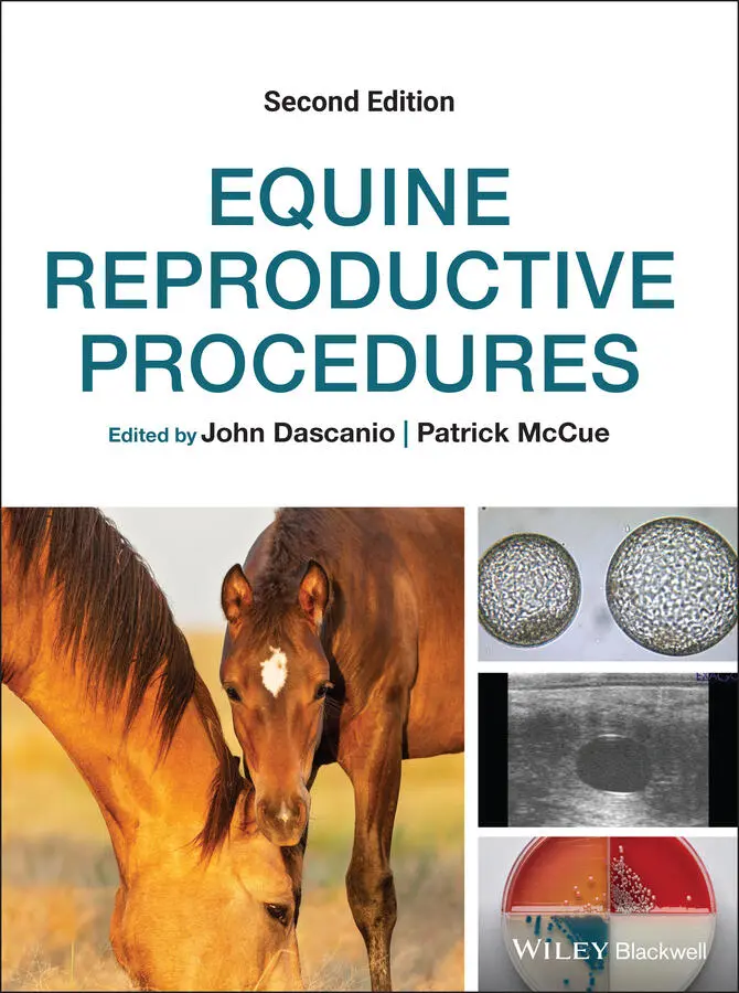
Equine Reproductive Procedures
Здесь есть возможность читать онлайн «Equine Reproductive Procedures» — ознакомительный отрывок электронной книги совершенно бесплатно, а после прочтения отрывка купить полную версию. В некоторых случаях можно слушать аудио, скачать через торрент в формате fb2 и присутствует краткое содержание. Жанр: unrecognised, на английском языке. Описание произведения, (предисловие) а так же отзывы посетителей доступны на портале библиотеки ЛибКат.
- Название:Equine Reproductive Procedures
- Автор:
- Жанр:
- Год:неизвестен
- ISBN:нет данных
- Рейтинг книги:4 / 5. Голосов: 1
-
Избранное:Добавить в избранное
- Отзывы:
-
Ваша оценка:
Equine Reproductive Procedures: краткое содержание, описание и аннотация
Предлагаем к чтению аннотацию, описание, краткое содержание или предисловие (зависит от того, что написал сам автор книги «Equine Reproductive Procedures»). Если вы не нашли необходимую информацию о книге — напишите в комментариях, мы постараемся отыскать её.
provides equine practitioners, veterinary students, and equine theriogenologists with a fully updated, practical guide to techniques in equine reproductive practice. This easy-to-use resource covers skills-based information with a clinical focus, taking an easy-to-follow step-by-step approach.
The book covers topics related to the reproductive management of horses, including diagnostic techniques and therapeutic procedures for stallions, mares, and foals. Procedures highlighted in the book are supported with clear descriptions and photographs. Readers will also find a list of required supplies for the procedure and a reference list.
· Provides step-by-step descriptions of techniques related to equine theriogenology
· Serves as a valuable practice tool
· Offers 39 new techniques not found in the first edition
· Includes key updates to existing techniques
Those studying equine reproduction and specialists in theriogenology will find this an essential ‘how-to’ guide for their practice library.
Equine Reproductive Procedures — читать онлайн ознакомительный отрывок
Ниже представлен текст книги, разбитый по страницам. Система сохранения места последней прочитанной страницы, позволяет с удобством читать онлайн бесплатно книгу «Equine Reproductive Procedures», без необходимости каждый раз заново искать на чём Вы остановились. Поставьте закладку, и сможете в любой момент перейти на страницу, на которой закончили чтение.
Интервал:
Закладка:
57 Chapter 71Figure 71.1 Cervix of a pregnant mare viewed through a speculum (day 45 of g...
58 Chapter 72Figure 72.1 Location and appearance of the genital tubercle (arrow) in a mal...Figure 72.2 Location and appearance of the genital tubercle (arrow) in a fem...Figure 72.3 Directions of ultrasound imaging for fetal sexing (planes I, II,...Figure 72.4 Genital tubercle (arrows) of a 68‐day‐old male equine fetus in p...Figure 72.5 (a–c) Genital tubercle (arrows) of a 65‐day‐old female equine fe...Figure 72.6 Ultrasound image of an equine fetus in plane III at day 100 of g...Figure 72.7 Ultrasound image of a male equine fetus in plane II at 95 days o...Figure 72.8 Female equine fetus at 112 days of gestation showing the hyperec...Figure 72.9 Ultrasound image of a female equine fetus at 125 days of gestati...Figure 72.10 Ultrasound image of a mammary gland (red arrow) and teats (blue...Figure 72.11 Ultrasound image of the mammary gland of a female equine fetus ...Figure 72.12 Glans penis (blue arrow) within the prepuce of a male equine fe...Figure 72.13 Gonads (testes) of a male equine fetus at 130 days of gestation...Figure 72.14 Gonads (ovaries) of a female equine fetus.
59 Chapter 73Figure 73.1 Portable pump sprayer for application of alcohol prior to ultras...Figure 73.2 Transabdominal ultrasonography of a late‐term pregnant mare.Figure 73.3 Transrectal ultrasonography of placental edema in a near‐term no...
60 Chapter 74Figure 74.1 Color Doppler ultrasound image demonstrating the uterine branch ...Figure 74.2 Ultrasound measurement of the combined thickness of the uterus a...Figure 74.3 Average combined thickness of the uterus and placenta in normal ...Figure 74.4 Ultrasound image of a placenta exhibiting increased thickness an...Figure 74.5 Ultrasound image showing separation (arrows) of the chorioallant...
61 Chapter 75Figure 75.1 Milk secretions in a mare with precocious mammary development.Figure 75.2 A late‐term mare with purulent vaginal discharge.Figure 75.3 A late‐term mare with purulent discharge in the ventral tail hai...Figure 75.4 Measurement of the combined thickness of the uterus and placenta...Figure 75.5 An area of separation of the chorioallantois from the uterine wa...
62 Chapter 76Figure 76.1 Pulsed Doppler used to obtain the heart rate during a transabdom...Figure 76.2 Color Doppler image of the cranial cervix and caudal uterus obta...Figure 76.3 (a) Upper transabdominal image demonstrating potential placental...
63 Chapter 77Figure 77.1 A graph of the maximum height (mm) of the embryonic vesicle vers...Figure 77.2 Aortic root diameter measurement of 22 mm in a mare 10 months’ p...Figure 77.3 Per rectum measurement of fetal eye diameter showing a vertical ...
64 Chapter 78Figure 78.1 Two embryonic vesicles at 12 days post‐ovulation located in clos...Figure 78.2 Irregular 14‐day embryonic vesicle just after manual disruption....Figure 78.3 Two embryos adjacent to each other prior to separation and reduc...
65 Chapter 79Figure 79.1 Hand, uterus, and transvaginal probe orientation for aspiration....Figure 79.2 Needle extended from the needle guide (small arrow). Needle exte...
66 Chapter 80Figure 80.1 Position of fingers for side to side manipulation for the disrup...Figure 80.2 Position of fingers to cause dislocation of the cranium from the...Figure 80.3 Placenta from a mare after cranio‐cervical dislocation to elimin...
67 Chapter 81Figure 81.1 (a) Static image captured from a transrectal ultrasound video sh...
68 Chapter 82Figure 82.1 Ultrasound needle guide made with a shortened 14 gauge catheter ...Figure 82.2 Transabdominal fetal cardiac puncture. The light white line depi...Figure 82.3 (a) Heart chambers (black arrows) filled with hyperechoic penici...
69 Chapter 84Figure 84.1 Abortion due to a twin pregnancy.Figure 84.2 Twisted umbilical cord as a cause of abortion in a mare. Note ed...
70 Chapter 85Figure 85.1 Reproductive physiology of the pregnant mare. CL, corpus luteum;...Figure 85.2 Concentrations of equine chorionic gonadotropin (pregnant mare s...
71 Chapter 86Figure 86.1 Mare with numerous varicose veins (arrows) near the vestibulo‐va...Figure 86.2 Mare with numerous varicose veins in the inner aspect of the ves...Figure 86.3 Laser endoscopic ablation of varicose veins surrounding the vest...Figure 86.4 The same mare shown in Figures 86.3 and 86.4, 1.5 years after la...
72 Chapter 87Figure 87.1 Palpation of the mare once recumbent to reorientate the location...Figure 87.2 Start of the rolling procedure with one or two people standing o...Figure 87.3 Rolling of the mare from right lateral recumbency to left latera...
73 Chapter 88Figure 88.1 Two fingers placed inside the vulva to separate the labia in pre...Figure 88.2 Metzenbaum scissors can be used to open a Caslick vulvoplasty.Figure 88.3 Final milk calcium levels in 120 mares prior to foaling.Figure 88.4 Relationship between the number of foalings and time of day. Mos...
74 Chapter 89Figure 89.1 Collection of mammary secretions.Figure 89.2 Titration instrument immersed in milk with dye. Gentle pushing o...Figure 89.3 Milk calcium content of 125 ppm using a milk titration test kit....Figure 89.4 Milk titration test kit results from a mare over 11 days startin...Figure 89.5 Dipping a test strip into a glass vial of diluted milk to evalua...Figure 89.6 All five boxes have changed color (upper strip) indicating a hig...
75 Chapter 90Figure 90.1 pH indicator strips (range 5.5 to 8.0) that may be used to deter...Figure 90.2 pH meter being used to determine the pH of a milk sample.Figure 90.3 Comparison of pH and calcium carbonate levels in the mammary flu...
76 Chapter 91Figure 91.1 Hard‐wired black and white camera monitoring a foaling stall.Figure 91.2 Web‐cam on a foaling stall as viewed on a smart phone.Figure 91.3 Mounted video camera.
77 Chapter 92Figure 92.1 Base receiver (left) and auto dialer (right) along with blue and...Figure 92.2 Foalert ®device in a mare at 300 days’ gestation with a pur...Figure 92.3 Foalert ®device; the pregnant mare partially rubbed out the...
78 Chapter 93Figure 93.1 Breeder Alert ®system with a main unit and autodialer, repe...Figure 93.2 Mare wearing a Breeder Alert ®transmitter (arrow).Figure 93.3 Breeder Alert ®transmitter.
79 Chapter 94Figure 94.1 Mare lifting her tail after the administration of oxytocin to in...Figure 94.2 Mare sweating after the administration of oxytocin to induce lab...Figure 94.3 Mare in stage 2 of parturition with the amnion presented at the ...
80 Chapter 95Figure 95.1 Ventral edema plaque in a later term pregnant mare.Figure 95.2 Mare in stage 1 of labor. Note the extended stance and steam ris...Figure 95.3 Mare in stage 1 labor. The mare is flagging her tail up and down...Figure 95.4 Mare in stage 2 of parturition with the amnion presented at the ...Figure 95.5 Mare in stage 2 labor. The first foot appears covered by amnion....Figure 95.6 Mare in stage 2 labor. The foal’s right front foot (small black ...Figure 95.7 Mare in stage 2 labor. (a) The foal’s nose is about even with th...Figure 95.8 Mare in stage 2 labor. Note that the foal’s chest has passed the...Figure 95.9 Mare in stage 2 labor. The foal is completely delivered with the...
81 Chapter 96Figure 96.1 Amnion protruding through the vulva during a normal foaling.Figure 96.2 Intact chorioallantoic membrane protruding through the vulva, in...Figure 96.3 Another example of premature placental separation. The brick‐red...Figure 96.4 Ultrasound showing slight separation of the chorioallantoic memb...Figure 96.5 Ultrasound showing massive separation of the chorioallantoic mem...
82 Chapter 97Figure 97.1 Premature placental separation (“red bag”) with the intact chori...Figure 97.2 Applying traction to a fetus in a mare exhibiting uterine inerti...Figure 97.3 Elevation of the hind end of a mare to facilitate examination an...
Читать дальшеИнтервал:
Закладка:
Похожие книги на «Equine Reproductive Procedures»
Представляем Вашему вниманию похожие книги на «Equine Reproductive Procedures» списком для выбора. Мы отобрали схожую по названию и смыслу литературу в надежде предоставить читателям больше вариантов отыскать новые, интересные, ещё непрочитанные произведения.


Обсуждение, отзывы о книге «Equine Reproductive Procedures» и просто собственные мнения читателей. Оставьте ваши комментарии, напишите, что Вы думаете о произведении, его смысле или главных героях. Укажите что конкретно понравилось, а что нет, и почему Вы так считаете.