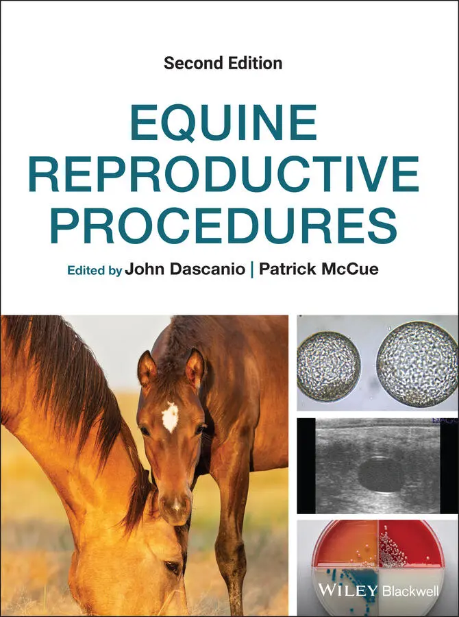
Equine Reproductive Procedures
Здесь есть возможность читать онлайн «Equine Reproductive Procedures» — ознакомительный отрывок электронной книги совершенно бесплатно, а после прочтения отрывка купить полную версию. В некоторых случаях можно слушать аудио, скачать через торрент в формате fb2 и присутствует краткое содержание. Жанр: unrecognised, на английском языке. Описание произведения, (предисловие) а так же отзывы посетителей доступны на портале библиотеки ЛибКат.
- Название:Equine Reproductive Procedures
- Автор:
- Жанр:
- Год:неизвестен
- ISBN:нет данных
- Рейтинг книги:4 / 5. Голосов: 1
-
Избранное:Добавить в избранное
- Отзывы:
-
Ваша оценка:
Equine Reproductive Procedures: краткое содержание, описание и аннотация
Предлагаем к чтению аннотацию, описание, краткое содержание или предисловие (зависит от того, что написал сам автор книги «Equine Reproductive Procedures»). Если вы не нашли необходимую информацию о книге — напишите в комментариях, мы постараемся отыскать её.
provides equine practitioners, veterinary students, and equine theriogenologists with a fully updated, practical guide to techniques in equine reproductive practice. This easy-to-use resource covers skills-based information with a clinical focus, taking an easy-to-follow step-by-step approach.
The book covers topics related to the reproductive management of horses, including diagnostic techniques and therapeutic procedures for stallions, mares, and foals. Procedures highlighted in the book are supported with clear descriptions and photographs. Readers will also find a list of required supplies for the procedure and a reference list.
· Provides step-by-step descriptions of techniques related to equine theriogenology
· Serves as a valuable practice tool
· Offers 39 new techniques not found in the first edition
· Includes key updates to existing techniques
Those studying equine reproduction and specialists in theriogenology will find this an essential ‘how-to’ guide for their practice library.
Equine Reproductive Procedures — читать онлайн ознакомительный отрывок
Ниже представлен текст книги, разбитый по страницам. Система сохранения места последней прочитанной страницы, позволяет с удобством читать онлайн бесплатно книгу «Equine Reproductive Procedures», без необходимости каждый раз заново искать на чём Вы остановились. Поставьте закладку, и сможете в любой момент перейти на страницу, на которой закончили чтение.
Интервал:
Закладка:
Technique
The self‐contained probe should be wiped with 70% isopropyl alcohol. The biopsy channel should be similarly cold sterilized and then rinsed with sterile saline or a sterile pipette should be inserted into the biopsy channel so the biopsy instrument does not contact an unclean surface.
If ultrasound probe housing is used it should be cold sterilized for a minimum of 10 minutes and the solution vigorously rinsed off with sterile water. The probe to be inserted in the housing should be wiped with 70% isopropyl alcohol and the unit assembled using sterile gloves.
The distal 10 cm (4 inch) of the biopsy instrument should be cold sterilized with the sample notch exposed. The immersed end of the biopsy instrument should be rinsed with sterile saline and the remaining needle length and handle wiped with 70% isopropyl alcohol or disinfectant wipes and placed on a sterile towel.
Place the mare in stocks to provide adequate restraint from lateral movement.
Wrap the tail and remove feces from the rectum (see Chapter 4). Administer N‐butylscopolammonium bromide at 0.15 mg/kg slowly IV to relax the rectum.
Tie the mare’s tail to the side, and remove the feces. Perform an ultrasound examination to determine the ovary or ovaries to be biopsied and the target location to be sampled.
Prepare the mare’s perineum by washing and then drying the perineum (see Chapter 3).
Administer sedation consisting of xylazine (0.3–0.4 mg/kg IV) combined with butorphanol (0.01–0.02 mg/kg IV), or detomidine (0.01–0.02 mg/kg IV) combined with butorphanol (0.01–0.02 mg/kg IV). Position the mare’s head to prevent airway obstruction when the head drops due to the sedation. Figure 33.1 Self‐contained transvaginal ultrasound probe (a), custom modified spring‐loaded 65 cm biopsy instrument (b), and handle of biopsy device when cocked (c) and when advanced to the first stop (d).
Sterile lubricant should be applied to the surface of the probe. The self‐contained vaginal probe or the assembled probe, may be placed into a sterile sleeve, and inserted into the vagina. Alternatively, the examiner may wear a sterile rectal sleeve and shield the end of the probe while placing it lateral to the cervix, ipsilateral to the ovary to be biopsied. The opening to the biopsy port is covered with a sterile towel to prevent fecal contamination.
The examiner’s hand is then placed in the rectum, the feces evacuated, and the ovary to be biopsied is positioned adjacent to the vaginal probe. This may be accomplished by moving the ovary deeper into the abdomen and then moving the ovary dorsally over the broad ligament. Figure 33.2 Biopsy device tip with sample notch exposed (arrows) revealing a core ovarian biopsy.
The ovary is examined using ultrasound and the target area to be biopsied is identified. The biopsy guide may be used to assist in obtaining the biopsy, by positioning the ovary so the biopsy guide lines bisect the area to be sampled.
The assistant, wearing surgical gloves, cocks the biopsy instrument and lubricates the tip with a small amount of sterile lubricant. The tip of the biopsy instrument is inserted into the biopsy channel of the self‐contained probe, or ultrasound housing, and advanced until resistance is felt. Slight intermittent pressure against the vaginal wall will help identify the position of the tip of the biopsy instrument in relationship to the target area on the ovary as viewed on the ultrasound.
The examiner, with their hand in the rectum, steadies the ovary firmly against the ultrasound probe. The assistant estimates the distance to the target area to be biopsied.
The needle is advanced into the ovary a smooth quick motion to the approximate depth needed to reach the target area.
The tip of the biopsy instrument may be positioned 2 cm (0.75 inch) behind the ovarian target area and fired (the one step method) or the tip of the biopsy instrument may be advanced to the first stop, which exposes the notch on the biopsy device’s needle, and then fired (two step method).
The biopsy instrument is removed from the ovary and placed on the sterile field. The biopsy instrument is cocked and advanced to the first stop to expose the sample notch. The sample is removed from the notch using small (22 or 25 gauge) needles. Any of the following may be performed with the sample: imprints may be made on a sterile slide for cytology, it may be placed in formalin for histopathology, or the sample may be snap frozen in liquid nitrogen for mRNA analysis. The biopsy instrument is returned to the shielded position, re‐lubricated, and the process repeated to obtain 3–5 good biopsy cores from the target area per ovary.
It is recommended that a physical examination (temperature, pulse, respiration rate) is performed on the mare daily for 3 days post‐procedure to monitor for any complications.
Interpretation
There is a slight risk of significant vaginal bleeding from the biopsy needle penetration. There is small risk of peritonitis arising due to the biopsy instrument penetrating the vaginal wall, which contains bacterial flora.
There is a slight risk of rectal puncture arising from overzealous penetration of the ovary, failure to position the biopsy instrument accurately, accidentally firing the biopsy instrument, or mare movement.
Antibiotics and anti‐inflammatory drugs are typically not administered following this procedure. If a rectal puncture has occurred then non‐steroidal anti‐inflammatory therapy for 3 days and trimethoprim sulfa (30 mg/kg by mouth) twice daily for 7–10 days, or other broad‐spectrum antibiotic therapy may be indicated.
Further Reading
1 Aerts JMJ, Oste M, Bols PEJ. 2005. Development and practical applications of a method for repeated transvaginal, ultrasound‐guided biopsy collection of the bovine ovary. Theriogenology 64: 947–57.
2 Diel de Amorim M, Nairn LD, Manning S, Dedden I, Ripley E, Card C. 2016. Evaluation of diagnostic utility, safety considerations, and effect on fertility of transvaginal ultrasound‐guided ovarian biopsy in mares. Theriogenology 5(6): 1030–6.
3 Gastal, GA, Alves BG, Alves, KA, et al. 2017. Ovarian fragment sizes affect viability and morphology of preantral follicles during storage at 4°C. Reproduction 153(5): 577–87.
4 Murase H, Ball B, Tangyuenyong S, et al. 2018. Serum anti‐müllerian hormone concentrations in mares with granulosa cell tumors versus other ovarian abnormalities. J Eq Vet Sci 60: 6–10.
5 Slough TL, Rispoli LA, Carnevale EM, Niswender GD, Bruemmer J. 2011. Temporal gene expression in equine corpora lutea based on serial biopsies in vivo. J Anim Sci 89: 389–96.
6 Velez IC, Arnold C, Jacobson CC, et al. 2012. Effects of repeated transvaginal aspiration of immature follicles on mare health and ovarian status. Eq Vet J Suppl 44: 78–83.
Конец ознакомительного фрагмента.
Текст предоставлен ООО «ЛитРес».
Прочитайте эту книгу целиком, купив полную легальную версию на ЛитРес.
Безопасно оплатить книгу можно банковской картой Visa, MasterCard, Maestro, со счета мобильного телефона, с платежного терминала, в салоне МТС или Связной, через PayPal, WebMoney, Яндекс.Деньги, QIWI Кошелек, бонусными картами или другим удобным Вам способом.
Интервал:
Закладка:
Похожие книги на «Equine Reproductive Procedures»
Представляем Вашему вниманию похожие книги на «Equine Reproductive Procedures» списком для выбора. Мы отобрали схожую по названию и смыслу литературу в надежде предоставить читателям больше вариантов отыскать новые, интересные, ещё непрочитанные произведения.


Обсуждение, отзывы о книге «Equine Reproductive Procedures» и просто собственные мнения читателей. Оставьте ваши комментарии, напишите, что Вы думаете о произведении, его смысле или главных героях. Укажите что конкретно понравилось, а что нет, и почему Вы так считаете.