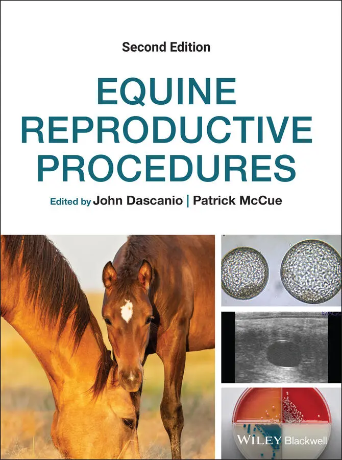
Equine Reproductive Procedures
Здесь есть возможность читать онлайн «Equine Reproductive Procedures» — ознакомительный отрывок электронной книги совершенно бесплатно, а после прочтения отрывка купить полную версию. В некоторых случаях можно слушать аудио, скачать через торрент в формате fb2 и присутствует краткое содержание. Жанр: unrecognised, на английском языке. Описание произведения, (предисловие) а так же отзывы посетителей доступны на портале библиотеки ЛибКат.
- Название:Equine Reproductive Procedures
- Автор:
- Жанр:
- Год:неизвестен
- ISBN:нет данных
- Рейтинг книги:4 / 5. Голосов: 1
-
Избранное:Добавить в избранное
- Отзывы:
-
Ваша оценка:
Equine Reproductive Procedures: краткое содержание, описание и аннотация
Предлагаем к чтению аннотацию, описание, краткое содержание или предисловие (зависит от того, что написал сам автор книги «Equine Reproductive Procedures»). Если вы не нашли необходимую информацию о книге — напишите в комментариях, мы постараемся отыскать её.
provides equine practitioners, veterinary students, and equine theriogenologists with a fully updated, practical guide to techniques in equine reproductive practice. This easy-to-use resource covers skills-based information with a clinical focus, taking an easy-to-follow step-by-step approach.
The book covers topics related to the reproductive management of horses, including diagnostic techniques and therapeutic procedures for stallions, mares, and foals. Procedures highlighted in the book are supported with clear descriptions and photographs. Readers will also find a list of required supplies for the procedure and a reference list.
· Provides step-by-step descriptions of techniques related to equine theriogenology
· Serves as a valuable practice tool
· Offers 39 new techniques not found in the first edition
· Includes key updates to existing techniques
Those studying equine reproduction and specialists in theriogenology will find this an essential ‘how-to’ guide for their practice library.
Equine Reproductive Procedures — читать онлайн ознакомительный отрывок
Ниже представлен текст книги, разбитый по страницам. Система сохранения места последней прочитанной страницы, позволяет с удобством читать онлайн бесплатно книгу «Equine Reproductive Procedures», без необходимости каждый раз заново искать на чём Вы остановились. Поставьте закладку, и сможете в любой момент перейти на страницу, на которой закончили чтение.
Интервал:
Закладка:
26 Chapter 33 Figure 33.1 Self‐contained transvaginal ultrasound probe (a), custom modifie... Figure 33.2 Biopsy device tip with sample notch exposed (arrows) revealing a...
27 Chapter 34Figure 34.1 Lavage solution hung next to a mare with the uterine lavage tubi...Figure 34.2 Double‐ended “Christmas tree” adapter attached to the uterine la...Figure 34.3 Inflated air cuff on the end of the uterine lavage tubing.Figure 34.4 Uterine lavage of a mare post‐breeding with “Y” tubing. The lava...Figure 34.5 Cloudy uterine lavage return solution.
28 Chapter 35Figure 35.1 Intrauterine ends of various types of pipettes. From left to rig...Figure 35.2 Ends of pipettes to which a syringe is attached. From left to ri...
29 Chapter 38Figure 38.1 Diagnostic and treatment protocol for the use of bActivate.
30 Chapter 40Figure 40.1 Rope wrapped around the pastern and forearm. The free ends of th...Figure 40.2 Rope wrapped around the pastern and forearm. One end is tied to ...Figure 40.3 Mare with hobbles and tail wrap in preparation for breeding.
31 Chapter 41Figure 41.1 Stallion (left) gathering his harem of mares.Figure 41.2 Stallion mating a mare with a foal at her side. The mare was par...
32 Chapter 42Figure 42.1 Evaluation of a mare for live‐cover breeding using a teaser stal...Figure 42.2 Mare wearing a neck apron and kick boots while restrained with a...Figure 42.3 Live cover of a mare by a stallion.
33 Chapter 43Figure 43.1 Schematic of a breeding stitch (arrow) encircling a region just ...Figure 43.2 Breeding stitch (arrow) just placed in a Thoroughbred mare after...
34 Chapter 45Figure 45.1 Placement of an insemination pipette into the hand for placement...Figure 45.2 External cervical os. The cervical canal is located in the cente...Figure 45.3 Introduction of an artificial insemination pipette along the ind...Figure 45.4 Vertical orientation of the syringe during insemination to allow...
35 Chapter 46Figure 46.1 Viewing the cervix through a disposable speculum during insemina...Figure 46.2 Position of the speculum prior to passage of the insemination pi...
36 Chapter 47Figure 47.1 End of the stylet that will be inserted into the straw inside th...Figure 47.2 The cone on the stylet shaft that will catch the semen straw. Up...Figure 47.3 Semen remaining in the syringe if semen is drawn into the syring...
37 Chapter 48Figure 48.1 Bending of an artificial insemination pipette after removing fro...Figure 48.2 Bend that remains in the pipette after curling. This bend aids i...
38 Chapter 49Figure 49.1 Catheter used to deposit semen on the utero‐tubular junction. Th...Figure 49.2 Uterine bifurcation as viewed through a videoendoscope.Figure 49.3 A dilated uterine horn as viewed through a videoendoscope.Figure 49.4 Utero‐tubal junction (arrow) viewed through a videoendoscope.
39 Chapter 50Figure 50.1 A sterile controlled flushing set is spiked into a pre‐warmed ba...Figure 50.2 Positioning the TVA handle encasing the ultrasound probe per vag...Figure 50.3 (a) Lab equipment (dissecting microscope with heated stage, 150 ...Figure 50.4 Pre‐warmed 75 μm embryo filtration cup with contents of immature...Figure 50.5 (a) Image of maturing oocyte recovered from a hormone‐stimulated...
40 Chapter 51Figure 51.1 Two examples of isothermic containers utilized for shipment of o...
41 Chapter 52Figure 52.1 Equine oocyte held in place with suction from a holding pipette ...
42 Chapter 53Figure 53.1 Grasping the dorsal commissure of the vulva in preparation for s...Figure 53.2 Placement of skin staples in the vulvar labia for staple vulvopl...Figure 53.3 Finished temporary vulvoplasty.Figure 53.4 Trimming mucosa from the inner labia.Figure 53.5 Suturing the labia together.Figure 53.6 Placement of fingers for the removal of the vulvoplasty.Figure 53.7 Cutting the co‐joined mucosa to remove the vulvoplasty.
43 Chapter 54Figure 54.1 Culture of the central clitoral sinus using an ultrafine culture...Figure 54.2 Culture of the clitoral fossa.Figure 54.3 Flushing the central clitoral fossa using a curved tip syringe....Figure 54.4 Packing the clitoris with 0.2% nitrofurazone antibiotic ointment...Figure 54.5 Culture locations on the distal penis: urethral sinus (thin arro...
44 Chapter 55Figure 55.1 Mare in box stall with a fluorescent light overhead.Figure 55.2 Mare wearing a mask emitting a blue light into one eye.
45 Chapter 57Figure 57.1 Insertion of an Ovuplant™ implant (arrow) in the vulvar mucosa....
46 Chapter 59Figure 59.1 Unsterilized 35 mm glass ball.Figure 59.2 Ultrasound image of an intrauterine glass ball. The arrow points...Figure 59.3 Disassembled iUPOD TMapplicator: outer tube (A), applicator plun...Figure 59.4 Disassembled iUPOD TMmagnetic retriever and applicator: applicat...Figure 59.5 Ultrasound image of an iUPOD TM(arrow) in the uterus. Note the a...Figure 59.6 Ultrasound image of a 16‐day embryonic vesicle (black arrow) adj...
47 Chapter 61Figure 61.1 Ovaries (small arrow) in a plastic bag are placed into the cente...
48 Chapter 62Figure 62.1 Non‐surgical embryo collection procedure.Figure 62.2 Embryo collection setup. The media bag is connected by “Y” tubin...Figure 62.3 Flush media passing through an embryo filter (open system).Figure 62.4 Flush media passing through an embryo filter (closed system).Figure 62.5 Straw (0.25 ml), connector and 1.0 ml syringe for handling small...Figure 62.6 Petri dish (35 mm) with holding medium (left side). Washing an e...
49 Chapter 63Figure 63.1 Morula stage embryo (grade 1). Note the thick zona pellucida and...Figure 63.2 Early blastocyst stage embryo (grade 1). Note the thinner zona p...Figure 63.3 Blastocyst stage embryo (grade 1). Note the thin zona pellucida,...Figure 63.4 Expanded blastocyst stage embryo (grade 1). The zona pellucida h...Figure 63.5 Unfertilized oocytes. Note the thick zona pellucida and lack of ...Figure 63.6 Grade 2 early blastocyst stage embryo. Note the extruded blastom...Figure 63.7 Measurement of embryo diameter using an eye piece micrometer.
50 Chapter 64Figure 64.1 Aspiration biopsy of an equine blastocyst embryo.
51 Chapter 65Figure 65.1 Left to right, 50 ml conical tube, 5 ml vial, and Parafilm® for ...Figure 65.2 Sealing the 5 ml vial with Parafilm®. Pull the Parafilm® so that...Figure 65.3 Placing the small 5 ml vial containing the embryo inside the lar...Figure 65.4 The50 ml conical tube sealed with Parafilm® with a sealed 5 ml t...Figure 65.5 Loading the 50 ml conical tube into a passive cooling system for...
52 Chapter 66Figure 66.1 Petri dish prepared for vitrification of an embryo. VS1, solutio...Figure 66.2 Thermos with a cane holding a goblet with the top suspended abov...Figure 66.3 Straw placed into a goblet that is suspended above liquid nitrog...Figure 66.4 Embryo plunged into liquid nitrogen.
53 Chapter 67Figure 67.1 Embryo in a column of holding medium (large arrow) inside a 0.25...
54 Chapter 68Figure 68.1 A filly successfully born from an in vitro produced embryo trans...Figure 68.2 A mature oocyte from an autotransfer donor undergoing the ICSI p...Figure 68.3 A 14‐day embryonic vesicle (yellow dotted circle) (a) following ...
55 Chapter 69Figure 69.1 Palpation of a uterine specimen demonstrating the technique for ...
56 Chapter 70Figure 70.1 Ultrasonographic image of a 12‐day pregnancy.Figure 70.2 Ultrasonographic image of a 16‐day pregnancy.Figure 70.3 Ultrasonographic image of a 25‐day pregnancy.Figure 70.4 Ultrasonographic image of a 35‐day pregnancy.Figure 70.5 Ultrasonographic image of twin pregnancies, with the embryonic v...Figure 70.6 Ultrasonographic image of twin pregnancies, with the embryos tou...Figure 70.7 Ultrasonographic image of a 25‐day pregnancy examination showing...Figure 70.8 Ultrasonographic image of a 25‐day pregnancy (arrow) and adjacen...Figure 70.9 Ultrasonographic image of an empty trophoblastic vesicle 28 days...Figure 70.10 Ultrasonographic image of multiple supplementary (accessory) co...
Читать дальшеИнтервал:
Закладка:
Похожие книги на «Equine Reproductive Procedures»
Представляем Вашему вниманию похожие книги на «Equine Reproductive Procedures» списком для выбора. Мы отобрали схожую по названию и смыслу литературу в надежде предоставить читателям больше вариантов отыскать новые, интересные, ещё непрочитанные произведения.


Обсуждение, отзывы о книге «Equine Reproductive Procedures» и просто собственные мнения читателей. Оставьте ваши комментарии, напишите, что Вы думаете о произведении, его смысле или главных героях. Укажите что конкретно понравилось, а что нет, и почему Вы так считаете.