Biological Mechanisms of Tooth Movement
Здесь есть возможность читать онлайн «Biological Mechanisms of Tooth Movement» — ознакомительный отрывок электронной книги совершенно бесплатно, а после прочтения отрывка купить полную версию. В некоторых случаях можно слушать аудио, скачать через торрент в формате fb2 и присутствует краткое содержание. Жанр: unrecognised, на английском языке. Описание произведения, (предисловие) а так же отзывы посетителей доступны на портале библиотеки ЛибКат.
- Название:Biological Mechanisms of Tooth Movement
- Автор:
- Жанр:
- Год:неизвестен
- ISBN:нет данных
- Рейтинг книги:5 / 5. Голосов: 1
-
Избранное:Добавить в избранное
- Отзывы:
-
Ваша оценка:
Biological Mechanisms of Tooth Movement: краткое содержание, описание и аннотация
Предлагаем к чтению аннотацию, описание, краткое содержание или предисловие (зависит от того, что написал сам автор книги «Biological Mechanisms of Tooth Movement»). Если вы не нашли необходимую информацию о книге — напишите в комментариях, мы постараемся отыскать её.
delivers a comprehensive reference for orthodontic trainees and specialists.
It is fully updated to include new chapters on personalized orthodontics as well as the inflammatory process occurring in the dental and paradental tissues. It is heavily illustrated throughout, making it easier for readers to understand and retain the information discussed within. The topics covered range from bone biology, the effects of mechanical loading on tissues and cells, genetics, tissue remodeling, and the effects of diet, drugs, and systemic diseases.
The Third Edition of
features seven sections that cover subjects such as:
The development of biological concepts in orthodontics, including the cellular and molecular biology behind orthodontic tooth movement Mechanics meets biology, including the effects of mechanical loading on hard and soft tissues and cells, and biological reactions to temporary anchorage devices Inflammation and orthodontics, including markers for tissue remodeling in the gingival crevicular fluid and saliva Personalized diagnosis and treatment based on genomic criteria, including the genetic influences on orthodontic tooth movement Rapid orthodontics, including methods to accelerate or decelerate orthodontic tooth movement Perfect for residents and PhD students of orthodontic and periodontal programs,
is also useful to academics, clinicians, bone biologists, and researchers with an interest in the mechanics and biology of tooth movement.
Biological Mechanisms of Tooth Movement — читать онлайн ознакомительный отрывок
Ниже представлен текст книги, разбитый по страницам. Система сохранения места последней прочитанной страницы, позволяет с удобством читать онлайн бесплатно книгу «Biological Mechanisms of Tooth Movement», без необходимости каждый раз заново искать на чём Вы остановились. Поставьте закладку, и сможете в любой момент перейти на страницу, на которой закончили чтение.
Интервал:
Закладка:
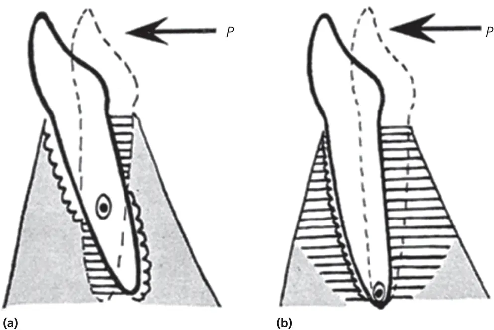
Figure 2.11 Comparative diagram of the theories put forward by Sandstedt (1904) and Oppenheim (1911) as drawn by Schwarz (1932). In (a), which depicts the theory of pressure (Sandstedt), the tooth moved by the force, P, tilts around an axis, O, lying a little apically from the center of the root. By this means two regions of pressure and pull arise, lying diametrically opposite. In the regions of pressure in the PDL, the old alveolar bone is resorbed (jagged line) and in the regions of pull, new bone is added (horizontal shading). Gray shading, alveolar bone without transformation. (b) This depicts the theory of transformation (Oppenheim, 1911, 1944). There is only one side of pressure and one side of pull. On both sides the alveolar bone opens into a transitional spongy bone, whose elements are arranged vertically to the surface of the tooth (horizontal shading). On the side of pressure, this newly formed transitional bone is resorbed (jagged line). On the side of pull, new bone is added. Gray shading indicates the old untransformed alveolar bone at a greater distance from the moved tooth,
(Source: Schwarz, 1932. Reproduced with permission of Elsevier.)
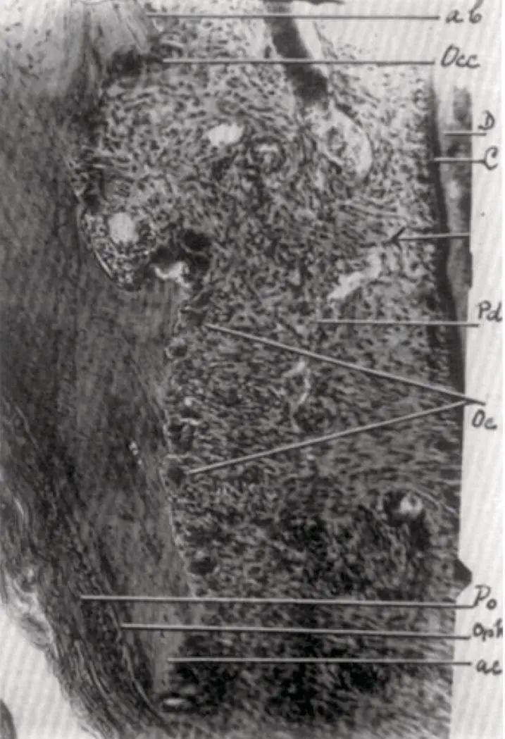
Figure 2.12 Higher magnification image from Oppenheim’s article (1944) showing labial alveolar crest. The aplastic zone facing the periodontium has for the greatest part disappeared, as has the crest itself. Where some aplastic bone is still present (ab), the secondary osteoclasts (Occ) are still at work removing it. No osteoclastic activity whatsoever is found at the periosteal smooth bone surface. The still remaining but decreased pressure caused the appearance of primary osteoclasts (Oc) and will be present for 2 days after force discontinuation. D, dentine; C, cementum; Pd, periodontal membrane; Po, formation of smooth periosteal bone surface; Opk, scarce osteophyte. ac, acellular cementum.
(Source: Oppenheim, 1944. Reproduced with permission of Elsevier.)
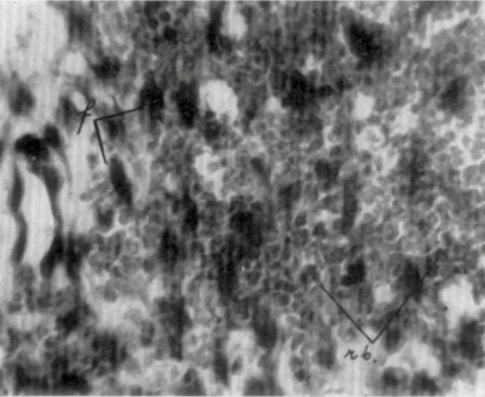
Figure 2.13 Higher magnification image of hemorrhage as portrayed in Oppenheim (1944).
(Source: Oppenheim, 1944. Reproduced with permission of Elsevier.)
In short, all three major researchers (Sandstedt, 1904, 1905; Oppenheim, 1911, 1944; Schwarz, 1932) exploring tissue reactions during OTM, agreed that there is a creation of pressure and tension sites in the PDL during OTM. Furthermore, it appears that cell replication is decreased in pressure sites owing to a decrease in vascular supply, whereas it is increased in tension sites due to PDL fiber stretching.
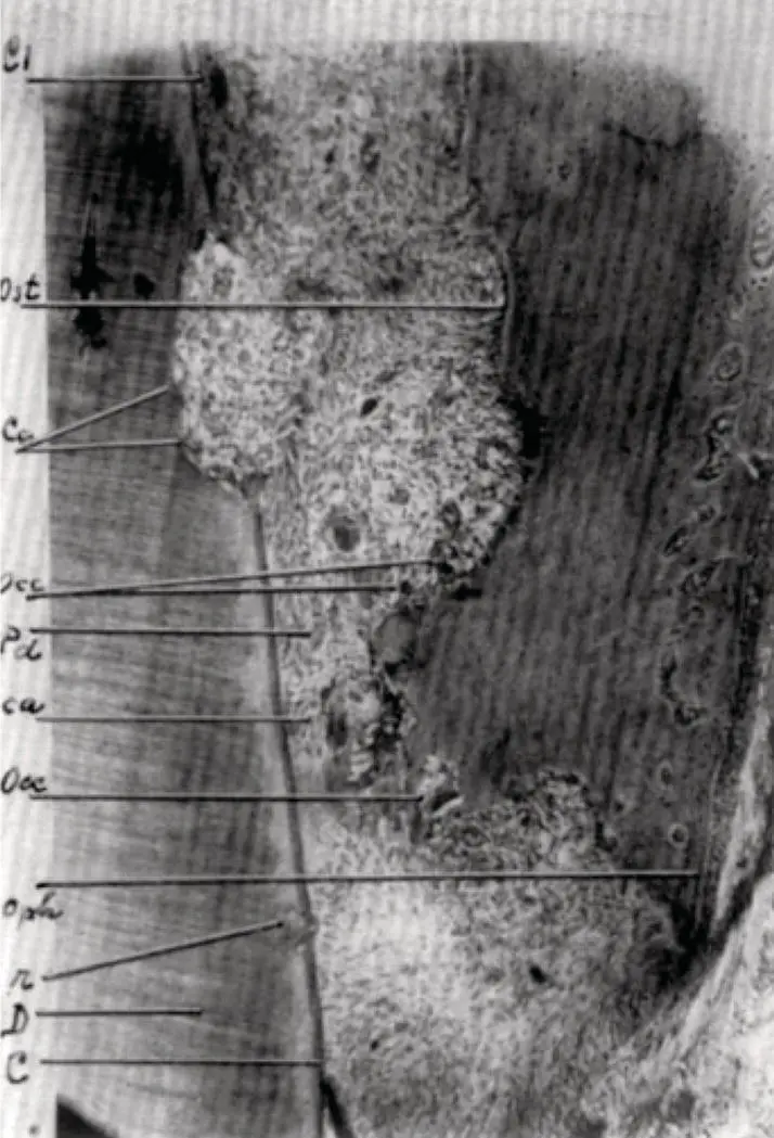
Figure 2.14 Hyalinization reaction as portrayed in Oppenheim (1944). The osteocytes are mostly normal; the osteophytic bone formation (Oph) is quite poor; no sign of any periosteal osteoclastic activity was found. The cementum within the compression area is aplastic, and again displays its signs of vitality (cementoblasts, cementoid seam) above the compression area (C). Within this area, we see a cementum resorption with cementoclasts (Cc) still present 4 days after force discontinuation. A proof that the lowering of the crest has really taken place is found in the presence of another small cementum resorption (r) in a region opposite which bone is no longer present. The larger resorption, though not deep, is already quite extended buccolingually. Above the crest we find the effect of the relapse movement of 4 days, the formation of an osteoid seam (Ost). C, Aplastic cementum; D, dentine; Pd, periodontal membrane; ca, crushed periodontal tissue with hyaline degeneration and debris; Occ, secondary osteoclasts: Oph, osteophytic apposition; r, small cementum resorption: cc, greater cementum resorption with cementoclasts still present; Ost, osteoid.
(Source: Oppenheim, 1944. Reproduced with permission of Elsevier.)
Reitan (1957, 1960), in his classic papers on histological changes during OTM, reported that hyalinization refers to cell‐free areas within the PDL, in which the normal tissue architecture and staining characteristics of collagen in the processed histological material have been lost ( Figures 2.15and 2.16). He observed that:
hyalinization occurred within the PDL following the application of even minimal force, meant to bring about a tipping movement;
a greater degree of hyalinization occurred following application of force, if a tooth had a short root;
during tooth translation, very little hyalinization was observed.
Reitan (1960) concluded that the tissue changes observed were those of degeneration related to force per unit area, and that attempts should be made to minimize these changes.
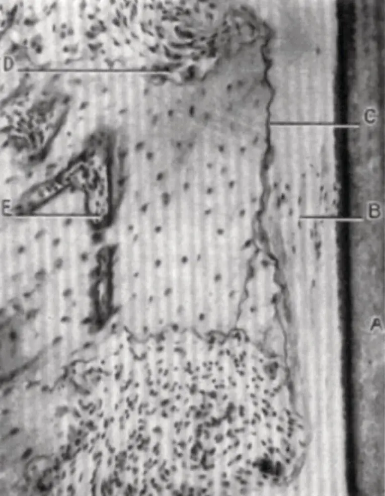
Figure 2.15 Cell free areas as shown by Reitan (1960). The figure shows pressure in the PDL during tooth movement, where cells gradually disappear in a circumscribed area. A, Root surface: B, compressed cell free fibers; C, border line between bone and hyalinized tissue: D, undermining bone resorption; E, small marrow space in dense, compact lamina dura.
(Source: Reitan, 1960. Reproduced with permission of Elsevier.)
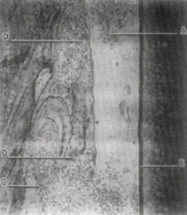
Figure 2.16 (A) Formation of cells and capillaries in hyalinized tissue after the force was released as shown by Reitan (1960). B, Root surface; C, direct resorption; D, undermining resorption.
(Source: Reitan, 1960. Reproduced with permission of Elsevier.)
The fluid dynamic hypothesis
Bien (1966), through his research on the effect of intrusive forces on mandibular incisors, recorded an oscillation of the force inside the dental socket, and named it the hydraulic damping effect. He identified three distinct but interactive fluid systems present in paradental tissues: the vascular system, interstitial fluids, and cellular fluids. All three are presumably involved in damping oscillations of the tooth. He used Reynold’s numbers to measure tooth oscillation and concluded that the low Reynold’s number observed for the tooth subjected to oscillations is due to predominance of viscous forces acting within the system. He observed an escape of extracellular fluids from the PDL to the marrow spaces through the minute perforations in the alveolar wall ( Figure 2.17). This phenomenon, occurring in the first stage of OTM, when PDL fibers are slack, depends mainly on size and number of alveolar bone perforations. The slack fibers become tightened once the extracellular fluids are exhausted. Owing to the presence of interstitial fluid or ground substance throughout the PDL, and the fact that the PDL is extremely thin, when compared with the sizes of the dental root and alveolus, he related the behavior of the PDL to that of the “squeeze film effect” proposed by Hays (1961). The presence of this film enables the tooth to withstand the heavy forces applied as part of orthodontic treatment or masticatory efforts. Masticatory forces, which are momentary in nature, will displace the fluid in the PDL space to its boundaries of the squeeze film (towards the apex and cervical areas of the dental root). Once the force is released, replenishment of fluid occurs through recirculation of interstitial fluid and diffusion through capillary walls, restoring the equilibrium. Likewise, a similar chain of events may occur following the application of low, sustained orthodontic forces.
Читать дальшеИнтервал:
Закладка:
Похожие книги на «Biological Mechanisms of Tooth Movement»
Представляем Вашему вниманию похожие книги на «Biological Mechanisms of Tooth Movement» списком для выбора. Мы отобрали схожую по названию и смыслу литературу в надежде предоставить читателям больше вариантов отыскать новые, интересные, ещё непрочитанные произведения.


Обсуждение, отзывы о книге «Biological Mechanisms of Tooth Movement» и просто собственные мнения читателей. Оставьте ваши комментарии, напишите, что Вы думаете о произведении, его смысле или главных героях. Укажите что конкретно понравилось, а что нет, и почему Вы так считаете.