Biological Mechanisms of Tooth Movement
Здесь есть возможность читать онлайн «Biological Mechanisms of Tooth Movement» — ознакомительный отрывок электронной книги совершенно бесплатно, а после прочтения отрывка купить полную версию. В некоторых случаях можно слушать аудио, скачать через торрент в формате fb2 и присутствует краткое содержание. Жанр: unrecognised, на английском языке. Описание произведения, (предисловие) а так же отзывы посетителей доступны на портале библиотеки ЛибКат.
- Название:Biological Mechanisms of Tooth Movement
- Автор:
- Жанр:
- Год:неизвестен
- ISBN:нет данных
- Рейтинг книги:5 / 5. Голосов: 1
-
Избранное:Добавить в избранное
- Отзывы:
-
Ваша оценка:
Biological Mechanisms of Tooth Movement: краткое содержание, описание и аннотация
Предлагаем к чтению аннотацию, описание, краткое содержание или предисловие (зависит от того, что написал сам автор книги «Biological Mechanisms of Tooth Movement»). Если вы не нашли необходимую информацию о книге — напишите в комментариях, мы постараемся отыскать её.
delivers a comprehensive reference for orthodontic trainees and specialists.
It is fully updated to include new chapters on personalized orthodontics as well as the inflammatory process occurring in the dental and paradental tissues. It is heavily illustrated throughout, making it easier for readers to understand and retain the information discussed within. The topics covered range from bone biology, the effects of mechanical loading on tissues and cells, genetics, tissue remodeling, and the effects of diet, drugs, and systemic diseases.
The Third Edition of
features seven sections that cover subjects such as:
The development of biological concepts in orthodontics, including the cellular and molecular biology behind orthodontic tooth movement Mechanics meets biology, including the effects of mechanical loading on hard and soft tissues and cells, and biological reactions to temporary anchorage devices Inflammation and orthodontics, including markers for tissue remodeling in the gingival crevicular fluid and saliva Personalized diagnosis and treatment based on genomic criteria, including the genetic influences on orthodontic tooth movement Rapid orthodontics, including methods to accelerate or decelerate orthodontic tooth movement Perfect for residents and PhD students of orthodontic and periodontal programs,
is also useful to academics, clinicians, bone biologists, and researchers with an interest in the mechanics and biology of tooth movement.
Biological Mechanisms of Tooth Movement — читать онлайн ознакомительный отрывок
Ниже представлен текст книги, разбитый по страницам. Система сохранения места последней прочитанной страницы, позволяет с удобством читать онлайн бесплатно книгу «Biological Mechanisms of Tooth Movement», без необходимости каждый раз заново искать на чём Вы остановились. Поставьте закладку, и сможете в любой момент перейти на страницу, на которой закончили чтение.
Интервал:
Закладка:
Experimenting with dog mandibles in vitro and in vivo , Zengo et al . (1973) demonstrated that orthodontic canine tipping bends the alveolar bone, creating on it concave and convex surfaces identical to those generated in bent long bones. In areas of PDL tension, the interfacing bone surface assumes a concave configuration in which the molecules are compressed, whereas in zones of compressed PDL the adjacent alveolar bone surface becomes convex ( Figure 2.19). Hence, there is no contradiction between the response of alveolar bone and other parts of the skeleton to mechanical loading. The confusion in this regard has resulted from the usage of the same descriptions for different tissues. While orthodontic “tension” refers to the PDL, an orthopedist may declare the area as being under “compression,” because the bone near the stretched PDL has become concave.
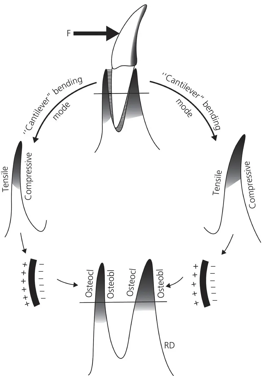
Figure 2.19 Behavior of bone during orthodontic tooth movement. The net force, compression, and tension applied by the “leading” edge of the tooth deforms the alveolar bone convexly toward the root. At the “trailing” edge, the periodontal fibers distort the alveolar bone, producing concavity toward the root. Areas that have been described as characterized by osteoblastic activity were electronegative and, conversely, areas of positivity of electrical neutrality were observed in regions characterized by osteoclasia.
(Source: Zengo et al ., 1973. Reproduced with permission of Elsevier.)
Bioelectric signals in orthodontic tooth movement
In 1957, Fukada and Yasuda published the results of their systematic investigation on dry specimens cut from human and bovine long bones, in an article titled “On the piezoelectric effect of bone,” which credited them with the discovery of the existence of piezoelectricity in bone. They demonstrated that dry bone under proper load application generates surface charges, called piezoelectric currents. They established that the piezoelectric effect appears only when shearing force is applied to the collagen fibers in the bone, which are highly oriented, to make them slip past each other. There exist two types of piezoelectric effects – positive and negative. The former is due to strains generated within the crystal lattice of a material, leading to the production of a potential difference across the faces of that crystal and the latter, when an electric charge is passed across a molecule or crystal and leads to an inherent strain within that molecule (Isaacs, 1987). Both effects involve the organic molecules of collagen and the inorganic crystals of hydroxyapatite (McDonald, 1993). Bassett and Becker (1962) extended that research and discovered that the charges emanating from the bone surface at the time of bending are proportional to the internal strains engendered by the bending. They also showed that the polarization sign always depended upon the type of stress – there was a positive sign where there is tension and a negative sign where there is compression. These experiments were further developed by Shamos et al . (1963) and Shamos and Lavine (1964), who reported finding this phenomenon in a number of different bones, in different anatomical sites and species. They suggested that local electric fields resulting from these surface changes influence the deposition of ions and polarizable molecules.
The first observations of the piezoelectric phenomenon in wet and living bone was made by Bassett (1968), and this finding has contributed to the working hypothesis that piezoelectricity leads to a physical explanation of Wolff ’s law. Following this discovery, the universal existence of piezoelectricity in biological tissues was demonstrated by Fukada and Hara (1969) through their experiments on trachea, aorta, intestines, ligaments, and venous vessels. Marino and Becker (1975) reported on the piezoelectric characteristics of collagen, and concluded that these effects originate in tropocollagen molecules, or in molecules no larger than 50 Å in diameter. However, the hypothesis claiming that piezoelectricity is a major determinant of bone remodeling is weakened by the following:
The generated electric potential is dependent on a strain gradient, and this was not taken into account when the hypothesis was proposed. Bone always experiences nonhomogenous deformation because of its centro‐symmetric nature, and because it can produce electrical polarization proportional to the strain gradient.
The modulus of elasticity (E) of cortical bone under physiologic conditions is frequency dependent. Hence, bone cannot be considered as an elastic‐plastic material.
End‐for‐end rotation of the sample in cantilever bending mode does not change the sign of generated potential as would be expected from classical piezoelectric material.
Proffit (2013) outlined two unusual properties of piezoelectricity, which do not seem to correlate well with OTM:
A quick decay rate, where the electron transfer from one area to another following force application reverts back when the force is removed, which does not or should not happen once orthodontic treatment is over.
Production of an equivalent signal in the opposite direction upon force removal.
Anderson and Erikkson (1968) challenged the piezoelectric hypothesis and reported that although dry collagen is strongly piezoelectric, full hydrated collagen is not, because of the structured water it contains. They argued that bone is a tissue with high symmetry, as the hydroxyapatite it contains is centrosymmetric in nature, and not piezoelectric. Follow‐up experiments conducted by these investigators (1970), using a similar apparatus to the one used by Fukada and Yasuda in 1957, proved that the piezoelectric coefficients varied with the state of hydration of bone, and that the variation decreased as the specimens were dried by evaporation. The loss of piezoelectricity in fully hydrated tendon collagen was explained as attainment of more symmetrical, nonpiezoelectric structure by absorption of water. Instead, the electrical signals generated when stress is applied to fully wet collagen are actually streaming potentials.
The electrokinetic phenomenon known as streaming potential or streaming current was described by many authors, like Glasstone, Overbeek, and Kortum (Gross and Williams, 1982). Anderson and Eriksson (1968) reintroduced the concept against the theoretical faults of the piezoelectric effect, and reported it to be present in bone as it is a porous tissue containing a fluid phase and calcified matrix (which is composed of inorganic (mainly hydroxyapatite) and organic (collagen) contents). Further, the porosity of bone contains membrane‐lined capillary vessels helping in the transportation of nutrition to the inside of bone. There exists a compartmental model for the bone fluid system whereby extracellular fluid in the calcified matrix is separated from membrane‐lined vascular channels. When the bone is mechanically stressed, as part of different loading conditions, or therapeutically induced as in orthodontic treatment, remodeling changes are initiated by the bioelectric potentials generated from inside of the bone. The presence of bioelectric potentials in bone matrix, which also contains extracellular ionic fluid, will generate a charge separation at the matrix–fluid interface. There will be attraction of opposite‐charged molecules at the matrix–fluid interface and repelling of similar charged molecules away from this interface. When the fluid is forced through a bone plug, the ionic charges in the fluid phase near the fluid–matrix interface are carried towards the low pressure end, which is otherwise known as Poiseuille flow. This flow constitutes the streaming current and accumulation of charges set up as an electrical field. This field will result in generation of conduction currents which flow in opposite directions through the bulk of liquid in the porous structure of bone. In steady state the conduction current is equal to the streaming current. The resulting electrostatic potential difference between these two sides of bone plug is known as streaming potential ( Figure 2.20) (Walsh and Guzelsu, 1993). Briefly, streaming potential is the electric potential developed between two components by an electrolyte flowing between the solid surfaces. The movement of the electrostatic double layer at the fluid bone interface, created through the net surface charge gained by the bone surface in contact with an ionic fluid, generates this effect (Anderson and Eriksson, 1968). In bone, streaming potentials can be observed in vascular channels, Haversian systems, canaliculi, and microporosities of the structure due to blood flow and interstitial fluid movement. Neutrality is restored following equilibration of ion distribution.
Читать дальшеИнтервал:
Закладка:
Похожие книги на «Biological Mechanisms of Tooth Movement»
Представляем Вашему вниманию похожие книги на «Biological Mechanisms of Tooth Movement» списком для выбора. Мы отобрали схожую по названию и смыслу литературу в надежде предоставить читателям больше вариантов отыскать новые, интересные, ещё непрочитанные произведения.


Обсуждение, отзывы о книге «Biological Mechanisms of Tooth Movement» и просто собственные мнения читателей. Оставьте ваши комментарии, напишите, что Вы думаете о произведении, его смысле или главных героях. Укажите что конкретно понравилось, а что нет, и почему Вы так считаете.