Biological Mechanisms of Tooth Movement
Здесь есть возможность читать онлайн «Biological Mechanisms of Tooth Movement» — ознакомительный отрывок электронной книги совершенно бесплатно, а после прочтения отрывка купить полную версию. В некоторых случаях можно слушать аудио, скачать через торрент в формате fb2 и присутствует краткое содержание. Жанр: unrecognised, на английском языке. Описание произведения, (предисловие) а так же отзывы посетителей доступны на портале библиотеки ЛибКат.
- Название:Biological Mechanisms of Tooth Movement
- Автор:
- Жанр:
- Год:неизвестен
- ISBN:нет данных
- Рейтинг книги:5 / 5. Голосов: 1
-
Избранное:Добавить в избранное
- Отзывы:
-
Ваша оценка:
Biological Mechanisms of Tooth Movement: краткое содержание, описание и аннотация
Предлагаем к чтению аннотацию, описание, краткое содержание или предисловие (зависит от того, что написал сам автор книги «Biological Mechanisms of Tooth Movement»). Если вы не нашли необходимую информацию о книге — напишите в комментариях, мы постараемся отыскать её.
delivers a comprehensive reference for orthodontic trainees and specialists.
It is fully updated to include new chapters on personalized orthodontics as well as the inflammatory process occurring in the dental and paradental tissues. It is heavily illustrated throughout, making it easier for readers to understand and retain the information discussed within. The topics covered range from bone biology, the effects of mechanical loading on tissues and cells, genetics, tissue remodeling, and the effects of diet, drugs, and systemic diseases.
The Third Edition of
features seven sections that cover subjects such as:
The development of biological concepts in orthodontics, including the cellular and molecular biology behind orthodontic tooth movement Mechanics meets biology, including the effects of mechanical loading on hard and soft tissues and cells, and biological reactions to temporary anchorage devices Inflammation and orthodontics, including markers for tissue remodeling in the gingival crevicular fluid and saliva Personalized diagnosis and treatment based on genomic criteria, including the genetic influences on orthodontic tooth movement Rapid orthodontics, including methods to accelerate or decelerate orthodontic tooth movement Perfect for residents and PhD students of orthodontic and periodontal programs,
is also useful to academics, clinicians, bone biologists, and researchers with an interest in the mechanics and biology of tooth movement.
Biological Mechanisms of Tooth Movement — читать онлайн ознакомительный отрывок
Ниже представлен текст книги, разбитый по страницам. Система сохранения места последней прочитанной страницы, позволяет с удобством читать онлайн бесплатно книгу «Biological Mechanisms of Tooth Movement», без необходимости каждый раз заново искать на чём Вы остановились. Поставьте закладку, и сможете в любой момент перейти на страницу, на которой закончили чтение.
Интервал:
Закладка:
Zeta potential, the common link among different electrokinetic potentials and the one used to allow comparison of different measuring techniques, is defined as average potential difference between the bulk and surface of shear. Surface of shear is an imaginary surface present in the area adjacent to electrically charged bone matrix, where the ions and fluid molecules remain stationary. The role of zeta potential is to separate the movement of ions bound to the solid surface from other ions that show normal viscous behavior under mechanical force application (Lech and Iwaniec, 2010). Zeta potential can be calculated from streaming potential experiments by knowing the applied pressure difference across the sample and generated streaming potentials (Hunter, 1981). Fluid conductivity and fluid viscosity determines the stress‐generated potentials in fluid‐filled bone, and it is possible to calculate the potential generated by the distortion of a fluid by the formula (McDonald, 1993):
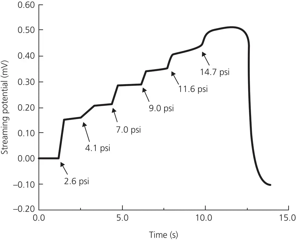
Figure 2.20 Results of a typical intact bone‐streaming potential (mV) in pH 7.3, 0.145 M ionic strength buffer (physiologic conditions) versus time at various pressures (kPa). The arrows indicate when an increase in pressure (nitrogen gas) was placed on the sample. Streaming potential magnitude increased with an increase in pressure and a stable streaming potential was obtained. A positive streaming potential versus pressure response corresponds to a negative zeta potential and an exposed organic interface. Streaming potentials were consistently positive throughout all pressure levels in 0.145–0.6 M NaCl.
(Source: Walsh and Guzelsu, 1993. Reproduced with permission of Elsevier.)

where z is the zeta potential; V is the magnitude of the potential; δ P is the pressure difference that forces the liquid through the channel; ε is the dielectric constant of the liquid; n is the viscosity of the liquid; σ is the specific conductance.
Scientists working in this field have concluded that piezoelectricity and streaming potentials are two coexisting phenomena and both play an important role in stress information transmission. They combined both phenomena as a bioelectric hypothesis, which states that structural alterations throughout the bone are conducted and/or triggered by ionic charge differences (Masella and Chung, 2008). Stress‐generated electric potentials were first reported in dog mandibles and teeth by Cochran, Pawluk and Bassett (1968). They postulated that these stress‐induced bioelectric potentials play a major role in regulating the orientation and functional demands of bone per se. Zengo et al . (1973) measured the electric potential in mechanically stressed dog alveolar bone during in vivo and in vitro experiments. They have demonstrated that the concave side of orthodontically treated bone is electronegative and favors osteoblastic activity, whereas the areas of positivity or electrical neutrality, i.e., convex surfaces, showed elevated osteoclastic activity ( Figure 2.19). Shapiro, Roeber, and Klempner (1979) suggested that application of piezoelectric charges as pulses of force to the teeth can accelerate the osteogenic response. Based on their experiment in one patient, with application of 8 ounces of force, they reported increase in the rate of movement of maxillary second molar with less pain perception than that of the control teeth.
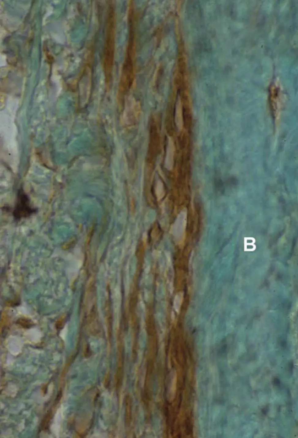
Figure 2.21 Transverse section, 6 μm thick, of a 1‐year‐old female cat’s mandible, after a 7‐day exposure to sham electrodes (control). Shown is the buccal periosteum of the second premolar opposite the sham cathode, stained immunohistochemically for cAMP. B, Alveolar bone. The bone surface lining cells are flat, and most stain lightly for cAMP.
It has been proposed by Davidovitch et al . (1980a, b) that a physical relationship exists between mechanical and electrical perturbation of bone. Their experiments in female cats with administration of exogenous electrical currents in conjunction with orthodontic forces demonstrated enhanced cellular activities in the PDL and alveolar bone, as well as rapid tooth movement (Figures ). Taken together, these findings led to the suggestion that bioelectric responses (piezoelectricity and streaming potentials) propagated by bone bending incident to orthodontic force application, might act as pivotal cellular first messengers.
Borgens (1984) investigated this phenomenon in bone fracture sites by inducing electric current for healing purposes. His experiments did not disclose any correlation with what had been proposed as piezoelectric effects and showed that the dispersion of current as it enters the lesion is unpredictable. He attributed this finding to the complexity of distribution of mineralized and nonmineralized matrices. However, he observed generation of endogenous ionic currents evoked in intact and damaged mouse bones, and classified these currents as stress‐generated potentials or streaming potentials, rather than piezoelectric currents. In contrast to piezoelectric spikes, the streaming potentials were having long decay periods. This finding led him to hypothesize that the mechanically stressed bone cells themselves, not the matrix, are the source of the electric current. His hypothesis received support from Pollack et al . (1984), who proposed a mechanism by which force‐evoked electric potentials may reach the surface of bone cells. Accordingly, an electric double layer surrounds bone, where electric charges flow in coordination with stress‐related fluid flow. These stress‐generated potentials may affect the charge of cell membranes, and of macromolecules present in the neighborhood. Davidovitch et al . (1980a, b) suggested that piezoelectric potentials result from distortion of fixed structures of the periodontium, such as collagen, hydroxyapatite, or bone cells’ surface. In hydrated tissues, however, streaming potentials (the electrokinetic effects that arise when an electrical double layer overlying a charged surface is displaced) predominate as the interstitial fluid moves. They further reported that mechanical perturbations of the order of about 1 min/day are apparently sufficient to cause an osteogenic response ( Figure 2.24), perhaps due to matrix proteoglycan related strain memory.
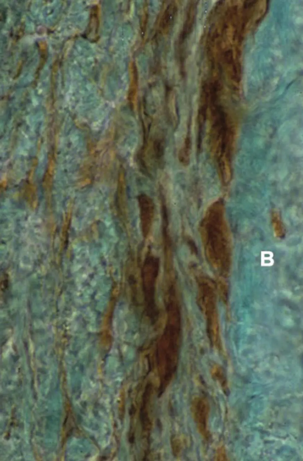
Figure 2.22 Transverse section, 6 μm thick, of a 1‐year‐old female cat’s mandible (the same animal as shown in Figure 2.21), after exposure for 7 days to a constant application of a 20 μA direct current to the gingival mucosa, noninvasively. Shown are the tissues near the stainless‐steel cathode, stained immunohistochemically for cAMP. B, Alveolar bone. Compared with the cells shown in Figure 2.21, the bone surface lining cells near the cathode are larger and more darkly stained for cAMP.
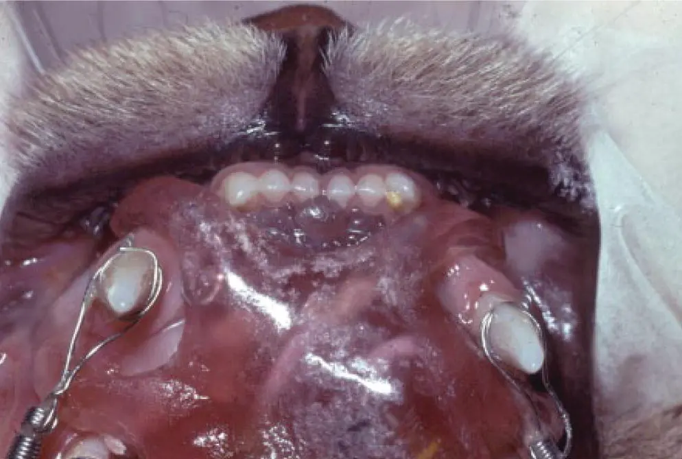
Figure 2.23 Constant direct current, 20 μA, noninvasively, to the gingival and oral mucosa labial to the left maxillary canine in a cat. The right canine (control) received the same electrodes, but without electrical current. Both canines were moved distally by an 80 g tipping force. The right canine, which had been subjected only to mechanical force, moved distally a smaller distance than the left canine, which had been administered a combination of mechanical force and electrical current.
Читать дальшеИнтервал:
Закладка:
Похожие книги на «Biological Mechanisms of Tooth Movement»
Представляем Вашему вниманию похожие книги на «Biological Mechanisms of Tooth Movement» списком для выбора. Мы отобрали схожую по названию и смыслу литературу в надежде предоставить читателям больше вариантов отыскать новые, интересные, ещё непрочитанные произведения.


Обсуждение, отзывы о книге «Biological Mechanisms of Tooth Movement» и просто собственные мнения читателей. Оставьте ваши комментарии, напишите, что Вы думаете о произведении, его смысле или главных героях. Укажите что конкретно понравилось, а что нет, и почему Вы так считаете.