Richard J. Miron - Understanding Platelet-Rich Fibrin
Здесь есть возможность читать онлайн «Richard J. Miron - Understanding Platelet-Rich Fibrin» — ознакомительный отрывок электронной книги совершенно бесплатно, а после прочтения отрывка купить полную версию. В некоторых случаях можно слушать аудио, скачать через торрент в формате fb2 и присутствует краткое содержание. Жанр: unrecognised, на английском языке. Описание произведения, (предисловие) а так же отзывы посетителей доступны на портале библиотеки ЛибКат.
- Название:Understanding Platelet-Rich Fibrin
- Автор:
- Жанр:
- Год:неизвестен
- ISBN:нет данных
- Рейтинг книги:5 / 5. Голосов: 1
-
Избранное:Добавить в избранное
- Отзывы:
-
Ваша оценка:
Understanding Platelet-Rich Fibrin: краткое содержание, описание и аннотация
Предлагаем к чтению аннотацию, описание, краткое содержание или предисловие (зависит от того, что написал сам автор книги «Understanding Platelet-Rich Fibrin»). Если вы не нашли необходимую информацию о книге — напишите в комментариях, мы постараемся отыскать её.
Understanding Platelet-Rich Fibrin — читать онлайн ознакомительный отрывок
Ниже представлен текст книги, разбитый по страницам. Система сохранения места последней прочитанной страницы, позволяет с удобством читать онлайн бесплатно книгу «Understanding Platelet-Rich Fibrin», без необходимости каждый раз заново искать на чём Вы остановились. Поставьте закладку, и сможете в любой момент перейти на страницу, на которой закончили чтение.
Интервал:
Закладка:
PRF, on the other hand, is a particularly simple and inexpensive way to utilize the three principles of tissue engineering by utilizing a 3D scaffold (fibrin) that incorporates both regenerative host cells (platelets and leukocytes) and various GFs. These include PDGF, TGF-β, and VEGF, each of which is crucial during the regeneration process. Furthermore, the concentrated leukocytes (as opposed to simply platelets) in PRF have been well implicated as key regulators of tissue healing and formation. 26–28,31,38
Snapshot of PRF
PRF is considered a second-generation platelet concentrate with a longer GF release profile.
Centrifugation protocols are shorter and do not need any chemical additives such as anticoagulants.
PRF falls more in line with tissue engineering principles in that it is not only an accumulation of cells and GFs but also a scaffold (fibrin matrix).
PRF incorporates leukocytes, which are key cells in pathogen defense and biomaterial integration.
A-PRF and i-PRF (2014–2018)
While much of the research performed in the late 2000s and early 2010s was dedicated to the clinical uses and indications of L-PRF discussed later in this textbook, major discoveries were made several years later from basic research laboratories. Following extensive clinical use and research with the original L-PRF protocol, it was discovered in 2014 by Dr Shahram Ghanaati that centrifugation carried out at relatively high centrifugation speeds (~700g) led to the great majority of leukocytes being located either at the buffy coat zone (between the red blood cell layer and the upper plasma layer) or more commonly at the bottom of centrifugation tubes ( Fig 1-6). 39It was expressed that the longer the centrifugation time is carried out, the more likely it is that cells get pushed further down the centrifugation tube. Similarly, the faster the spin centrifugation speed (higher g-force), the greater the proportion of cells found in the lower levels of centrifugation tubes.
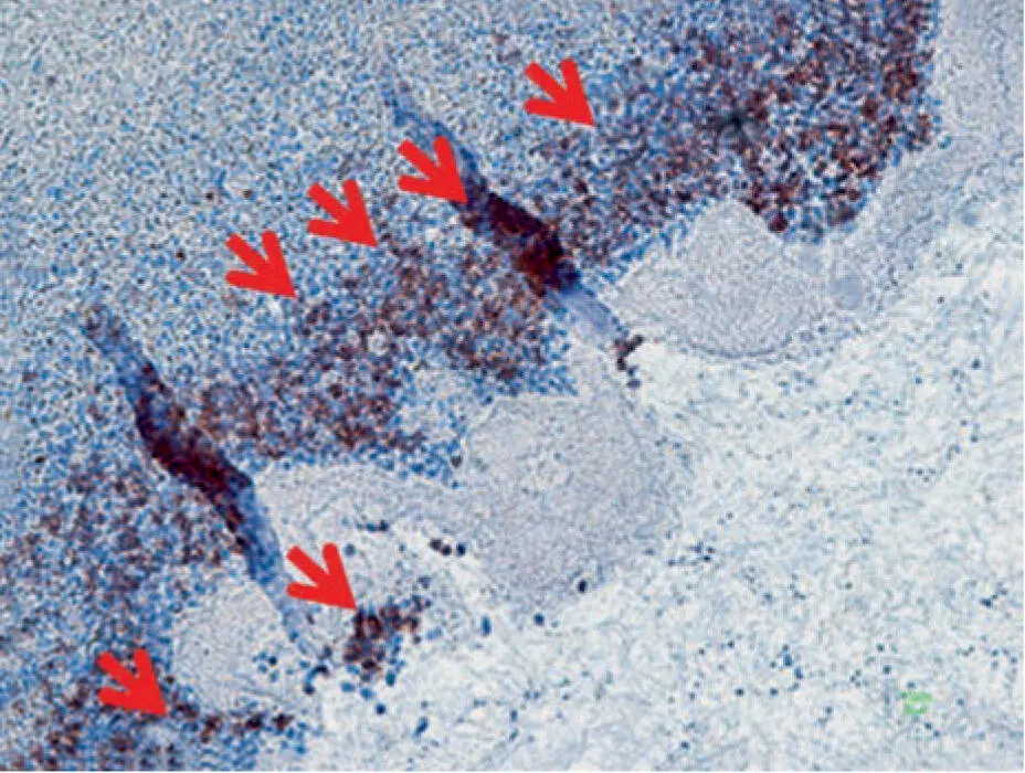
Fig 1-6Histologic observation of leukocytes following centrifugation. Resulting white blood cells have been shown to be contained basically in the layers between the plasma PRF layer and the red blood cell clot. This finding demonstrated quite clearly that the g-force was excessive, necessitating the development of newer protocols aimed to improve the retention of leukocytes within the PRF matrix. (Reprinted with permission from Ghanaati et al. 39)
Pioneering research within his laboratory led to the development of an advanced PRF (A-PRF) whereby lower centri-fugation speeds (~200g) led to a higher accumulation of platelets and leukocytes more evenly distributed throughout the upper PRF layers. These newer protocols more favorably led to a higher release and concentration of GFs over a 10-day period when compared to PRP or L-PRF. 19In 2015 to 2017, our research team further demonstrated that optimization of PRF could be achieved by reducing not only centrifugation speed but also the time involved. The A-PRF protocol was therefore modified from 14 minutes at 200g as originally described in 2014 down to an 8-minute protocol. 19
Following an array of basic research studies on this topic, it was observed that by further reducing the g-force and also the time, it was possible to obtain a plasma layer that had not yet converted into fibrin (ie, scientifically liquid fibrinogen but often referred to as liquid-PRF for simplicity). In a study titled “Injectable platelet rich fibrin (i-PRF): Opportunities in regenerative dentistry?”, 20it was demonstrated that at lower centrifugation speeds and times (~60g for 3 minutes), a liquid-PRF (termed injectable-PRF or i-PRF ) could be obtained. While these protocols typically produced minimal volumes (~1.0–1.5 mL), it was shown that both platelets and leukocytes were even more highly concentrated when compared to L-PRF or A-PRF ( Fig 1-7). 40This liquid-PRF layer could be utilized clinically for approximately 15 to 20 minutes, during which time fibrinogen and thrombin had not yet converted to a fibrin matrix (ie, remained liquid). This has since been utilized for injection into various joints/spaces similar to PRP, however with the reported advantages of a longer GF release time. Furthermore, the concept of “sticky” bone was also developed. Importantly, a different type of tube (plastic) was needed to minimize clotting, as will be discussed in detail in chapter 5.
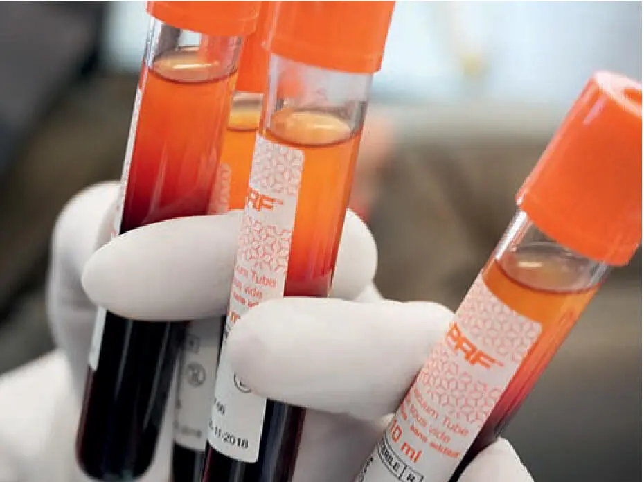
Fig 1-7Newer centrifugation protocols allow production of a liquid formulation of PRF found in the top 1- to 2-mL layer of centrifugation tubes following a 3- to 5-minute protocol. This liquid can be collected in a syringe and reinjected into defect sites or mixed with biomaterials to improve their bioactive properties. (Reprinted with permission from Davies and Miron. 40)
Snapshot of A-PRF and i-PRF
Original L-PRF protocols were shown to be too fast, leading to all the cells being accumulated only at the buffy coat zone, with the majority of leukocytes found within the red blood cell layer.
The low-speed centrifugation concept was shown in 2014 to favor a higher concentration of cells within PRF membranes.
By further lowering speed and time, a liquid-PRF formulation became available, commonly known as injectable-PRF (or i-PRF).
H-PRF and C-PRF (2019–Present)
Very recently, our research group discovered through a series of basic laboratory experiments that horizontal centrifugation led to significantly greater concentrations of platelets and leukocytes when compared to currently available fixed-angle centrifugation devices most commonly utilized to produce L-PRF and A-PRF. Simply, horizontal centrifuges are routinely utilized in high-end research laboratories as well as in medical hospitals because of their greater ability to separate layers based on density ( Fig 1-8; see also chapters 2and 3). Unlike fixed-angle centrifugation systems whereby the tubes are actually inserted at a 45-degree angle, in horizontal centrifugation systems (often referred to as swing-out bucket centrifugation ), the tubes have the ability to swing out to 90 degrees once they are in rotation ( Video 1-2). Amazingly, the original PRP systems developed by Harvest and Marx utilized and still use this technology.
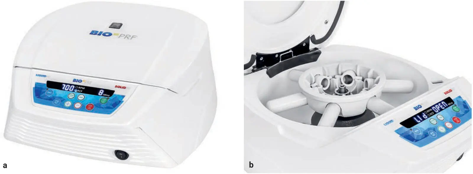
Fig 1-8 (a) Clinical photograph of a Bio-PRF centrifuge. (b) Photograph demonstrating the horizontal centrifugation concept. The tubes are inserted vertically (up and down), but once the device begins to rotate, the tubes swing out completely horizontally. This favors better blood cell layer separation with higher platelet and GF concentrations.

 Video 1-2
In 2019, an article on the topic demonstrated clearly that horizontal centrifugation could lead to up to a four-times greater cell content when compared to fixed-angle centrifugation. 41This represented a marked ability to greatly concentrate cells found within PRF, which were primarily being accumulated on the back distal surfaces of PRF tubes ( Fig 1-9). The major disadvantage of fixed-angle centrifugation is that during the spin cycle, cells are typically driven along the back wall of the centrifugation tubes at high g-forces ( Fig 1-10). This also exposes cells to higher compressive forces against the back wall, and cells must then separate by traveling either up or down the inclined centrifugation slope based on their respective cell density differences. Because red blood cells are larger and heavier than platelets and leukocytes, they travel downward, whereas lighter platelets travel toward the top of the tube where PRF is collected. This makes it relatively difficult for the small cell types such as platelets and leukocytes to reach the upper layer, especially granted that red blood cells outnumber in particular white blood cells typically by 1,000-fold (see chapter 2). Therefore, it is not possible to reach optimal accumulation of platelets or leukocytes using a fixed-angle centrifuge.
Читать дальшеИнтервал:
Закладка:
Похожие книги на «Understanding Platelet-Rich Fibrin»
Представляем Вашему вниманию похожие книги на «Understanding Platelet-Rich Fibrin» списком для выбора. Мы отобрали схожую по названию и смыслу литературу в надежде предоставить читателям больше вариантов отыскать новые, интересные, ещё непрочитанные произведения.


Обсуждение, отзывы о книге «Understanding Platelet-Rich Fibrin» и просто собственные мнения читателей. Оставьте ваши комментарии, напишите, что Вы думаете о произведении, его смысле или главных героях. Укажите что конкретно понравилось, а что нет, и почему Вы так считаете.